J Korean Soc Radiol 2016;74(6):412-416 http://dx.doi.org/10.3348/jksr.2016.74.6.412
INTRODUCTION
Retroperitoneal fibrosis (RPF) is an uncommon fibrotic reac- tion in the retroperitoneum. There is an idiopathic form and a secondary form associated with IgG4-related disease, drugs (me- thysergide, pergolide, bromocriptine, ergotamine, methyldopa, hydralazine), infections (tuberculosis, histoplasmosis, actinomy- cosis), malignancies (carcinoid tumor, Hodgkin’s and non-Hodg- kin’s lymphomas, sarcomas, carcinomas of the colon, prostate, breast, and stomach), prior surgeries, and radiation therapy. The idiopathic form accounts for more than two-thirds of cases and it was thought to result from a local inflammatory response to anti- gens in the atherosclerotic plaques in the abdominal aorta, com- bined with auto-immunologic factors. Thus, the fibroinflamma- tory tissue usually encases the infrarenal portion of the abdominal aorta, inferior vena cava, and ureters (1-3). Here, we report two rare cases of idiopathic RPF involving only a unilateral renal pel-
vis and sinus; thus mimicking renal pelvic cancer.
CASE REPORT
Case 1
A 66-year-old female presented with an incidental finding at computed tomography (CT) screening. Her previous medical history revealed that she had undergone hysterectomy for an un- known cause 10 years ago. Contrast-enhanced genitourinary CT showed a homogeneously enhancing soft tissue mass replacing the left renal pelvis, which was suspicious for renal pelvic cancer.
The renal pelvis was slightly dilated, but ureteric dilatation was not present (Fig. 1A, B).
Urine cytologies of voided urine and washed urine from the left renal pelvis were negative.
On retrograde pyelography (RGP) performed by urologists, there was no mass-like lesion and biopsy of the left renal pelvis
Idiopathic Retroperitoneal Fibrosis Involving a Unilateral Renal Sinus:
A Case Report and Literature Review
일측 신장 신우만 침범한 특발성 복막후섬유증: 증례 보고와 문헌 고찰
Seul-Bi Lee, MD, Jung-Hee Yoon, MD*, Seung-Ho Kim, MD, Yedaun Lee, MD, Suk-Jung Kim, MD, Yun-Jung Lim, MD, Hyun-Kyung Jung, MD, Jin-Soo Lee, PhD
Department of Radiology, Haeundae Paik Hospital, Inje University College of Medicine, Busan, Korea
Idiopathic retroperitoneal fibrosis (RPF) is a rare disease entity and its etiology is uncertain. We report two similar cases which showed an uncommon presentation of idiopathic RPF. A 66-year-old woman and an 80-year-old man presented with incidental findings of left renal pelvic mass-like lesions. Computed tomography re- vealed a soft tissue density mass replacing the left renal pelvis, which was suspi- cious for renal pelvic cancer, and the diagnosis of idiopathic RPF was surgically confirmed. To the best of our knowledge, a few cases of idiopathic RPF presenting with features of a localized unilateral renal pelvic mass mimicking renal pelvic can- cer have been reported.
Index terms
Retroperitoneal Fibrosis Tomography, X-Ray Computed Kidney
Received November 13, 2015 Revised December 5, 2015 Accepted January 28, 2016
*Corresponding author: Jung-Hee Yoon, MD Department of Radiology, Haeundae Paik Hospital, Inje University College of Medicine, 875 Haeun-daero, Haeundae-gu, Busan 48108, Korea.
Tel. 82-51-797-0355 Fax. 82-51-797-0379 E-mail: [email protected]
This is an Open Access article distributed under the terms of the Creative Commons Attribution Non-Commercial License (http://creativecommons.org/licenses/by-nc/3.0) which permits unrestricted non-commercial use, distri- bution, and reproduction in any medium, provided the original work is properly cited.
also yielded unsatisfactory results. She underwent laparoscopic radical nephrectomy. Histopathologic findings of the resected kidney were negative for malignancy. Fibrosis and chronic in- flammation with myofibroblast proliferation and lymphoplasma cell infiltration were observed. The lesion was mainly located in the renal sinus fat tissue around renal vessels, suggesting an in- flammatory or immunologic process such as idiopathic RPF (Fig.
1C). Immunochemistry of IgG and IgG4 was performed to con- firm the presence of IgG4+ plasma cells in the tissue. The ratio of IgG4 to IgG plasma cells was within the normal range (< 5%), and these results were inconsistent with the diagnosis of IgG4-re- lated disease, which shows an IgG4 to IgG ratio of more than 30- 40%. Pathology report was compatible with idiopathic RPF.
Case 2
An 80-year-old man presented with fever and cough for one week. His past history was nonspecific except for hypertension.
There was no abnormal finding on physical examination. The re- sults of peripheral blood smear examination showed slightly ele- vated C-reactive protein (2.93 mg/dL). On chest CT, mild bron- chitis and the incidental finding of a left renal pelvic mass were detected. Additional contrast enhanced genitourinary CT was performed for further evaluation. CT showed an enhancing soft tissue mass in the left renal pelvis and sinus with mild pelvic dila- tation. Periureteric fat infiltration in the left proximal ureter without definite wall thickening was observed (Fig. 2A, B).
Two urine cytologies of washed urine from the left renal pelvis
were negative, and on RGP, there was no filling defect but mild luminal narrowing of the renal pelvis and mild calyectasia were detected (Fig. 2C). He underwent laparoscopic radical nephrec- tomy. Histopathologic findings were consistent with idiopathic RPF (Fig. 2D). Stromal fibrosis with lymphoplasma cell infiltra- tion mainly into the renal sinus was observed. On immuno- chemistry of the tissue, the ratio of IgG4 to IgG plasma cells was less than 5%, which was within the normal range, and on labora- tory examination, serum levels of IgG and IgG4 were within their normal range [1056.6 mg/dL (range, 700-1600 mg/dL) and 1020.0 mg/L (range, 30-2010 mg/L), respectively].
DISCUSSION
RPF is a rare autoimmune, inflammatory disorder that devel- ops in about one in 200000 people (4). It is most common in in- dividuals 40 to 60 years of age (5).
Men are affected by this disorder two to three times more often than women (1). In typical RPF, a mass of fibrous tissue develops around the abdominal aorta with an epicenter at the L4-5 level and it also encases the inferior vena cava and the ureter. Ureteral involvement is reported in 80-100% of cases at presentation (4).
At presentation, such ureteral involvement is often bilateral, but in patients with an apparently unilateral obstruction, contralater- al disease can develop even within a short period (1).
CT is the modality of choice for imaging diagnosis of RPF and for evaluating the extent of the process. On CT scan, RPF usually
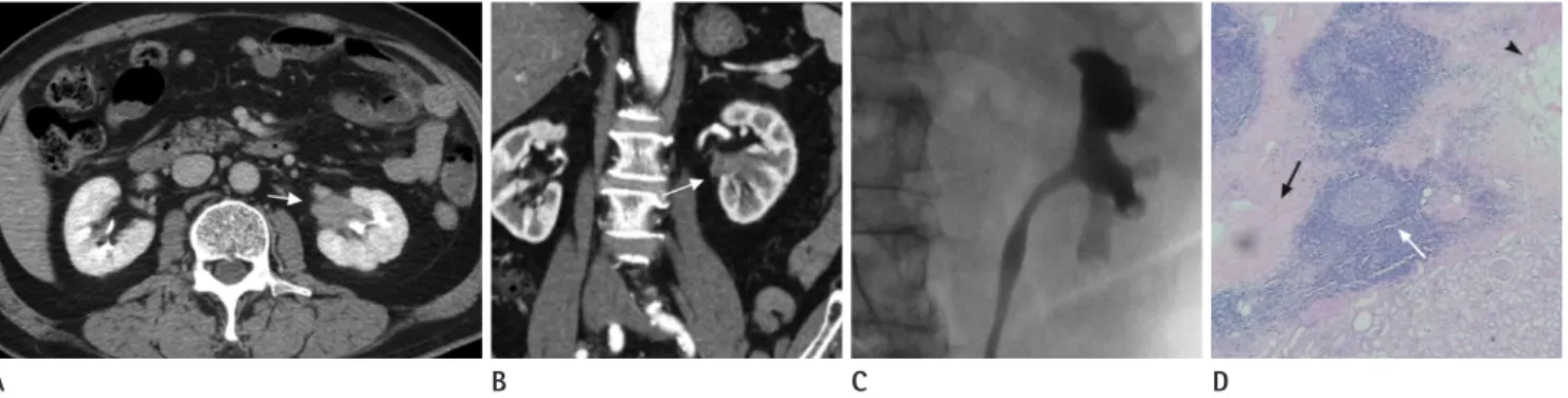
Fig. 1. A 66-year-old woman who underwent left nephrectomy due to idiopathic retroperitoneal fibrosis mimicking urothelial carcinoma.
A, B. Contrast enhanced CT scan axial image in the nephrographic phase (A), and coronal image in the corticomedullary phase (B) show a ho- mogeneously enhancing soft tissue mass (arrows) replacing the left renal pelvis.
C. Microscopic examination of the resected kidney specimen shows diffuse fibrosis (black arrow) and chronic inflammation with myofibroblast proliferation (arrowhead) and lymphoplasma cell infiltrates (white arrow) mainly located in the renal sinus fat tissue (hematoxylin and eosin stain, × 40).
A B C
appears as a homogeneous plaque, which is isodense to the adja- cent muscle, surrounding the lower abdominal aorta and the iliac arteries, and often enveloping the ureters and the inferior vena cava (4). The degree of contrast enhancement is related to in- flammation and process activity.
Intense enhancement indicates fibrous tissue in an initial phase with associated inflammation (6). The advantage of MR imaging is avoidance of nephrotoxicity of iodinated contrast media in pa- tients with compromised renal function and better definition against the surrounding tissues, mainly when fat-saturation im- ages are used.
Idiopathic RPF is hypointense in T1-weighted MR images. In T2-weighted MR images, its intensity is variable; high signal in- tensity in the active inflammatory stage because of tissue edema and hypercellularity and low signal intensity in the late fibrotic stage (5-7).
In rare cases, RPF presents as an asymmetric poorly circum- scribed retroperitoneal mass with atypical localization, and thus it is not characterized by aortic involvement as observed in our cases, and differentiation from a primary retroperitoneal tumor or malignant lymphadenopathy is very difficult. Atypical sites of RPF involvement include the small bowel mesentery, duodenum,
Fig. 2. An 80-year-old man with the incidental finding of a left renal pelvic mass-like lesion.
A, B. Contrast enhanced CT scan axial images in the nephrographic phase (A), and coronal image in the corticomedullary phase (B) show an en- hancing soft tissue mass (arrows) in the left renal pelvis and sinus with mild pelvic dilatation. Periureteric fat infiltration in the left proximal ure- ter without definite wall thickening is noted.
C. Retrograde pyelography shows suspected luminal narrowing of the renal pelvis and mild calyectasia, but there is no definite abnormal filling defect.
D. Microscopic examination of the resected kidney specimen shows diffuse stromal fibrosis (black arrow) with lymphoplasma cell infiltration and reactive lymphoid follicle formation (white arrow), which is compatible with retroperitoneal fibrosis, mainly located in the renal sinus fat tissue (arrowhead) (hematoxylin and eosin stain, × 40).
A B C D
Table 1. Previous Reports of Perirenal Extension of RPF
Year Authors (Ref. No.) Age Sex Perirenal Involvement Periaortic, Pericaval Involvement
1937 Erischar - - Bilateral -
1941–1943 Lebbin - - -
1988 Yancey 35 F Bilateral No involvement
1992 Rominger 60 F Unilateral Involvement
1995 Barret 68 M Unilateral Involvement
1990 Brooks 14 F Unilateral Involvement
1996 Bechtold - - Bilateral No involvement
1999 Ayuso et al. (7) 53 F Bilateral No involvement
2002 Triantopoulou 51 F Unilateral Involvement
2005 Ergen et al. (6) 51 M Unilateral Involvement
2012 Ellimoottil 62 F Bilateral No involvement
2013 Sofiane 64 M Bilateral No involvement
2013 Yoshino et al. (8) 71 M Unilateral No involvement
2014 Weining 51 M Unilateral No involvement
2015 Lang et al. (9) 45 M Unilateral No involvement
RPF = retroperitoneal fibrosis
colon, bladder, and periduodenal, peripancreatic, pelvic, periure- teral and epidural space (1, 4).
Perirenal extension of RPF is rarely reported (6-10), whereas in only a few cases, the disease was unilaterally localized. To the best of our knowledge, three cases of idiopathic RPF presenting with features of a localized unilateral renal pelvic mass mimicking re- nal pelvic cancer (8, 9) without periaortic or pericaval involve- ment have been reported (Table 1).
IgG4-related kidney disease is an important part of the spec- trum of IgG4-related sclerosing disease, which manifests as a sec- ondary form of RPF. IgG4-related kidney disease is usually ac- companied by other organ involvement, and isolated IgG4-related kidney disease without other organ involvement is very rare; only 6% of cases have been reported as renal lesions alone (10).
Although CT and MR imaging have an important role in diag- nosis of RPF, histological tissue examination is needed, especially in cases with atypical location of a retroperitoneal mass to rule out malignancy. In addition to malignancy (urothelial carcino- ma), differential diagnosis includes retroperitoneal fibromatosis, which is characterized by uniform proliferation of fibroblasts and is associated with Gardner’s syndrome. Another differential diag- nosis is inflammatory pseudotumor, which mainly affects chil- dren, and its appearance is that of a huge mass with infiltrative borders. It is histologically characterized by myofibroblast prolif- eration with myxoid and inflammatory areas (1).
REFERENCES
1. Vaglio A, Salvarani C, Buzio C. Retroperitoneal fibrosis. Lan-
cet 2006;367:241-251
2. Amis ES Jr. Retroperitoneal fibrosis. AJR Am J Roentgenol 1991;157:321-329
3. Vaglio A, Buzio C. Chronic periaortitis: a spectrum of dis- eases. Curr Opin Rheumatol 2005;17:34-40
4. Kottra JJ, Dunnick NR. Retroperitoneal fibrosis. Radiol Clin North Am 1996;34:1259-1275
5. Warakaulle DR, Prematilleke I, Moore NR. Retroperitoneal fibrosis mimicking retrocrural lymphadenopathy. Clin Ra- diol 2004;59:292-293
6. Ergen FB, Arslan EB, Turkbey B, Akinci D, Akata D. Unilateral perirenal fibrosis. J Comput Assist Tomogr 2005;29:477-480 7. Ayuso JR, Garcia-Criado A, Caralt TM, Ayuso C, Torras A,
Ribalta T. Atypical retroperitoneal fibrosis: MRI findings.
Eur Radiol 1999;9:937-939
8. Yoshino T, Moriyama H, Fukushima M, Sanda N. A case of IgG4-related retroperitoneal fibrosis mimicking renal pel- vic cancer. Urol Int 2013;90:365-368
9. Lang JT, Kang N, Zhang JH, Xing NZ. Unilateral perirenal fi- brosis without aorta involvement. Eur Rev Med Pharmacol Sci 2015;19:732-735
10. Seo N, Kim JH, Byun JH, Lee SS, Kim HJ, Lee MG. Immuno- globulin G4-related kidney disease: a comprehensive picto- rial review of the imaging spectrum, mimickers, and clinico- pathological characteristics. Korean J Radiol 2015;16:
1056-1067
일측 신장 신우만 침범한 특발성 복막후섬유증: 증례 보고와 문헌 고찰
이슬비 · 윤정희* · 김승호 · 이예다운 · 김숙정 · 임윤정 · 정현경 · 이진수
특발성 복막후섬유증은 드문 질환으로 그 원인은 아직 잘 알려져 있지 않다. 저자들은 66세 여자 환자와 80세 남자 환자 에서 우연히 발견된 좌측 신장 신우만을 침범한 종괴 형태로 발현된 비전형적 특발성 복막후섬유증을 보고한다. 전산화단 층촬영(computed tomography)에서 좌측 신장의 신우를 대치하는 연조직 음영으로 신장암을 의심하게 하는 종괴였고 수술 로 특발성 복막후섬유증으로 확진되었다. 이와 같이 일측 신장 신우만을 침범하여 신장암과 유사한 형태로 발현되는 특발성 복막후섬유증은 매우 드문 형태로 문헌 고찰과 함께 보고한다.
인제대학교 의과대학 해운대백병원 영상의학과교실