INTRODUCTION
Occupational asthma is induced by many agents in work- places. A number of herbal medicines, e.g., Sanyak (1), Chun- kung (2), Banha (3), and Brazilian ginseng (4) have been report- ed to be causative agents of occupational asthma. Recently, one case report of Sanyak and Korean ginseng-induced occupa- tional asthma was reported (confirmed by bronchial provo- cation testing) (5). Although skin prick testing with Kore- an ginseng extract was weakly positive in this reported case, enzyme-linked immunosorbent assay (ELISA) result for serum anti-Korean ginseng-specific immunoglobulin (IgE) and no IgE binding component was detected by immunoblotting (5). Here, we report a case of Korean ginseng-induced occu- pational asthma with a strong positive skin prick test to ginseng extract and an elevated serum anti-Korean ginseng- specific IgE level. The IgE binding components of Korean ginseng were also identified in this case. To the best of our knowledge, this is the first report to reveal the pathogenic mechanism of Korean ginseng-induced occupational asthma.
MATERIALS AND METHODS Study subjects
In May 2005, a 34-yr-old woman was referred for recur- rent dyspnea and nasal symptoms. The patient had allergic rhinitis for 9 yr. She presented with abdominal pain and diarrhea after eating many different foods (e.g., tomatoes, bananas, oranges, and oysters) and experienced generalized urticaria and angioedema after eating persimmon. Five years previously, she had started working at a Korean ginseng whole- sale premise, where she was exposed to dried ginseng and gin- seng dust. After starting the work, she developed frequent rhinorrhea, sneezing, and nasal obstruction. Six months pre- viously, she had developed dyspnea and wheezing on a daily basis, and these were aggravated at work, but improved after a weekend break. She had no allergic symptoms after ingest- ing non-dried ginseng or steamed red ginseng. During her first visit, she had no symptoms, presumably because she had not been working for a week. Her physical examination was normal, and according to laboratory tests, her peripheral eo-
232
Kyung-Mook Kim*,�, Hyouk-Soo Kwon*,�, Sung-Gyu Jeon�, Chang-Han Park*,�, Seong-Wook Sohn*,�, Duck-In Kim*,�, Sun-Sin Kim*,�, Yoon-Seok Chang*,�,�, Yoon-Keun Kim*,�, Sang-Heon Cho*,�, Kyung-Up Min*,�, and You-Young Kim*,� Department of Internal Medicine*, Seoul National University College of Medicine; Institute of Allergy and Clinical Immunology�, Seoul National University Medical Research Center, Seoul; Department of Internal Medicine�, Seoul National University Bundang Hospital, Seongnam, Korea
Address for correspondence You-Young Kim, M.D.
Department of Internal Medicine, Seoul National University College of Medicine, 28 Yeongeon-dong, Jongno-gu, Seoul 110-744, Korea
Tel : +82.2-2072-3291, Fax : +82.2-742-2912 E-mail : [email protected]
*This study was supported by a grant from the Korean Ministry of Health & Welfare (#03-PJ01-PJ13-GD01-0002
& 0412-CR03-0704-001).
J Korean Med Sci 2008; 23: 232-5 ISSN 1011-8934
DOI: 10.3346/jkms.2008.23.2.232
Copyright � The Korean Academy of Medical Sciences
Korean Ginseng-Induced Occupational Asthma and Determination of IgE Binding Components
A number of case reports on occupational asthma caused by herbal medicines have been issued, for example, on Sanyak, Chunkung, Banha, and Brazilian ginseng.
Recently, cases of occupational asthma induced by Sanyak and Korean ginseng have been reported, but the pathogenic mechanisms involved are unknown. This study was carried out to evaluate the immunologic mechanism underlying Korean ginseng-induced occupational asthma. A patient engaged in Korean ginseng whole- sale was referred for recurrent dyspnea, wheezing, and nasal symptoms, which were aggravated at work. Allergen bronchial provocation testing to Korean ginseng extract showed a typical immediate response, and skin prick testing to Korean ginseng extract also showed a strong positive response. Moreover, serum-specific IgE levels to Korean ginseng extract were significantly higher than in controls. Enzyme- linked immunosorbent assay (ELISA) inhibition tests showed a dose-dependent inhibition by Korean ginseng, but not by Dermatophagoides farinae, wheat flour, or Chinese balloon flower. Sodium dodecylsulfate-poly-acrylamide gel electrophoresis (SDS-PAGE) and immunoblotting revealed four specific Immunoglobulin E (IgE) binding components at 26, 30, 47, and 60 kDa, which were not bound by control sera. These results strongly suggest that occupation asthma induced by Korean ginseng is induced via an IgE-mediated mechanism.
Key Words : Asthma; Hypersensitivity, Immediate; Panax; Immunoglobulin E; Occupational Diseases
Received : 10 January 2007 Accepted : 14 August 2007
Korean Ginseng-Induced Occupational Asthma 233
sinophil count was 572/ L and total IgE 1,050 U/mL. Forced expiratory volume in 1 sec (FEV1) was 3,010 mL (106%) and forced vital capacity (FVC) 3,150 mL (96%), with an FEV1/
FVC ratio of 95.5%. Skin prick testing results were as fol- lows: Dermophagoides farinae (6×7 mm), cat (7×14 mm), wheat flour (3.5×5 mm), barley (4×4 mm), Chinese bal- loon flower (7×14 mm), wheat bran (5×5 mm), alder (3×
5 mm), beech (2×3 mm), oak (2×3 mm), orchard (2×3 mm), and positive control-histamine (4×4.5 mm).
Airway hyperresponsiveness was confirmed by bronchial provocation testing to methacholine (PC20=14.77 mg/mL).
Preparation of ginseng extract and skin prick testing
Ginseng was obtained from the patient’s employer and was prepared as previously described (6). In brief, ginseng was cut into small pieces and extracted into phosphate-buffered saline (PBS; pH 7.5) 1:5 wt/vol at 4℃for 24 hr. The extract was then centrifuged at 15,000 rpm at 4℃for 30 min. The supernatant obtained was dialyzed (at a molecular weight cut-off of 6 kDa) against 4 L of normal saline at 4℃for 48 hr, and this was then used as a crude extract. Extract protein concentrations were determined using the bicinchoninic acid (BCA) assay, accord- ing to the manufacturer’s instructions (Pierce, Rockford, IL, U.S.A.). Extract was used for skin prick tests and specific br- onchial challenge testing.
Specific bronchial challenge with ginseng extract
Normal saline (1 mL) was administered using a DeVil-biss 646 nebulizer (CS & M Instrument Co., Doylestown, PA, U.S.A.) connected to a dosimeter. The subject was asked to breathe the aerosol with tidal breathing, and this was followed by ginseng extract. FEV1 and FVC were measured using a spirometer (Multispiro-SX, Irvine, CA, U.S.A.) before and 10 min after each inhalation, every 10 min during the first hour, and then hourly for the next 8 hr after challenge.
ELISA for specific IgE to ginseng extract and ELISA inhibition testing
The presence of specific IgE antibody to ginseng extract was determined by ELISA, as previously described (6). In brief, microtiter plates (NUNC; immunoplates, Roskilde, Denmark) were coated with 50 L of Korean ginseng extract (100 g/
mL), and then incubated with 50 L of either the patient s serum or undiluted sera from five asthmatics with negative skin prick test responses to common aeroallergens and to gin- seng extract. After washing, the immunoplate was incubat- ed with 1:1,000 vol/vol biotin-labeled goat anti-human IgE antibody (Sigma, St. Louis, MO, U.S.A.) and then with 1:
1,000 vol/vol streptavidin-peroxidase (Sigma). After wash- ing, 75 L of TMB solution (one tablet of 3,3,5,5-tetram- ethylbenzidine in 10 mL of phosphate citrate buffer contain-
ing 2 L of 30% hydrogen peroxide) was added as substrate, and 75 L of 2.5 NH2SO4was added to stop the reaction 5 min later. The calorimetric reaction was quantified by mea- suring absorbance at 450 nm using an ELISA reader. All assays were performed in triplicate.
Competitive ELISA inhibition tests were performed to deter- mine the specificity of IgE binding to ginseng antigen. In brief, 50 L of patient serum was preincubated with ginseng extract, Dermatophagoides farinae, wheat flour, or Chinese balloon flower extracts for 1 hr at room temperature. The mixtures were then incubated on a ginseng-coated microtiter plate for 2 hr. The same steps were then followed as for ELISA. After studying control samples, where equal volumes of PBS were preincu- bated instead of inhibitors, the inhibition of the specific IgE binding was expressed as: 100- (absorbance of samples prein- cubated with allergens/absorbance of samples preincubated with PBS) ×100 (%).
Sodium dodecyl sulfate-poly-acrylamide gel
electrophoresis (SDS-PAGE) and immunoblot analysis
The protein composition pattern and specific IgE binding components of ginseng were analyzed by SDS-PAGE and by immunoblotting using the patient’s serum. In brief, 25 g of ginseng extract was loaded and separated by 12% SDS- PAGE. After electrophoresis, the gel was stained with Coo- massie Brilliant Blue R-250 solution (Bio-Rad, Hercules, CA, U.S.A.) and analyzed. To identify the specific IgE bind- ing components of ginseng extracts, immunoblot analysis was performed using the patient’s serum. After separating the proteins by SDS-PAGE, they were electrophoretically transferred from the gel to a nitrocellulose membrane using a Bio-Rad Trans-Blot system. Blocking was done by incuba- tion the membrane in a solution of 10% non-fat dried milk in 0.05% TBS-T buffer at pH 7.5 for 1 hr at room temper- ature. The nitrocellulose membrane was then washed, cut into strips, and separately incubated overnight at 4℃with the patient’s serum that had been diluted 1:10 with block- ing solution. The membrane was then washed and incubat- ed with goat anti-human IgE-conjugated HRP (Sigma), in the presence of blocking solution, for 1 hr at room tempera- ture. After further washing, the membrane was incubated in SuperSignal West Pico chemiluminescent substrate (Pierce, Rockford, IL, U.S.A.) for 5 min. Fluorescence signals were detected by autoradiography using Kodak Biomax Light ML film (Eastman Kodak Company, Rochester, NY, U.S.A.).
RESULTS
Skin prick testing and specific bronchial challenge testing with ginseng extract
Skin prick testing with a 1:100 dilution of Korean gin-
234 K.-M. Kim, H.-S. Kwon, S.-G. Jeon, et al.
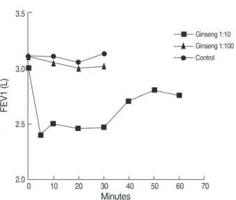
seng extract (0.01 mg/mL) induced a strong positive response to ginseng extract (histamine 4×4.5 mm, Korean ginseng extract 7×14 mm). An allergen bronchial provocation test with a 1:100 dilution of Korean ginseng extract induced no asthmatic reaction, but an early asthmatic reaction with severe coughing and dyspnea was observed 5 min after first inhaling a 1: 10 dilution of Korean ginseng extract. A physi- cal examination revealed expiratory wheezing through- out the lung field, and FEV1 was significantly reduced ver- sus baseline (2,430 mL vs. 3,010 mL, a 22.6% reduction) (Fig. 1).
Serum specific IgE levels to ginseng and ELISA inhibition testing
Serum specific IgE levels to ginseng extract were signifi- cantly elevated as compared with those of 5 atopic and 5 non- atopic controls, as shown in Fig. 2 (p<0.05, O.D.=patient 1.980, atopic controls 0.110±0.170, and non-atopic con- trols 0.008±0.006). ELISA inhibition tests showed dose- dependent inhibition by Korean ginseng, but not by Der- matophagoides farinae, wheat flour, or Chinese balloon flower (Fig. 3).
SDS-PAGE and immunoblotting
To identify IgE-producing components in ginseng extract, it was analyzed by 12% SDS-PAGE (Fig. 3). Immunoblot anal- ysis revealed four specific IgE binding components in extract
Fig. 1. Allergen bronchial provocation test using a 1:10 and 1:100 dilution of Korean ginseng extract. Allergen bronchial challenge testing induced a typical immediate response.
FEV1 (L)
3.5
3.0
2.5
2.00 10 20 30 40 50 60 70
Ginseng 1:10 Ginseng 1:100 Control
Minutes
Fig. 3. ELISA inhibition test using Korean ginseng, Dermatophagoid- es farinae, wheat flour, or Chinese balloon flower extracts. ELISA inhibition testing showed dose-dependent inhibition by Korean gin- seng, not by Dermatophagoides farinae, wheat flour, or Chinese balloon flower extract.
Percent Inhibition
100
75
50
25
0
0 0.1 1 10
Panax ginseng Wheat flour Balloon flower Dermatophagoides farinae
Inhibitor ( g/mL)
Fig. 2. Measurement of serum specific IgE antibody to ginseng.
The patient’s serum specific IgE levels to ginseng extract were significantly higher versus those of controls.
Absorbance
2.5
2.0
1.5
1.0
0.5
0.0
Atopic control Nonatopic control Patient
Group
Fig. 4. SDS-PAGE (left) and immunoblot analysis (right) for Kore- an ginseng. Indicating four IgE binding components at 26, 30, 47, and 60 kDa. NAC, non-atopic controls; AC, atopic controls.
170
60 47
3026 130
100 72 55 40 33
24
Patient NAC AC
Korean Ginseng-Induced Occupational Asthma 235
at 26, 30, 47, and 60 kDa, which were not present in con- trol sera (Fig. 4).
DISCUSSION
The word Ginseng is actually used to refer to the roots of several plant species in the Araliaceae family, which include Korean ginseng (or Panax ginseng, Asian ginseng), Siberian ginseng (Eleutherococcus senticosus), and American ginseng (Panax quinquefolius) (7). Korean ginseng is the most com- monly used form worldwide as an ‘adaptogen’. Moreover, the ginsenosides are the main active components of ginseng and have several beneficial effects, e.g., antioxidant, anti-inflam- matory, and anticancer effects (7).
A small number of cases of ginseng-induced hypersensitiv- ity have been reported, but the IgE binding components in- volved have not been identified. In 1991, Brazilian ginseng was first reported to be a possible cause of respiratory hyper- sensitivity (4). An IgE-mediated mechanism, as confirmed by immediate skin test reactivity, a positive bronchial challenge, and by the presence of specific IgE detected by ELISA, was implicated in this case. More recently, a case of occupational asthma and rhinitis caused by Sanyak and Korean ginseng was reported (5). In this case, a 29-yr-old female herbal mer- chant presented with asthmatic symptoms 14 months after starting work and had also experienced angioedema of the lip, tongue, and throat after ingesting ginseng. She was diagnosed as having Sanyak and Korean ginseng-induced occupational asthma and rhinitis, which was confirmed by bronchial chal- lenge testing. However, in this case only the Sanyak IgE bind- ing component was detected by immunoblotting.
In the present case, ginseng-induced occupational asthma was also confirmed by bronchial challenge testing. The sub- ject was a Korean ginseng wholesale merchant and her asth- matic symptoms were aggravated by inhaling ginseng dust, which is in line with previous cases. However, unlike previ-
ous cases, she did not develop allergic symptoms after ingest- ing Korean ginseng, and her duration of exposure was longer than that of the previous case (four and a half years). In the pre- sent case, skin prick testing to ginseng extract produced a strong positive response, and specific IgE to Korean ginseng was significantly elevated. Immunoblot analysis revealed four specific IgE binding components at 26, 30, 47, and 60 kDa, which were not bound by control sera. These results strongly suggest that occupational asthma induced by Korean ginseng in this subject was induced via an IgE-mediated mechanism.
This is the first report to reveal the IgE binding components involved in Korean ginseng induced occupational asthma.
REFERENCES
1. Park HS, Kim MJ, Moon HB. Occupational asthma caused by two herb materials Dioscorea batatas and Pinellia ternata. Clin Exp Allergy 1994; 24: 575-81.
2. Lee SK, Cho HK, Cho SH, Kim SS, Nahm DH, Park HS. Occupation- al asthma and rhinitis caused by multiple herbal agents in a pharma- cist. Ann Allergy Asthma Immunol 2001; 86: 469-74.
3. Kim SH, Jeong H, Kim YK, Cho SH, Min KU, Kim YY. IgE-medi- ated occupational asthma induced by herbal medicine, Banha (Pinel- lia ternata). Clin Exp Allergy 2001; 31: 779-81.
4. Subiza J, Subiza JL, Escribano PM, Hinojosa M, Garcia R, Jerez M, Subiza E. Occupational asthma caused by Brazil ginseng dust. J Aller- gy Clin Immunol 1991; 88: 731-6.
5. Lee JY, Lee YD, Bahn JW, Park HS. A case of occupational asthma and rhinitis caused by Sanyak and Korean ginseng dusts. Allergy 2006;
61: 392-3.
6. Park HK, Jeon SG, Kim TB, Kang HR, Chang YS, Kim YK, Cho SH, Min KU, Kim YY. Occupational asthma and rhinitis induced by a herbal medicine, Wonji. J Korean Med Sci 2005; 20: 46-9.
7. Kiefer D, Pantuso T. Panax ginseng. Am Fam Physician 2003; 68:
1539-42.