보 문
Evaluation of the anti-Helicobacter pylori and cytotoxic properties of the antimicrobial substances from Lactobacillus acidophilus BK13 and Lactobacillus paracasei BK57
Eun-Seo Lim*
Department of Food Science & Nutrition, Tongmyong University, Busan 608-735, Republic of Korea
Lactobacillus acidophilus BK13 and Lactobacillus paracasei BK57 균주가 생산한 항균물질의 anti-Helicobacter pylori 활성 및
위장상피세포에 대한 세포독성 평가
임은서*
동명대학교 식품영양학과
(Received June 3, 2015; Accepted June 25, 2015)
ABSTRACT: The objective of this study is to investigate the anti-Helicobacter pylori and anti-cancer activities of the live cells (LC), cell-free culture supernatants (CFCS), and bacteriocin solution (BS) obtained from Lactobacillus acidophilus BK13 and Lactobacillus paracasei BK57 strains. After incubation for 30 h in MRS broth, the concentration of lactic acid produced by L. paracasei BK57 (155.9±10.2 mM) was higher than in MRS broth using L. acidophilus BK13 (126.8±7.9 mM). Maximum bacteriocin activity (128 AU/ml) of BK13 strain was observed after 30 h of cultivation at 37°C, however its magnitude was significantly lower than that of BK57 strain (256 AU/ml). The LC of L. acidophilus BK13 and L. paracasei BK57 were able to inhibit the growth of H. pylori ATCC 43504 at different incubation times, depending on the initial inoculum of the LAB. These CFCS and BS obtained from BK13 and BK57 strains dramatically inhibited the growth, adhesive ability, and enzymatic activity of H. pylori. Meanwhile, the anti-cancer effect of the lactic acid from L. acidophilus BK13 and L. paracasei BK57 strains on AGS cells had significant differences with the control group. Therefore, these antagonistic substances- producing strains are potentially useful as new potential antimicrobial agents for the management and prevention of H. pylori infections.
Key words: Helicobacter pylori, adhesion, anti-cancer activity, antimicrobial activity, urease
*For correspondence. E-mail: [email protected];
Tel.: +82-51-629-1714; Fax: +82-51-629-1709
Helicobacter pylori is a helix-shaped Gram-negative found in the upper gastrointestinal tract and is present in more than 50% of the world’s population. As a general rule, infection rate appears to be higher in developing than in developed countries and it seems to be decreasing with improvements in hygiene practices (Brown, 2000). Possible routes of infection include either oral-oral or fecal-oral contact, use of unsterile endoscopes, and crowed or high-density living conditions (Cave, 1997).
Chronic infection with H. pylori causes gastric mucous
membrane damage and atrophic and even metaplastic changes
in the stomach, thereby inducing inflammation of the gastric
mucosa. Furthermore, gastric colonization with H. pylori can
lead to variety of upper gastrointestinal disorders, such as
nonulcer dyspepsia, peptic ulcer disease, distal gastric
adenocarcinomas, gastric mucosa-associated lymphoid tissue
(MALT) lymphoma, and gastric cancer. Bacterial virulence
factors such as the cytotoxin-associated gene pathogenicity
island-encoded protein CagA and the vacuolating cytotoxin
VacA can cause inflammation, damage of gastric epithelial
cells, and/or apoptosis and are potentially a carcinogen (Kusters
et al., 2006).
Medications used to eradicate H. pylori include antibiotics (amoxicillan, clarithromycin, metronidazole or tetracycline), proton pump inhibitor (PPI, lansoprarzole and omeprazole), and anti-diarrheal medicine (bismuth subsalicylate). Although these standard therapies are commonly used, increasing antimicrobial resistance and side effects and falling eradication rates are the result of the widespread use of antibiotics (Egan et al., 2007).
For this reason, antibiotic therapy is not recommended in patients infected with H. pylori, therefore the use of probiotics as alternative strategies has been suggested. According to previous studies, probiotics which is defined as a living microbial species have been proven to be useful in the treatment of H. pylori-associated disorders and diseases (Sgouras et al., 2004; Lesbros-Pantoflickova et al., 2007). Ingesting lactic acid bacteria (LAB) exerted a suppressive effect on H. pylori infection in both animals and humans, and probiotic preparations of yogurt containing Lactobacillus acidophilus La5 and Bifidobacterium lactis Bb12 were effective in reducing the rates of eradication of H. pylori in humans (Wang et al., 2004).
Some probiotic strains are able to inhibit H. pylori growth through the release of the antimicrobial agent called bacteriocins or organic acids (lactic, acetic, and butyric acid) and the competition for the limited nutrients required for bacterial growth or the receptors on epithelial cells (Boirivant and Strober, 2007). In addition, they prevent the adherence of the pathogenic bacteria to the host cells by enhancing the barrier function of the intestinal mucosa, stimulating the immune response, and releasing gut-protective metabolites such as arginine, glutamine, short-chain fatty acids, and conjugated linoleic acids (Hemaiswarya et al., 2013). Probiotic treatment reduced the inflammatory effects of infection by this pathogen and the side effects caused by traditional anti-H. pylori therapy, so adjuvant therapy with probiotics is currently becoming more popular than the traditional eradication methods.
L. acidophilus BK13 and L. paracasei BK57 strains used in this study were originally isolated from Baikkimchi and these strains showed relatively good survival property in artificial gastric juice and the antimicrobial activity against H. pylori in the previous experiment (Lim, 2014). The objective of this study is to investigate the anti-H. pylori and anti-cancer activities of the live cells (LC), cell-free culture supernatants (CFCS), and
bacteriocin solution (BS) obtained from L. acidophilus BK13 and L. paracasei BK57 strains.
Materials and Methods
Bacterial strains and culture conditions
LAB strains were grown in Lactobacilli MRS broth (Difco) for 24 h at 37°C. The tested strains were stored in Lactobacilli MRS broth (Difco) with 20% glycerol at -80°C. These strains were recovered from frozen stock and cultivated twice on MRS agar aerobically at 37°C for 24 h.
Culture conditions of H. pylori
H. pylori ATCC 43504 strain used in this study was obtained from American Type Culture Collection (ATCC). H. pylori was cultured under microaerophilic conditions (10% CO
2, Anoxomat system, MART Co.) on Brucella agar (Difco) plates supplemented with 5% (v/v) fetal bovine serum (FBS, Gibco BRL), 0.2%
(w/v) 2,6-di-ο-methyl-β-cyclodextrin (CD), and antibiotics (cefsulodine, vancomycin, trimethoprim, and amphotericin B, Sigma-Aldrich) at 37°C for 48 h. The strain was stored in Brucella broth (Difco) containing 20% (v/v) glycerol at -80°C.
And then, it was recovered from frozen stock and sub-cultured twice in Brucella broth supplemented with 5% FBS at 37°C for 48 h with agitation on a rotary shaker at 100 rpm in a microaerobic condition prior to experimental use.
Microbiological and chemical analysis during the incubation
The tested LAB strains were propagated in MRS for 30 h at
37°C aerobically. At convenient time intervals, culture samples
were aseptically withdrawn from the fermentation vessel to
determine viable cell number, pH, and titratable acidity. Viable
cell counts of LAB culture were determined by the standard
plate count (SPC) method with MRS medium at 37°C after 48 h
of incubation. The pH value of samples was measured separately
by a digital pH meter (Metrohm 744). Finally, titratable acidity
of the culture was determined by titration with 0.1 N NaOH and
expressed in terms of milliliters of NaOH.
Preparation of antimicrobial substances
Lactic acid: Cultures of LAB strains were grown in MRS broth at 37°C under the optimum conditions. After incubation, the CFCS was derived from fresh overnight cultures, centrifuged at 7,000 × g for 10 min at 4°C, and freezed until used. The CFCS was filtered using a 0.22 μm membrane filter (Millipore) and precipitated protein by the addition of HClO
4(1 M). And then, lactic acid was determined by high-pressure liquid chromato- graphy (HPLC, Shimadzu), with Aminex HPX-87H column (300 mm by 7.8 mm: Bio-Rad) and a refractive index detector (GBC Scientific Equipment Pty Ltd.). The operating conditions were the following: H
2SO
4solution (5 mM) was used as eluent at a flow rate of 0.5 ml/min and column temperature of 35°C.
The sample injection volume was 50 μl. Lactic acid was monitored by measuring the optical density at 220 nm and its concentration was calculated from a standard curve of known
L
-lactate concentration.
Crude bacteriocin: The obtained CFCS was filter-sterilized using 0.45 μm pore size filter to eliminate the possible presence of viable cells and their pH was adjusted to 6.5 by means of 1 N NaOH to exclude the antimicrobial effect of organic acid.
Inhibitory activity from hydrogen peroxide was eliminated by the addition of 1 mg/ml catalase (Sigma-Aldrich). The CFCS were treated with ammonium sulfate to 50% saturation and agitated at 4°C for overnight. The crude BS was then precipitated from the CFCS by centrifugation (12,000 × g for 30 min at 4°C) and the precipitates were re-suspended in 20 mM sodium phosphate buffer (pH 6.5). Dialysis was followed in a dialysis bag (Spectrum Medical Industries, Inc.) with a molecular weight cut-off of 1,000 Da against the same buffer for 24 h at 4°C.
Bacteriocin activity
Bacteriocin activity was measured using a microtiter plate assay (Hole et al., 1991) described previously against H. pylori as the target organism. Cells of H. pylori ATCC 43504 were obtained by centrifugation (7,000 × g, 4°C, 10 min) from 48 h-old cultures, washed twice with sterile phosphate buffer saline (PBS, pH 7.0), and resuspended in the same buffer. The bacterial suspension (1.0 × 10
5CFU/ml), the diluted BS of different concentrations, and Brucella broth were individually placed in microtiter plate wells (Falcon). Plates were incubated
at 37°C for 24 h and the growth inhibition of the indicator organism was measured spectrophotometrically at 600 nm with a microplate reader (BioTek, Inc.). The bacteriocin activity (arbitrary units, AU) was defined as the reciprocal of the highest dilution inhibiting the growth of the indicator strain by 50%.
Growth of H. pylori co-cultured with LC, CFCS, and BS LAB strains were grown in MRS broth at 37°C overnight.
The LC of LAB strain were prepared by centrifuging (7,000 × g, 10 min, 4°C) the active cultures and washing two times by PBS (pH 7.0) and diluted to a concentration of 10
6, 10
7, and 10
8CFU/ml. Meanwhile, 48 h-old H. pylori cultures were resuspended in sterile PBS (pH 7.0) and centrifuged at 7,000 × g for 10 min at 4°C. The cell pellet was collected, and the fresh H.
pylori cells (1.0 × 10
6CFU/ml) suspended in antibiotic-free Brucella broth containing 5% FBS were incubated under microaerophilic conditions for 48 h at 37°C in the presence of the LC, CFCS or BS at different concentration. Initially and then at predetermined intervals, aliquots of the cultures were removed, serially diluted, and cultured at 37°C under micro- aerophilic conditions for 48 h on Brucella agar containing 5%
FBS to determine the viable cell counts of H. pylori. Finally, the inhibition (%) of viability of H. pylori by the LC, CFCS, and BS treatment was calculated as follows; Inhibition (%) = (1-cell counts of H. pylori after each treatment/cell counts of H. pylori after PBS treatment) × 100.
Cell culture
Human gastric cancer cell lines (AGS and SNU-1) were from American Type Culture Collection (USA). Cells were routinely grown in RPMI 1640 medium (Gibco, BRL), supplemented with 10% (v/v) inactivated (30 min, 56°C) FBS, l-glutamine, NaHCO
3, kanamycin (60 μg/ml), and streptomycin (20 μg/ml) (Nikken bio medical). Monolayers of cells were grown in six-well tissue culture plates (Falcon) at the density of 5 × 10
4cells/well. Experiments and maintenance of the cells were carried out at 37°C in humidified atmosphere of 5% CO
2and 95% air. Culture medium was replaced every other day.
Cultures were used at late post-confluence, i.e., after 15 days in
culture.
Adhesion of H. pylori against AGS cells in the presence of LC, CFCS, and BS
The inhibition of adhesion of H. pylori by the tested LAB strains were examined as previously described by Wang et al.
(2014) with some modifications. AGS cells (4 × 10
5cells/ml) were seeded into six-well tissue culture plates (Gibco) and cultured at 37°C in an incubator with 5% CO
2-95% air until monolayers of the cells was nearly confluent. Monolayers were washed twice with PBS (pH 7.0) before inhibition assays. The LAB and H. pylori cultures propagated in MRS broth and Brucella broth containing 5% FBS, respectively, were harvested by centrifugation at 7,000 × g for 10 min at 4°C and washed two times with PBS (pH 7.0).
In the exclusion assay, 1 ml of the LC (1.0 × 10
8CFU/ml) suspended in antibiotic-free RPMI 1640 medium were added to each well seeded with AGS cells and incubated for 2 h at 37°C in 5% CO
2-95% air atmosphere. And then 1 ml of H. pylori (1.0
× 10
8CFU/ml) suspended in cell culture medium was added to wells and left to incubate for an additional 2 h. In the competition assays, 1 ml of the LC suspension (1.0 × 10
8CFU/ml) of LAB strains and 1 ml H. pylori suspension (1.0 × 10
8CFU/ml) were added to each well seeded with AGS cells simultaneously and incubated for 4 h at 37°C in 5% CO
2-95%
air. In the displacement assay, 1 ml of H. pylori (1.0 × 10
8CFU/ml) suspended in antibiotic-free cell culture medium were added to wells seeded with AGS cells. After incubation for 2 h at 37°C in 5% CO
2-95% air, 1 ml of LAB-LC suspension (1.0 × 10
8CFU/ml) prepared in antibiotic-free cell culture medium were added to wells and left to incubate for an additional 2 h.
Wells containing H. pylori alone served as controls.
Meanwhile, 1 ml aliquot of fresh cell culture medium containing the CFCS or BS adjusted to a suitable concentration was added to each wells seeded with AGS cells and allowed to incubate for 2 h to remove adherent H. pylori from the epithelial cells. In all assays, monolayers were washed three times with PBS (pH 7.0) to release unbound bacteria after incubation. To determine the number of adherent H. pylori, AGS cells were lysed with 0.1% Triton X-100 for 5 min. And then, aliquots of serial 10-fold dilutions were plated onto antibiotic-selective agar for 48 h at 37°C.
Urease assay of H. pylori co-cultured with CFCS or BS Urease activity in H. pylori adhering to AGS cells was determined by measuring the release of ammonia by a modification of phenol red method (Sgouras et al., 2004). The incubation conditions for the adherence of H. pylori to AGS cells in the presence of PBS, CFCS, and BS were the same as those described previously for the adhesion assay. The incubated cell cultures were washed five times with PBS (pH 7.0), and then urease reaction buffer (200 g/L urea and 0.12 g/L phenol red in PBS, pH adjusted to 6.5) was added to each well of the microtiter plate. The well plates were incubated for 3 h at 37°C in order to induce the production of ammonia from H. pylori adhering to AGS cells. After incubation, the absorbance at 550 nm was measured with a microtiter plate reader.
Cytotoxicity assay for gastric cancer cell lines Cytotoxic effect of the CFCS and BS from L. acidophilus BK13 and L. paracasei BK57 against AGS and SNU-1 cells was determined using an MTT assay described by Zheng et al.
(1996). A cell suspension (with cell density of 1.0 × 10
4cell/well) was seeded into each well of the 96-well culture plates (Falcon) and incubated for 24 h at 37°C in a 5% CO
2humidified incubator. After obtaining a semi confluent cell layer, the exhausted media were discarded. And then, the cultivated cells were exposed to various concentrations of the CFCS (50, 100, and 150 μl/ml) and BS (100, 200, and 300 AU/ml) prepared in RPMI 1640 medium and incubated at 37°C in humidified 5%
CO
2for 24 h. The wells containing the same amount of the cell culture medium and having no the antimicrobial substance were used as control. The medium of each well after incubation was replaced with equally fresh media containing 3-(4,5- dimethylthiazol-2-yl)-2,5-diphenyltetrazolium bromide (MTT) solution (2 mg/ml) and samples were incubated under dark conditions for 4 h at 37°C. After discarding all media from the plates, 100 μl of dimethyl sulfoxide (DMSO) was added to all wells. The plates were held for 5 min at room temperature with shaking to achieve complete dissolution of formazan. The absorbance of the MTT formazan was determined at 540 nm using a spectrophotometric plate reader. The percentage cell viability was determined based on the formula: Viability (%)=
(optical density of sample/optical density of control) × 100.
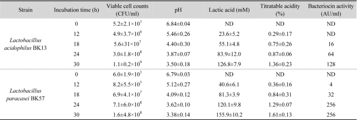
Table 1. Changes of viable cell counts, pH, titratable acidity, lactic acid content, and bacteriocin activity during incubation by the selected L. acidophilus BK 13 and L. paracasei BK57 in MRS broth at 37°C
Strain Incubation time (h) Viable cell counts
(CFU/ml) pH Lactic acid (mM) Titratable acidity
(%)
Bacteriocin activity (AU/ml)
Lactobacillus acidophilus BK13
0 5.2±2.1×10³ 6.84±0.04 ND ND ND
12 4.9±3.7×10⁶ 5.46±0.26 23.6±5.2 0.29±0.17 ND
18 5.6±31×107 4.40±0.30 55.1±4.8 0.75±0.26 16
24 3.0±1.8×10⁸ 3.87±0.07 83.9±12.0 0.87±0.06 64
30 1.1±0.2×10⁹ 3.50±0.18 126.8±7.9 1.36±0.23 128
Lactobacillus paracasei BK57
0 6.0±1.9×10³ 6.79±0.03 ND ND ND
12 8.2±5.5×10⁵ 5.12±0.27 40.6±6.1 0.36±0.16 4
18 6.9±4.1×10⁷ 4.09±0.12 81.3±3.9 0.84±0.31 32
24 7.1±6.0×10⁸ 3.62±0.10 120.1±9.8 1.29±0.07 256
30 1.6±4.8×10⁸ 3.38±0.14 155.9±10.2 1.61±0.13 256
ND, not detected.
Statistical analysis
All experiments were performed in triplicate and results were expressed as mean±standard deviations (SD). Differences between the means of the test and control groups were examined for significance by Student’s t-test and P<0.05 was considered to be statistically significant. In particular, the results on cytotoxicity for gastric cancer cell lines were subjected to one-way analysis of variance (ANOVA) and means were separated by Duncan’s multiple range test (P<0.05) using the software package SPSS 12.0 Window Program (SPSS Inc.).
Results and Discussion
Changes of cell number, pH, titratable acidity, lactic acid content, and bacteriocin activity during incubation period
Changes of the viable cell counts, pH, titratable acidity, lactic acid content, and bacteriocin activity during incubation by the selected L. acidophilus BK 13 and L. paracasei BK57 in MRS broth at 37°C are shown in Table 1. The cell growth rate of the two LAB varied slightly depending on the species. L.
acidophilus BK13 reached 1.1±0.2 × 10
9CFU/ml after 30 h at 37°C. The cell population of L. paracasei BK57 rapidly increased and reached 10
8CFU/ml in MRS broth after 24 h of fermentation and constant to 30 h fermentation. After 30 h of incubation, the pH values of L. acidophilus BK13 and L. paracasei BK57
cultures were 3.50±0.18 and 3.38±0.14, respectively. The two strains produced lactic acid followed by the decreasing of pH.
After incubation for 30 h, the concentration of lactic acid produced by L. paracasei BK57 (155.9±10.2 mM) was higher than in MRS broth using L. acidophilus BK13 (126.8±7.9 mM).
L. acidophilus BK13 and L. paracasei BK57 cultures showed a time-dependent decrease and increase in the pH and titratable acidity, respectively, when grown on MRS medium. The production of titratable acid was different among the two strains at different times of incubation. After 30 h incubation, the titratable acidity of BK13 and BK57 strains reached 1.36±0.23 and 1.61±0.13%, respectively. The bacteriocin activity of L.
acidophilus BK13 and L. paracasei BK57 started in the log phase of growth and increased with increase in incubation time.
Maximum bacteriocin activity (128 AU/ml) of BK13 strain was observed after 30 h of cultivation at 37°C. Although BK13 strain demonstrated similar antimicrobial activity on H. pylori, its magnitude was significantly lower than those of BK57 strain (P<0.05). The production of the bacteriocin from BK57 strain was first detected after 12 h of incubation (4 AU/ml), whereas the peak of bacteriocin production was observed at 24-30 h (256 AU/ml), during the late exponential growth phase.
In recent decades, it has become clear that the overall
inhibitory action of LAB is due to a variety of antagonistic
factors that include organic (lactic, acetic, and formic) acids,
ethanol, hydrogen peroxide, diacetyl, acetoin, 2,3-butanediol,
acetaldehyde, benzoate, and bacteriolytic enzymes (Klaenhammer,
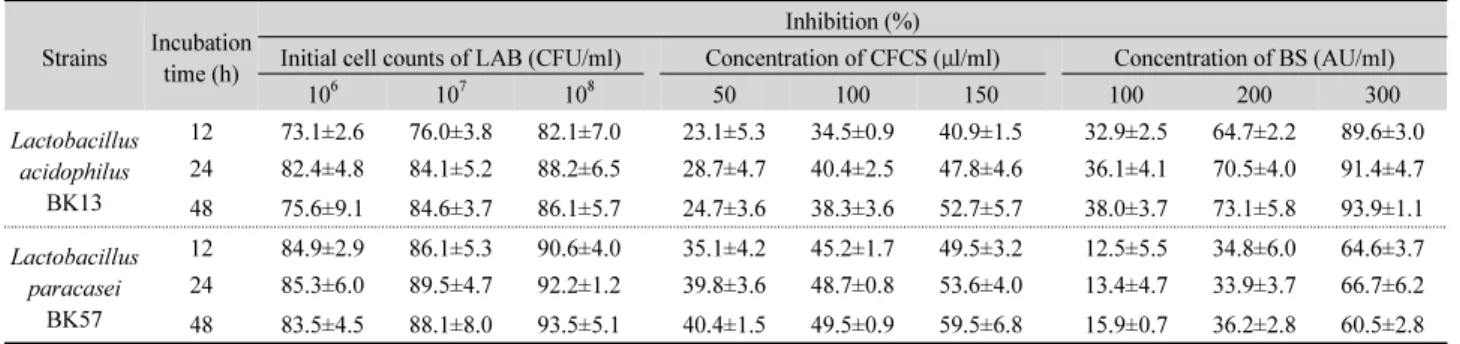
Table 2. Time-kill assay of H. pylori ATCC 43504 with LC, CFCS, and BS obtained from the selected LAB
Strains Incubation time (h)
Inhibition (%)
Initial cell counts of LAB (CFU/ml) Concentration of CFCS (μl/ml) Concentration of BS (AU/ml)
106 107 108 50 100 150 100 200 300
Lactobacillus acidophilus
BK13
12 73.1±2.6 76.0±3.8 82.1±7.0 23.1±5.3 34.5±0.9 40.9±1.5 32.9±2.5 64.7±2.2 89.6±3.0 24 82.4±4.8 84.1±5.2 88.2±6.5 28.7±4.7 40.4±2.5 47.8±4.6 36.1±4.1 70.5±4.0 91.4±4.7 48 75.6±9.1 84.6±3.7 86.1±5.7 24.7±3.6 38.3±3.6 52.7±5.7 38.0±3.7 73.1±5.8 93.9±1.1 Lactobacillus
paracasei BK57
12 84.9±2.9 86.1±5.3 90.6±4.0 35.1±4.2 45.2±1.7 49.5±3.2 12.5±5.5 34.8±6.0 64.6±3.7 24 85.3±6.0 89.5±4.7 92.2±1.2 39.8±3.6 48.7±0.8 53.6±4.0 13.4±4.7 33.9±3.7 66.7±6.2 48 83.5±4.5 88.1±8.0 93.5±5.1 40.4±1.5 49.5±0.9 59.5±6.8 15.9±0.7 36.2±2.8 60.5±2.8 Data were presented as mean±SD from three independent experiments.
*Statistical analysis preformed with s Student t-test between control and treated H. pylori with the bacteriocin showed a highly significant difference (P<0.05).
1988). In addition, certain strains of LAB are further known to produce bactericidal proteins, termed bacteriocins, which are antagonistic to a wide spectrum of microorganisms, and thus can make significant contributions to food preservation and intestinal ecology (De Vuyst and Leroy, 2007). Several studies have shown that the bacteriocin production in LAB is dependent on the biomass (Powell et al., 2007; Settanni et al., 2008). The bacteriocin biosynthesis is affected by type and level of the carbon, nitrogen and phosphate sources, cations surfactants, and inhibitors. Primary metabolite kinetics of the bacteriocin production with a peak activity usually occur at the end of exponential growth phase, followed by a decrease during the stationary phase (Savadogo et al., 2006). The decrease in bacteriocin activity at the end of exponential growth phase might be explained by the degradation of the bacteriocin by extracellular proteolytic enzymes (Todorov and Dicks, 2005).
New proteinaceous active substance produced by L. paracasei subsp. paracasei strain M3 displayed the bactericidal activity against H. pylori NCIPD 230. The synthesis of this anti- microbial substance was detected in the late logarithmic growth phase during batch fermentation (Atanassova et al., 2003).
LAB have their ability to produce lactic acid as the major product of sugar (e.g., glucose) fermentation by either the homo- or heterofermentative pathway (Wee et al., 2006). Chen et al. (2011) showed that the main organic acid present in the CFCS of Lactobacillus gasseri Chen and Lactobacillus plantarum 18 having significant anti-H. pylori activity was lactic acid with the range of content from 114 to 150 mM.
Among the strains showing antagonistic activity against H.
pylori, lactic acid levels were 13.4 to 18.6 mg/ml for Lactobacillus delbrueckii subsp. bulgaricus and 0.6 to 11.5
mg/ml for Streptococcus thermophilus (Aslim et al., 2011).
Antagonistic activity of LC, CFCS, and BS obtained from the LAB against H. pylori ATCC 43504
The antagonistic activity of the LC, CFCS, and BS obtained from L. acidophilus BK13 and L. paracasei BK57 against H.
pylori ATCC 43504 are presented in Table 2. The LC of L.
acidophilus BK13 and L. paracasei BK57 were able to inhibit the growth of H. pylori ATCC 43504 at different incubation times, depending on the initial inoculum of the LAB. Specially, the LC of BK57 strain exhibited higher inhibition capacity on H.
pylori than that of BK13 strain. When the inoculum of L.
paracasei BK57 was 10
8CFU/ml, 93.5±5.1% inhibition of the
pathogen growth was observed after 48 h. Furthermore, these
CFCS and BS obtained from BK13 and BK57 strains exhibited
varying degrees of inhibitory activity against H. pylori strain
and inhibited the growth of H. pylori dramatically after 48 h of
incubation. The CFCS and BS of the two LAB tested
significantly reduced the viable cell counts of H. pylori in
dose-dependent manner. At the same concentration, the CFCS
of BK13 strain showed a weaker inhibitory effect compared to
BK57-CFCS. These results noticed that the high degree of the
antimicrobial activity of L. paracasei BK57 may be partly
attributed to their higher acid production. Using time-kill
studies, the inhibition of H. pylori observed in the presence of
the BS (300 AU/ml) of L. acidophilus BK13 ranged between
89.6±3.0 and 93.9±1.1%, while 60.5±2.8 – 64.6±3.7% reduction
was observed in the BS of BK57 strain during incubation. This
implied that low pH values and high titratable acidity are
important for anti-H. pylori activity. In a study by Boyanova et
al. (2009), the anti-Helicobacter activity of L. delbrueckii subsp. bulgaricus cultures was strain-dependent and these tested strains secreted metabolic products such as lactic acid and bacteriocin which exert activity against H. pylori.
Several probiotic strains affect the host beneficially, have exhibited antagonistic properties against H. pylori in vitro without antibiotic-associated gastrointestinal side-effects (Pacifico et al., 2014). In previous study, BK13 and BK57 strains showed relatively high tolerance to artificial gastric juice (Lim, 2014) and good adhesive properties to the human epithelial cells (data not shown), so these isolates met partially the selection criteria for potential probiotics. These are several putative mechanisms for probiotic efficacy against H. pylori; enhancement of barrier function, competition for the limited nutrients, inhibition of the epithelial and mucosal adherence of pathogens, production of inhibitory compounds (hydrogen peroxide, diacetyl, organic acids, and bacteriocin-like substances), and stimulation of the immune response (Hamilton-Miller, 2003).
Two main types of substances such as short chain fatty acids (SCFAs) and bacteriocin proteins have been implicated in the inhibition of H. pylori by LAB. SCFAs such as formic, acetic, propionic, butyric and lactic acids are produced as a result of the metabolism of carbohydrates by probiotics and play an important role in decreasing the pH in vitro. Their antimicrobial activity could be due to the inhibition of urease activity by high lactic acid producers, such as Lactobacillus salivarius and Lactobacillus casei Shirota (Gotteland et al., 2006). Lactic acid is produced in largest amount during the metabolism of carbohydrate Lactobacillus and have an important role in decreasing pH, which is related with respect to H
+ions, important in the inhibition of H. pylori in vitro (Midolo et al., 1995). The antimicrobial activity occurs through the diffusion of lactic molecules into microbial cells until equilibrium is reached. Acids are generally thought to exert their antimicrobial effect by interfering with the maintenance of cell membrane potential, active transports, and a variety of metabolic reactions, causing membrane disruption, reducing intracellular pH homeostasis, and accumulating of toxic anions (Brul and Coote, 1999). Previous studies have indicated that the inhibitory role of L. acidophilus against H. pylori was related to the acid production and the low pH attained (Lorca et al., 2001). The supernatant of L. acidophilus culture has been shown to greatly
suppress the viability of H. pylori both in vitro and in vivo, dependent on pH and lactic acid level (Coconnier et al., 1998).
However, the antimicrobial activity of certain probiotic lactobacilli may even be based on the production of bacteriocins.
The proteinaceous bacteriocins with anti-H. pylori activity were synthesized by probiotic strains of Lactobacillus, Enterococcus faecium, Bacillus subtilis, and Bifidobacterium (Gotteland et al., 2006). In the mode of action, all types of bacteriocins show their antimicrobial effects on the target cell surface via various mechanisms including the deficiencies in the cell wall synthesis, changes in the membrane permeability, and/or formation of pores causing the death of the target cells (Moll et al., 1999).
Chen et al. (2011) reported that the count of H. pylori decreased in the co-culturing live lactobacilli and LAB-CFCS.
At 48 h, the count of the pathogen was reduced only in co-cultures with the L. gasseri Chen and L. plantarum 18 and their CFCS; the cells of live Lactobacillus rhamnosus GG could not inhibit the growth of H. pylori. The CFCS of L. plantarum 18 had a greater ability to inhibit the growth of H. pylori than the other two strains. Michetti et al. (1999) and Aiba et al. (1998) showed that the supernatants from L. johnsonii La1 and Lactobacillus salivarius cultures inhibit the growth of H. pylori in vitro whether or not H. pylori is bound to epithelial cells. This effect was due to the production of a large amount of lactic acid since the inhibitory effect could be reproduced after the incubation of H. pylori in the presence of lactic acid.
Furthermore, the urease activity became almost undetectable at a lactic acid concentration of 10 mM. Midolo et al. (1995) reported that lactic, acetic, and hydrochloric acids demonstrated an inhibition of H. pylori growth in a concentration-dependent manner, with lactic acid resulting in the highest inhibition.
Lorca et al. (2001) demonstrated that the antibacterial activity of
the spent broth obtained from 17 strains of lactobacilli against
10 strains of H. pylori was associated with organic acids or
intracellular proteinaceous component. De Vuyst et al. (2004)
demonstrated that the specific activity of the CFCS (pH 6.5) of
Lactobacillus johnsonii La1 was also observed in time-kill
assays. Also, the concentrated CFCS, containing no organic
acids, of L. johnsonii La1, Lactobacillus casei Shirota, and
Lactobacillus amylovorus DCE471 inhibited H. pylori ATCC
43504 in a killing assay. These data indicate that the antibacterial
substance(s) other than lactic acid or acetic acid is (are)
Fig. 1. Effects of the LC [A], CFCS [B], and BS [C] obtained from L.
acidophilus (■) BK13 and L. paracasei (■) BK57 strains on the adhesion of H. pylori ATCC 43504 to AGS cells. Experimental conditions were described in the text. Each value shown was the mean±standard error of the three experiments. *Statistical analysis preformed with Student’s t-test between control and treated H. pylori with the antimicrobial substance showed a highly significant difference (P<0.05).
responsible for the inhibitory activity. Kim et al. (2003) suggested that lacticins A164 and BH5 produced by Lactococcus lactis subsp. lactis A164 and L. lactis BH5, respectively, showed the anti-H. pylori activity of lacticin A164 was dependent on initial inoculum size as well as concentration of the bacteriocin added.
Inhibition of the adhesion of H. pylori by LC, CFCS, and BS obtained from the LAB
The inhibition capacities of two Lactobacillus strains on adhesion of H. pylori to AGS cells were examined. The adhesion of the pathogen to AGS cells without interruption by lactobacilli was assigned as 100%. As shown in Fig. 1, H. pylori adhesion was reduced significantly by the LC of the two lactobacilli strains in all assays. The level of inhibition of adhesion performed in exclusion assay was stronger than the competition and displacement assays. In the exclusion assay, L. acidophilus BK13 was found to significantly reduce the adherence of H.
pylori to 53.8%, and this strain also showed better inhibition activity in adherence of the pathogen than L. paracasei BK57 in competition and displacement assays. Meanwhile, the incubation of H. pylori with each of the active compounds (lactic acid and bacteriocin) produced by the tested strains inhibited its adhesion to AGS cells, but there were some differences between the compounds in their H. pylori inhibition kinetics. The CFCSs of these strains showed the concentration-dependent anti-H. pylori activity, however, the number of H. pylori adhering to the AGS cells of the CFCS (> 100 μl/ml) of BK13 strain was significantly lower than that of the CFCS prepared from the culture of BK57 strain. In addition, the adhesion of H. pylori decreased by about 50% from the control after exposure to 300 AU/ml of the BS produced by BK13 strain, whereas, there was no significant difference (P>0.05) between the control and the treatment with the BS (300 AU/ml) of BK57 strain. These results suggested that their antimicrobial substances in the cultures as well as viable cells of the selected L. acidophilus BK13 and L.
paracasei BK57 strains play a key role in inhibiting the adhesion of H. pylori to AGS cells and could help to prevent infection in an early stage of colonization of the gastric mucosa by this pathogen.
Numerous studies have investigated the binding properties of H. pylori. In vitro experiments have provided evidence that the
adhesion of H. pylori to epithelial cell lines and to mucins may involve sialic acid and sulfated oligosaccharides (Sutton, 2001).
This has been supported in vivo by Genta et al. (1996), who have
found that the adhesion of H. pylori to areas of intestinal
metaplasia is associated with the expression of sulfomucins on
the gastric tissue. In the gastric mucosa, the adhesion of H.
Fig. 2. Urease activity of H. pylori ATCC 43504 in the co-culture with the CFCS (A) and BS (B) obtained from L. acidophilus BK13 (■) and L. paracasei BK57 (■) strains. Experimental conditions were described in the text. Each value shown was the mean±standard error of the three experiments. *Statistical analysis preformed with Student’s t-test between control and treated H. pylori with the antimicrobial substance showed a highly significant difference (P<0.05).
pylori which interacts with epithelial cells via multiple bacterial surface components is important in determining the outcome in H. pylori-associated gastrointestinal diseases (Lesbros-Pantoflickova et al., 2007).
However, some probiotic bacteria with beneficial health effects are able to reduce the bacterial load and inflammation of H. pylori in animal and human studies. Two mechanisms of probiotic action have been identified to mediate the maintenance of the gastrointestinal microbial balance: production of antibacterial substances and competitive inhibition of pathogen and toxin adherence to the intestinal epithelium, and the suppression effect of probiotic strains is strain dependent (Servin and Coconnier, 2003). In vitro studies showed that certain lactobacilli including L. johnsonii La1 or L. acidophilus LB inhibit the attachment of H. pylori to intestinal HT-29 cells or to MKN-45 gastric cell lines by secreting the antimicrobial substances (Lesbros-Pantoflickova et al., 2007). In addition, the anti-adhesive effect might be the result of competition between probiotic strains such as Lactobacillus reuteri or Weissella confuse and pathogen for the same receptor or the induction of mucin production by probiotics. The ability to inhibit the adhesion of pathogens to immobilized human mucus appears to depend on both the specific probiotic strains and the pathogens (Oelschlaeger, 2010). Furthermore, a non-specific rather than a specific blockage of receptor sites is the most likely mechanism because lactobacilli can inhibit adhesion of the large varieties of pathogenic bacteria, although each adheres to its particular receptor on the cells (Pacifico et al., 2014).
Similar results were noted in other studies; certain lactobacilli such as L. johnsonii La1 or L. acidophilus LB can exert their anti-adhesion activity by secreting the antimicrobial substances (Lesbros-Pantoflickova et al., 2007). Lin et al. (2009) reported that the spent culture supernatants from LAB inhibit H. pylori infection and adhesion to AGS cells. In addition, treatment by the bacteriocin or the cells of E. faecium TM39 significantly reduced the binding of H. pylori to monolayers of AGS cell line (Tsai et al., 2004).
Inhibition of H. pylori urease activity of antimicrobial substances obtained from the LAB
The effect of the CFCS and BS of L. acidophilus BK13 and L.
paracasei BK57 strains on the urease activity of H. pylori
ATCC 43504 adhered against AGS cells was examined. As
shown in Fig. 2, the urease activity of H. pylori was inhibited
prominently by the CFCS from BK13 and BK57 strains. After
treatment with the CFCS (150 μl/ml) of BK13 strain, the urease
activity of the H. pylori cells decreased to 50% of the control
level and no significant difference in the inhibitory activity of
these CFCSs was observed between these strains. Furthermore,
the urease activity in H. pylori culture was significantly
inhibited by the presence of the BSs of the tested strains. The BS
(300 AU/ml) of L. acidophilus BK13 exhibited high inhibitory
effect on the urease activity of H. pylori (0.25±0.02), while the
BS (300 AU/ml) of L. paracasei BK57 was ineffective to inhibit
the urease activity of H. pylori. Our in vitro study found that the
antimicrobial substances of the LAB inhibited the urease
Fig. 3. Proliferation of AGS [A] and SNU-1 [B] cells in the presence of the CFCS obtained from L. acidophilus BK13 (■) and L. paracasei BK57 (■) strains.
Experimental conditions were described in the text. Each value shown was the mean±standard error of the three experiments. Means with different superscript letters indicate statistically significant differences as determined by ANOVA (P < 0.05).
activity of H. pylori in a dose-dependent manner, therefore, the amount of antimicrobial substance released by the lactobacilli strains correlated with the intensity of the inhibitory effect against H. pylori. These findings suggested that the lactic acid in the CFCS prepared from the two selected lactobacilli strains possess effective activity to inhibit the urease of H. pylori adhered to AGS cells.
The spiral morphology and flagellar motility of H. pylori allow this bacterium to penetrate in the viscous mucus layer of the stomach. Meanwhile, H. pylori urease catalyzes the hydrolysis of urea into bicarbonate and ammonia to neutralize the acidic environment of the gastrointestinal tract (Luo et al., 1999). This enzyme plays a central role in H. pylori pathogenesis and is critical for bacterial colonization of the human gastric mucosa. The ammonia produced by urease can damage the gastric mucosa through the disruption of tight junctions and the alteration of permeability of gastric epithelium. Moreover, urease stimulates activation of mononuclear phagocytes and production of inflammatory cytokines (Cellini and Donelli, 2000).
Lin et al. (2009) found that there was an inverse relationship between the exclusion rate and urease activity; that is the point when the exclusion rate of LAB-SCS against H. pylori infection was highest (41.1%), the urease activity of H. pylori was lowest.
Therefore, LAB-SCS is able to inhibit H. pylori infection in AGS cells. Examining whether lactic acid participates in inhibition of the H. pylori urease activity, it was found that a concentration of 200 mM
D, L-lactic acid totally inhibited the H.
pylori urease activity, whereas a range of concentrations from
60 to 100 mM, similar to that determined in the L. acidophilus LB-SCS (84 mM), failed to inhibit urease activity of H. pylori.
These results demonstrate that lactic acid does not participate in the action of LB-SCS against the H. pylori urease (Coconnier et al., 1998). A similar inhibition pattern was found in the other studies. Yoon and Won (2002) suggested that both Lactobacillus helveticus CU631 and CFCS obtained from this strain had strong inhibitory activities in urease of H. pylori NCTC 11637 and CJH12. Tsai et al. (2004) demonstrated that after 2 h contact of the H. pylori cells with the bacteriocin from E. faecium TM39, the urease activity of H. pylori adhered to AGS cells decreased to one tenth of its original level. Thus, the inhibitory activity of the antibacterial compound(s) secreted from strain TM39 could result in the inhibition on H. pylori urease activity.
Cytotoxicity of antimicrobial substances obtained from the LAB
The cytotoxic activity of the antimicrobial substances from
the LAB strains against human gastric cancer cells was
measured using the MTT method. Inhibition of AGS and
SNU-1 cells proliferation by the CFCSs from L. acidophilus
BK13 and L. paracasei BK57 is shown in Fig. 3. The CFCSs of
the tested Lactobacillus strains potently inhibited the viability
of each cell line, in a dose-dependent manner. According to
these results, the strongest effect of BK13 strain was found at a
concentration of 150 μl/ml of the CFCS at 24 h of incubation
(70.0±4.8% inhibition). Whereas, the cytotoxicity inhibition
rate of BK57-CFCS was 56.6±5.3% for a concentration of 150
μl/ml at 24 h of incubation. In addition, the viability of SNU-1
carcinoma cell was inhibited by the CFCSs from L. acidophilus BK13 and L. paracasei BK57 strains after 24 h incubation.
However, the anti-proliferative effect of these strains on SNU-1 carcinoma cell was lower than that on AGS cell. In contrast, the BSs from the two lactobacilli strains did not have any effect on the cell viability of the tested cancer cells (data not shown). Our findings demonstrated that the anti-cancer effect of the lactic acid from L. acidophilus BK13 and L. paracasei BK57 strains on AGS cells had significant differences in the concentration of the lactic acid (P<0.05).
Lactobacilli and bifidobacteria which are the most prominent probiotic bacteria have been reported to possess certain anti-cancer properties. The precise mechanisms by which LAB may inhibit cancer are presently unknown. However, the anti-carcinogenic effect of probiotics may be attributable to alteration of the metabolic activities of intestinal microflora, binding and degrading potential carcinogens, quantitative and/or qualitative alterations in the intestinal microflora incriminated in producing putative carcinogen(s) and promoters, production of anti-carcinogenic compounds, enhancing the host's immune response, and effects on physiology of the host (Rafter, 2002).
Sevda et al. (2015) demonstrated that the CFCS and the cell-free lyophilized filtrate of Pediococcus pentosaceus, L.
plantarum, and W. confuse were found to inhibit the growth of colon cancer cells in a dose-dependent manner as detected by the MTT assay. Anti-cancer activities were found in pepti- doglycans isolated from L. casei (Fichera and Giese, 1994).
Furthermore, it has been reported that the polysaccharide fractions originating from Lactobacillus cultures and glyco- proteins found in the supernatants of Lactobacillus cultures have the same effect (Manjunath and Ranganathan, 1989).
Sadeghi-Aliabadi et al. (2014) reported that L. plantarum A7 (2.5, 5, and 10 mg/ml) and L. rhamnosus GG (5 and 10 mg/ml) CFCSs that displayed significant (P<0.05) inhibitory effects on Caco-2 cells compared with the control groups could be considered as colon cancer biological product, most likely due to its advantages in significant organic acid production.
Our results disagree with a previous study by Joo et al.
(2012); they reported that nisin, a bacteriocin and commonly used food preservative, may serve as a novel potential therapeutic for treating head and neck squamous cell carcinoma
(HNSCC), as it induces preferential apoptosis, cell cycle arrest, and reduces cell proliferation in HNSCC cells, compared with primary keratinocytes. Fermenticin HV6b, a class Ⅱ anti- microbial peptide produced by Lactobacillus fermentum HV6b MTCC 10770 isolated from human vaginal ecosystem induced apoptosis in cancerous cells (Kaur et al., 2013). Shaikh et al.
(2012) revealed that L. salivarius bacteriocins possessed functional properties very similar to that of Azurin, a bacteriocin with proven cytostatic and apoptotic effect against human cancer cell, therefore, these bacteriocins may be an effective therapeutic candidate to control the cancer cells.
In conclusion, the antimicrobial substances obtained from L.
acidophilus BK13 and L. paracasei BK57 could inhibit the viability, adhesive ability against human gastric epithelial cells, and urease activity of H. pylori ATCC43504. Therefore, these antagonistic substances-producing strains are potentially useful as new potential antimicrobial agents for the management and prevention of H. pylori infections. In future, we will undertake further microbiological and clinical trials to evaluate whether these two strains can be used as an alternative or complementary therapy for the treatment of H. pylori infection without causing side effects.
적 요
Lactobacillus acidophilus BK13과 Lactobacillus paracasei
BK57 균주로부터 얻은 세포, 배양상등액 및 박테리오신 용액
의 anti-Helicobacter pylori 활성과 위장상피세포에 대한 세포 독성을 평가하였다. 실험균주를 MRS 배지 상에서 30시간 배 양한 결과, L. acidophilus BK57 (126.8±7.9 mM) 보다 L.
paracasei BK57 (155.9±7.9 mM)가 더 많은 양의 유산을 생산 하였다. 또한, BK13 균주의 최대 박테리오신 활성(128 AU/ml) 은 37°C에서 30시간 배양 후 관찰되었으나, 이는 BK57의 활성 (256 AU/ml) 보다는 낮았다. BK13 및 BK57 균주의 살아있는 세포를 H. pylori와 혼합 배양한 결과, 유산균의 초기균수에 의 존하여 H. pylori의 저해효과가 나타났다. 게다가 BK13과
BK57 로부터 얻은 배양상등액과 박테리오신은 H. pylori의 성
장을 억제할 뿐만 아니라 위장상피세포에 대한 부착력과
urease 활성도 저해하였다. 한편, 이러한 균주들이 생산한 유
산은 위암세포에 대한 세포독성 효과가 대조구보다 유의한 수
준으로 높게 나타났다. 따라서 BK13과 BK57 균주의 항균물
질은 위장질환의 원인균인 H. pylori를 저해시키는데 효과적 이므로 이들 유산균은 H. pylori 감염으로부터 위장을 보호하 는데 유용할 것으로 사료된다.
References
Aiba, Y., Suzuki, N., Kabir, A.M., Takagi, A., and Koga, Y. 1998.
Lactic acid-mediated suppression of Helicobacter pylori by the oral administration of Lactobacillus salivarius as a probiotic in a gnotobiotic murine model. Am. J. Gastroenterol. 93, 2097– 2101.
Aslim, B., Onbasili, D., and Yuksekdag, Z. 2011. Determination of lactic acid production and antagonistic activity against Helicobacter pylori in L. delbrueckii subsp. bulgaricus and S.
thermophilus strains. Kafkas Univ. Vet. Fak. Derg. 17, 609–614.
Atanassova, M., Choiset, Y., Dalgalarrondo, M., Chobert, J.M., Dousset, X., Ivanova, I., and Haertle, T. 2003. Isolation and partial biochemical characterization of a proteinaceouos anti- bacteria and anti-yeast compound produced by Lactobacillus paracasei subsp. paracasei strain M3. Int. J. Food Microbiol.
87, 63–73.
Boirivant, M. and Strober, W. 2007. The mechanisms of action of probiotics. Curr. Opin. Gastroenterol. 23, 679–692.
Boyanova, L., Stephanova-Kondratenko, M., and Mitov, I. 2009.
Anti-Helicobacter pylori activity of Lactobacillus delbrueckii subsp. bulgaricus strains: preliminary report. Lett. Appl.
Microbiol. 48, 579–584.
Brown, L.M. 2000. Helicobacter pylori: epidemiology and routes of transmission. Epidemiol. Rev. 22, 283–297.
Brul, S. and Coote, P. 1999. Preservative agents in foods: mode of action and microbiol resistance mechanisms. Int. J. Food Microbiol. 50, 1–17.
Cave, D.R. 1997. How is Helicobacter pylori transmitted? Gasroenterology 113, S9–S14.
Cellini, L. and Donelli, G. 2000. Virulence factors of Helicobacter pylori. Microb. Ecol. Health. D. 2, 259–262.
Chen, X., Liu, X.M., Tian, F., Zhang, Q., Zhang, H.P., Zhang, H., and Chen, W. 2011. Antagonistic activities of lactobacilli against Helicobacter pylori growth and infection in human gastric epithelial cells. J. Food Sci. 71, M9–M14.
Coconnier, M.H,, Lievin, V., Hemery, E., and Servin, A. 1998.
Antagonistic activity against Helicobacter pylori infection in vitro and in vivo by the human Lactobacillus acidophilus strain LB. Appl. Environ. Microbiol. 64, 4573–4580.
De Vuyst, L. and Leroy, F. 2007. Bacteriocins from lactic acid bacteria: Production, purification, and food applications. J. Mol.
Microbiol. Biotechnol. 13, 194–199.
De Vuyst, L., Makras, L., Avonts, L., Holo, H., Yi, Q., Servin, A., Fayol-Messaoudi, D., Gerger, C., Zoumpopoulou, G., Tsakalidou,
E., et al. 2004. Antimicrobial potential of probiotic or potentially probiotic lactic acid bacteria, the first results of the international European research project PROPATH of the PROEUHEALTH cluster. Microb. Ecol. Health. D. 16, 125-130.
Egan, B.J., Katicic, M., O’Connor, H.J., and O’Morain, C.A. 2007.
Treatment of Helicobacter pylori. Helicobacter 12, 31–37.
Fichera, G.A. and Giese, G. 1994. Non-immunologically-mediated cytotoxicity of Lactobacillus casei and its derivative pepti- doglycan against tumor cell lines. Cancer Lett. 85, 93–103.
Genta, R.M., Gurer, I.E., Graham. D.Y., Krishnan, B., Sequra, A.M., Gutierrez, O., Kim, J.G., and Burchette, J.L. 1996. Adherence of Helicobacter pylori to areas of incomplete intestinal metaplasia in the gatric mucosa. Gastroenterology 111, 1206–1211.
Gotteland, M., Brunser, O., and Cruchet, S. 2006. Systematic review:
are probiotics useful in controlling gastric colonization by Helicobacter pylori? Aliment Pharmacol. Ther. 23, 1077–1086.
Hamilton-Miller, J.M.T. 2003. The role of probiotics in the treatment and prevention of Helicobacter pylori infection. Int. J.
Antimicrob. Agents 22, 360–366.
Hemaiswarya, S., Raja, R., Ravikumar, R., and Carvalho, I.S. 2013.
Mechanism of action of probiotics. Braz. Arch. Biol. Technol.
56, 113–119.
Hole, H., Nilssen, O., and Nes, I.F. 1991. Lactococcin A, a new bacteriocin from Lactococcus lactis subsp. cremoris: Isolation and characterization of the protein and its gene. J. Bacteriol. 173, 3879–3887.
Joo, N.E., Ritchie, K., Kamarajan, P., Miao, D., and Kapila, Y.L. 2012.
Nisin, an apoptogenic bacteriocin and food preservative, attenuates HNSCC tumorigenesis via CHAC1. Cancer Med. 1, 295–305.
Kaur, B., Balgir, P.P., Mittu, B., Kumar, B., and Garg, N. 2013.
Biomedical applications of fermenticin HV6b isolated from Lactobacillus feremtum HV6b MTCC 10770. BioMed Res. Int.
2013, 1–8.
Kim, T.S., Hur, J.W., Yu, M.A., Cheigh, C.I., Kim, K.N., Hwang, J.K., and Pyun, Y.R. 2003. Antagonism of Helicobacter pylori by bacteriocins of lactic acid bacteria. J. Food Prot. 66, 3–12.
Klaenhammer, T.R. 1988. Bacteirocins of lactic acid bacteria. Biochimie 70, 337–349.
Kusters, J.G., Van Vliet, A.H.M., and Kuipers, E.J. 2006. Pathogenesis of Helicobacter pylori infection. Clin. Microbiol. Rev. 19, 449– 490.
Lesbros-Pantoflickova, D., Corthesy-Theulaz, I., and Blum, A.L.
2007. Helicobacter pylori and probiotics. J. Nutr. 137, 812S– 818S.
Lim, S.M. 2014. Anti-Helicobacter pylori activity of antimicrobial substances produced by lactic acid bacteria isolated from Baikkimchi. J. Kor. Soc. Appl. Biol. Chem. 57, 621–630.
Lin, W.H., Lin, C.K., Sheu, S.J., Hwang, C.F., Ye, W.T., Hwang, W.Z., and Tsen, H.Y. 2009. Antagonistic activity of spent culture supernatants of lactic acid bacteria against Helicobacter pylori growth and infection in human gastric epithelial AGS
cells. J. Food Sci. 74, M225–M230.
Lorca, G.L., Wadstrom, T., Valdez, G.F., and Ljungh, A. 2001.
Lactobacillus acidophilus autolysins inhibit Helicobacter pylori in vitro. Curr. Microbiol. 42, 39–44.
Luo, Y.Q., Teng, J.B., Pan, B.R., and Zhang, X.Y. 1999. Liver disease and Helicobacter. World J. Gastroenterol. 5, 334–338.
Manjunath, N. and Ranganathan, B. 1989. A cytotoxic substance produced by a wild culture of Lactobacillus casei D-34 against tumor cells. Indian J. Exp. Biol. 27, 141–145.
Michetti, P., Dorta, G., Wiesel, P.H., Brassart, D., Verdu, E., Herranz, M., Felley, C., Porta, N., Felley, C., Porta, N., et al. 1999. Effect of whey-based culture supernatant of Lactobacillus acidophilus (johnsonii) La1 on Helicobacter pylori infection in humans.
Digestion 60, 203–209.
Midolo, P.D., Lambert, J.R., Hull, R., Luo, F., and Grayson, M.L.
1995. In vitro inhibition of Helicobacter pylori NCTC 11637 by organic acids and lactic acid bacteria. J. Appl. Bacteriol. 79, 475– 479.
Moll, G.N., Konings, W.N., and Driessen, A.J.M. 1999. Bacteriocins:
mechanism of membrane insertion and pore formation. Antonie van Leeuwenhoek 76, 185–198.
Oelschlaeger, T.A. 2010. Mechanisms of probiotic actions - A review.
Int. J. Med. Microbiol. 300, 57–62.
Pacifico, L., Osborn, J.F., Bonci, E., Romaggioli, S., Baldini, R., and Chiesa, C. 2014. Probiotics for the treatment of Helicobacter pylori infection in children. World J. Gastroenterol. 20, 673– 683.
Powell, J.E., Witthuhn, R.C., Todorov, D.S., and Dicks, L.M.T. 2007.
Characterization of bacteriocin ST8KF produced by a kefir isolate Lactobacillus planatrum ST8KF. Int. Dairy J. 17, 190– 198.
Rafter, J. 2002. Lactic acid bacteria and cancer: mechanistic perspective.
Br. J. Nutr. 88, S89–S94.
Sadeghi-Aliabadi, H., Mohammadi, F., Fazeli, H., and Mirlohi, M.
2014. Effects of Lactobacillus planatrum A7 with probiotic potential on colon cancer and normal cells proliferation in comparison with a commercial strain. Iran J. Basic Med. Sci. 17, 815–819.
Savadogo, A., Ouattara, C.A.T., Bassole, I.H.N., and Traore, S.A.
2006. Bacteriocins and lactic acid bacteria - a minireview. Afr. J.
Biotechnol. 5, 678–683.
Servin, A.L. and Coconnier, M.H. 2003. Adhesion of probiotic strains to the intestinal mucosa and interaction with pathogens. Best Pract. Res. Cl. Ga. 17, 741–754.
Settanni, L., Valmorri, S., Suzzi, G., and Corsetti, A. 2008. The role of environmental factors and medium composition on bacteriocin like inhibitory substances (BLIS) production by Enterococcus mundtii strains. Food Microbiol. 25, 722–728.
Sevda, E.R., Koparal, A.T., and Kivang, M. 2015. Cytotoxic effects of various lactic acid bacteria on Caco-2 cells. Turk. J. Biol. 39, 23– 30.
Sgouras, D., Maragkoudakis, P., Petraki, K., Martinez-Gonzalez, B., Eriotou, E., Michopoulos, S., Kalantzopoulos, G., Tsakalidou, E., and Mentis, A. 2004. In vitro and in vivo inhibition of Helicobacter pylori by Lactobacillus casei strain Shirota. Appl.
Environ. Microbiol. 70, 518–526.
Shaikh, F., Abhinand, P.A., and Ragunath, P.K. 2012. Identification and characterization of Lactobacillus salavarius bacteriocins and its relevance in cancer therapeutics. Bioinformation 8, 589– 594.
Sutton, P. 2001. Helicobacter pylori vaccines and mechanisms of effective immunity: Is mucus the key? Immunol. Cell Biol. 79, 67–73.
Todorov, S.D. and Dicks, L.M. 2005. Optimization of bacteriocin ST311LD production by Enterococcus faecium ST311LD, isolated from spoiled black olives. J. Microbiol. 43, 370–374.
Tsai, C.C., Huang, L.F., Lin, C.C., and Tsen, H.Y. 2004. Antagonistic activity against Helicobacter pylori infection in vitro by a strain of Enterococcus faecium TM39. Int. J. Food Microbiol. 96, 1– 12.
Wang, K.Y., Li, S.N., Kiu, C.S., Perng, D.S., Su, Y.C., Wu, D.C., Jan, C.M., Lai, C.H., Wang, T.N., and Wang, W.M. 2004. Effects of ingesting Lactobacillus- and Bifidobacterium-containing yogurt in subjects with colonized Helicobacter pylori. Am. J. Clin. Nutr.
80, 737–741.
Wang, G., Zhao, Y., Tian, F., Jin, X., Chen, H., Liu, X., Zhang, Q., Zhao, J., Chen, Y., Zhang, H., et al. 2014. Screening of adhesive lactobacilli with antagonistic activity against Campylobacter jejuni. Food Control 44, 49–57.
Wee, Y.J., Kim, J.N., and Ryu, H.W. 2006. Biotechnological production of lactic acid and its recent applications. Food Technol. Biotechnol. 44, 163–172.
Yoon, Y.H. and Won, B.R. 2002. Antagonism against Helicobacter pylori and proteolysis of Lactobacillus helveticus CU631 and strain identification. Asian-Aust J. Anim. Sci. 15, 1057–1065.
Zheng, H., Shah, P.K., and Audus, K.L. 1996. Evaluation of antiulcer agents with a human adenocarcinoma cell line (AGS). Int. J.
Pharm. 129, 103–112.
![Fig. 1. Effects of the LC [A], CFCS [B], and BS [C] obtained from L.](https://thumb-ap.123doks.com/thumbv2/123dokinfo/5523521.460530/8.892.478.786.122.805/fig-effects-lc-cfcs-b-bs-c-obtained.webp)

![Fig. 3. Proliferation of AGS [A] and SNU-1 [B] cells in the presence of the CFCS obtained from L](https://thumb-ap.123doks.com/thumbv2/123dokinfo/5523521.460530/10.892.138.757.822.1026/fig-proliferation-ags-snu-cells-presence-cfcs-obtained.webp)
