Identification of Inhibitory Effect on Streptococcus mutans by Oleanolic Acid
Yohan Yoon and Kyoung-Hee Choi
1,2*
Team for Radiation Food Science & Biotechnology, Advanced Radiation Technology Institute, Korea Atomic Energy Research Institute, Jeongeup, Jeonbuk, 580-185, South Korea
1
Department of Oral Microbiology, College of Dentistry, Wonkwang University, Iksan, Jeonbuk 570-749, Korea
2
Institute of Biotechnology, Wonkwang University, Iksan, Jeonbuk, 570-749, Korea
Received November 23, 2009 /Accepted February 18, 2010Among endogenous oral microflora, Streptococcus mutans plays a critical role in dental plaque for- mation, which mainly contributes to the development of caries and periodontal disease.
Phytochemicals are plant-derived chemical compounds that have been studied as beneficial nutrients to human health. The purpose of this study was to determine the effects of phytochemicals against S. mutans. Among them, oleanolic acid (OA) and 5-(hydroxymethyl)-2-furfural (HF) from Thomson seedless raisins were tested for anti-microbial effects against various clinically important bacteria. OA inhibited the growth of Gram-positive bacteria, but not Gram-negative bacteria. However, HF did not display any antibacterial effect against any of the strains tested. OA also exhibited inhibitory effects in surface adherence and biofilm formation of S. mutans. The results suggest that OA can be utilized as a potential anti-plaque and anti-caries agent by controlling the physiological characteristics of S.
mutans on teeth.
Key words : Streptococcus mutans, phytochemicals, oleanolic acid, biofilm
*Corresponding author
*Tel:+82-63-850-6911, Fax:+82-63-850-7313
*E-mail : [email protected]
Introduction
Streptococcus mutans is the principal causative agent of dental caries, in which lactic acids are produced by bacterial fermentation of dietary carbohydrates, resulting in the de- mineralization of the tooth enamel and further dental caries (Fig. 1). It produces glucosyltransferases (GTFs) that catalyze the transfer of glucosyl groups from one compound to an- other, resulting in the formation of glucan which provides binding site for the adherence of the bacteria to tooth surfa- ces and is a major contributor of persistant biofilm formation [1,17].
Phytochemicals, aromatic compound produced by most fruits and vegetables, include phenols, alkaloids, and terpenes. Among them, oleanolic acid, an important penta- cyclic triterpenoid, has been considered as a key pharma- ceutical substance. This compound and its derivatives have been known to exhibit a variety of biological activities, in- cluding anti-inflammatory [5], anti-HIV [9], anti-angio- genesis [18], anti-mutagenic [2], and gastroprotective and ul- cer-healing [16] activities. In addition, oleanolic acid was suggested to display an antibacterial or bactericidal effect
against different bacteria including Mycobacterium tuber- culosis [8], Streptococcus pneumoniae and methicillin-resistant Staphylococcus aureus (MRSA) [6].
Recently, researchers showed that highly-concentrated cranberry polyphenol extract (at the concentration of 500 μg /ml) inhibited the growth of S. mutans [22]. This finding brought to launch dental products containing the cranberry extract as an agent for preventing dental caries in the U.S.
More recently, Thomson seedless raisins were tested for the possible use as a beneficial food that prevents oral diseases [25]. Five phytochemicals extracted from the raisins included oleanolic acid, oleanolic aldehyde, linoleic acid, linolenic acid, betulin, betulinic acid, and 5-(hydroxymethyl)-2-furfural [25].
Therefore, the aim of the present study was to investigate the effect of the phytochemicals found in the raisins on S.
mutans and further to emphasize the development to a po- tential therapeutic agent which controls the growth of vari- ous oral pathogens.
Materials and Methods Bacterial strains and culture conditions
Bacterial strains used in this study are listed as follows:
Gram-negative bacteria (Acinetobacter baumannii ATCC19606,
Burkholderia thailandensis E264, Escherichia coli DH5α, Klebsiella
n sucrose → n glucose + n fructose
Biofilm formation on tooth Glucan production GTF
B, C, and D fermentation
Lactic acid production
Tooth demineralization (Dental decay)
n sucrose → n glucose + n fructose
Biofilm formation on tooth Glucan production GTF
B, C, and D GTF
B, C, and D fermentation
Lactic acid production
Tooth demineralization (Dental decay)
Fig. 1. Mechanism of generation of dental plaque and decay by
S. mutans
.S. mutans
metabolizes sucrose to two mon- osaccharides, glucose and fructose. Glucans are synthe- sized from glucoses by glucosyltransferases (GTFs) en- coded by thegtfB
,gtfC
andgtfD
genes, resulting in facili- tating bacterial adherence to the tooth surface, followed by dental biofilm formation. In addition, lactic acid is produced by the fermentation process of the fructose, thus lowering pH of oral environment, which causes the tooth demineralization and decay.pneumoniae ATCC25306 and Pseudomonas aeruginosa PA14) and Gram-positive bacteria (Enterococcus faecalis ATCC35038, Enterococcus faecium ATCC19434, Listeria monocytogenes ATCC15313, Streptococcus mutans ATCC25175 and Streptococcus sanguinis CUG). The strains were routinely maintained on brain heart infusion medium (BHI, Difco). Stock solutions of oleanolic acid (OA, Sigma) and 5-(hydroxymethyl)-2-furfural (HF, Sigma) were freshly prepared at 4.567 mg/ml (10
-2mol/l) in 100% dimethyl sulfoxide. These solutions were di- luted in BHI medium to 1,024 μg/ml for further experiments.
Determination of minimum inhibitory concentrations (MICs) of phytochemicals against various clinically important bacteria
Two-fold serial dilutions of OA and HF were prepared in fresh BHI medium in 96-well plates. An equal volume of bacterial inoculum of 5×10
7CFU was added to each well on the 96-well plate containing 100 μl of the serially-diluted drugs. After incubation for 24 hr at 37
oC, the MICs were obtained by observing the optical density at 600 nm by spectrophotometer.
Biofilm assay
Biofilm formation was assessed by using the slightly modified protocol of Loo et al [11]. BHI medium containing 1% sucrose was used for biofilm assay. Attached cells were stained using 1% crystal violet (v/v) for 15 min followed by rinsing the wells three times with 200 μl of H
2O and sol-
ublizing the stained cells in 95% ethanol. Biofilm formation was quantified by measuring the optical density at 575 nm by spectrophotometer.
Adherence assay
The bacteria were grown at an angle of 30
oin a glass tube containing 10 ml of BHI medium supplemented with 1% sucrose in the presence of various concentrations of OA.
After growth, unattached cells were removed and attached cells were collected by adding 0.5 M of sodium hydroxide.
The adherence ability was quantified by measuring the opti- cal density at 600 nm by spectrophotometer [7].
Statistical Analysis
Optical density data were analyzed by the general linear model of SAS
®version 9.2 (SAS Institute, Cary, NC, USA), and all mean comparisons were performed with the Tukey’s method at alpha=0.05.
Results and Discussion OA inhibits the growth of Gram-positive bacteria
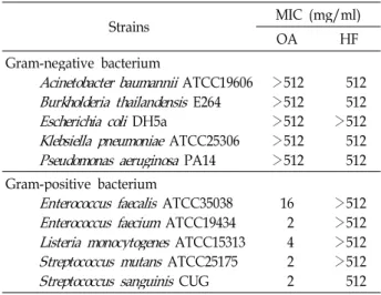
The results of MIC values against various bacteria were shown in Table 1. OA exhibited antimicrobial effects against all Gram-positive bacteria tested at low concentration of the compound, but not Gram-negative bacteria. However, HF did not display any antibacterial activity against all strains tested in this study. Horiuch research group suggested that the compounds are not active on Gram-negative bacterium, including E. coli, P. aeruginosa and Serratia marcescens, due to the existence of the outer membrane which is equipped
Table 1. MICs of OA and HF against several Gram-negative and Gram-positive bacteria
Strains MIC (mg/ml)
OA HF
Gram-negative bacterium
Acinetobacter baumannii
ATCC19606Burkholderia thailandensis
E264Escherichia coli
DH5aKlebsiella pneumoniae
ATCC25306Pseudomonas aeruginosa
PA14>512>512
>512>512
>512
512512
>512512 512 Gram-positive bacterium
Enterococcus faecalis
ATCC35038Enterococcus faecium
ATCC19434Listeria monocytogenes
ATCC15313Streptococcus mutans
ATCC25175Streptococcus sanguinis
CUG162 42 2
>512>512
>512>512 512
with several multidrug efflux pumps [6]. Of the Gram-pos- itive strains tested in this study, E. faecalis and E. faecium are commonly isolated from human infections and resistant to many antimicrobial agents in many cases [19]. Listeria monocytogenes is a clinically important human pathogen causing listeriosis with a high fatality rate [21] and has been reported as an antimicrobial-resistant strain [14]. Therefore, the discovery of a novel drug will be indispensable to treat patients suffering with the infection of the drug-resistant en- terococcal species and L. monocytogenes.
OA significantly suppressed the growth of two common endogenous oral pathogens, Streptococcus mutans and Streptococcus sanguinis, suggesting the possibility of the de- velopment to a potential therapeutic substance originated from plants to prevent serious dental caries. The results sug- gest that OA can be utilized as a valuable medical drug to eliminate the proliferation of clinically important human pathogens, particularly Gram-positive bacteria.
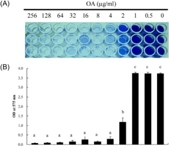
OA inhibits surface adherence and biofilm formation of S. mutans
To determine if OA hinders the biofilm formation of S.
mutans on surface, cells were grown on the 96-well poly- styrene plate containing BHI medium supplemented with 1% sucrose in the presence of various amounts of OA. In this experiment, sucrose was added to the medium for pro- viding enough substrate of GTFs, which synthesizes glucan polymers, key factors in the development of a biofilm [1].
OA solutions ranging from 0 to 256 μg/ml were prepared by routine two-fold serial dilution method. As shown in Fig.
2, OA inhibited biofilm formation in a dose-dependent man- ner, that was completely repressed at the concentration of 4 μg/ml of OA.
The level of bacterial adherence to glass surface was ex- amined in the presence of OA if the compound affects in vitro adherence of S. mutans to surface. The effects of differ- ent concentrations of OA on adherence of S. mutans to glass tubes are shown in Fig. 3. OA significantly reduced the bac- terial adherence to the glass surface at the concentration of 8 μg/ml by 80%. Adherence ability to glass surface was slightly higher than the one to polystyrene plates in the ad- herence assay. The initial adhesion between bacteria and the surface substrate is governed by the physical properties of the bacteria and the surface material. Charged bacteria adhere in more amounts to hydrophilic glass surface than to hydrophobic polystyrene surface. In addition, adhesion
(A)
(B)
Fig. 2. Inhibitory effect on biofilm formation of
S. mutans
by OA. A 5×10⁷CFU amount ofS. mutans
cells was added to each well on the 96-well plate containing different amounts of OA ranging from 0 to 256 μg/ml. The degree of biofilm formation was performed by staining the cells with crystal violet (A) and measuring the optical density at 575 nm (B).abc: means with different letters are differ- ent (p
<0.05)0.0 0.2 0.4 0.6 0.8 1.0 1.2
16 OA (ug/ml)
OD at 600 nm
b b b
a a
0 2 4 8 16
Fig. 3. Anti-adherence activity of OA against
S. mutans
. The bac- teria cells were grown at an angle of 30oin a glass tube in presence of different concentrations of OA ranging from 0 to 16 μg/ml. Attached cells to the tube were sol- ublized by adding sodium hydroxide, followed by meas- uring the optical density of the cells at 600 nm.ab: means with different letters are different (p
<0.05)increases with increasing contact angle [15,20].
Taken together, the data suggest that OA, quite common in nature, is capable of suppressing S. mutans associated with dental caries or periodontal diseases by hindering the growth of bacterial cells, the dental plaque formation or the bacterial adherence on the tooth surface.
Previously, it has been reported that OA inhibited pepti-
doglycan metabolism in L. monocytogenes, resulting in the im-
paired proliferation of the bacterial cells [10]. Therefore, it is necessary to examine if same mechanism applies to the case of S. mutans by OA addition.
The GTFs produced by S. mutans are responsible for syn- thesizing glucan polymers, which plays a critical role in bio- film formation and cell adhesion [1,17]. Among three GTFs, GtfB, GtfC and GtfD, the amount of GtfB and GtfC controls biofilm formation [12]. The expression of the gtfBC operon encoding two glucosyltransferase enzymes is negatively governed by a global regulator, CovR [4]. Besides glucan polymers, glucan-binding proteins (GbpA, GbpB, GbpC, and GbpD) synthesized by S. mutans have been found to influ- ence biofilm formation or adhesion [3].
S. mutans also produces autoinducer 2 (AI-2) by which virulence characteristics of the pathogen are regulated. The synthesis of the molecule is known to be carried out by the LuxS enzyme [24]. The loss of the LuxS enzyme was shown to inhibit biofilm growth in S. mutans [13,23].
Based on the findings explained above, it also will be nec- essary to determine what molecular mechanism(s) is/are de- scribed for inhibitory effect of OA on biofilm formation and/or cell adhesion to surface of S. mutans.
In conclusion, this work represents considerable possi- bility that OA, found in natural foods, is able to be utilized as an alternative for prevention and medical treatment of dental diseases. However, further experiments are required to understand how OA works on extirpating S. mutans on tooth.
Acknowledgements
This study was supported by a grant of the Korea Healthcare technology R&D Project, Ministry for Health, Welfare & Family Affairs, Republic of Korea (A084717).
References
1. Aoki, H., T. Shiroza, M. Hayakawa, S. Sato, and H. K.
Kuramitsu. 1986. Cloning of a Streptococcus mutans gluco- syltransferase gene coding for insoluble glucan synthesis.
Infect Immun. 3, 587-594.
2. Aparecida Resende, F., C. A. de Andrade Barcala, M.
C. da Silva Faria, F. H. Kato, W. R. Cunha, and D. C.
Tavares. 2006. Antimutagenicity of ursolic acid and ole- anolic acid against doxorubicin-induced clastogenesis in Balb/c mice. Life Sci. 79, 1268-1273.
3. Banas, J. A. and M. M. Vickerman. 2003. Glucan-binding
proteins of the oral streptococci. Crit. Rev. Oral Biol. Med.
14, 89-99.
4. Biswas, S. and I. Biswas. 2006. Regulation of the gluco- syltransferase (gtfBC) operon by CovR in Streptococcus mutans. J. Bacteriol. 188, 988-998.
5. Giner-Larza, E. M., S. Máñez, M. C. Recio, R. M. Giner, J. M. Prieto, M. Cerdá-Nicolás, and J. L. Ríos. 2001.
Oleanonic acid, a 3-oxotriterpene from Pistacia, inhibits leukotriene synthesis and has anti-inflammatory activity.
Eur. J. Pharmacol. 428, 137-143.
6. Horiuchi, K., S. Shiota, T. Hatano, T. Yoshida, T. Kuroda, and T. Tsuchiya. 2007. Antimicrobial activity of oleanolic acid from Salvia officinalis and related compounds on vancomycin-resistant enterococci (VRE). Biol. Pharm. Bull.
30, 1147-1149.
7. Islam, B., S. N. Khan, I. Haque, M. Alam, M. Mushfiq, and A. U. Khan. 2008. Novel anti-adherence activity of mulberry leaves: inhibition of Streptococcus mutans bio- film by 1-deoxynojirimycin isolated from Morus alba. J.
Antimicrob. Chemother. 62, 751-757.
8. Jiménez-Arellanes, A., M. Meckes, J. Torres, and J.
Luna-Herrera. 2007. Antimycobacterial triterpenoids from Lantana hispida (Verbenaceae). J. Ethnopharmacol. 111, 202-205.
9. Kashiwada, Y., H. K. Wang, T. Nagao, S. Kitanaka, I.
Yasuda, T. Fujioka, T. Yamagishi, L. M. Cosentino, M.
Kozuka, H. Okabe, Y. Ikeshiro, C. Q. Hu, E. Yeh, and K. H. Lee. 1998. Anti-AIDS agents. 30. Anti-HIV activity of oleanolic acid, pomolic acid, and structurally related triterpenoids. J. Nat. Prod. 61, 1090-1095.
10. Kurek, A., A. M. Grudniak, M. Szwed, A. Klicka, L.
Samluk, K. I. Wolska, W. Janiszowska, and M.
Popowska. 2009. Oleanolic acid and ursolic acid affect peptidoglycan metabolism in Listeria monocytogenes.
Antonie Van Leeuwenhoek in press.
11. Loo, C. Y., D. A. Corliss, and N. Ganeshkumar. 2000.
Streptococcus gordonii biofilm formation: identification of genes that code for biofilm phenotypes. J. Bacteriol. 182, 1374-1382.
12. Mattos-Graner, R. O., M. H. Napimoga, K. Fukushima, M. J. Duncan, and D. J. Smith. 2004. Comparative analy- sis of Gtf isozyme production and diversity in isolates of Streptococcus mutans with different biofilm growth phenotypes. J. Clin. Microbiol. 42, 4586-4592.
13. Merritt, J., F. Qi, S. D. Goodman, M. H. Anderson, and W. Shi. 2003. Mutation of luxS affects biofilm formation in Streptococcus mutans. Infect. Immun. 71, 1972-1979.
14. Poroś-Głuchowska, J. and Z. Markiewicz. 2003.
Antimicrobial resistance of Listeria monocytogenes. Acta Microbiol. Pol. 52, 113-129.
15. Rutter, P. R. and A. Abbott. 1978. A study of the inter- action between oral Streptococci and hard surfaces. J.
Gen. Microbiol. 105, 219-226.
16. Sánchez, M., C. Theoduloz, G. Schmeda-Hirschmann, I.
Razmilic, T. Yáñez, and J. A. Rodríguez. 2006.
Gastroprotective and ulcer-healing activity of oleanolic acid derivatives: in vitro-in vivo relationships. Life Sci. 79, 1349-1356.
17. Schilling, K., M. and W. H. Bowen. 1992. Glucans synthe- sized in situ in experimental salivary pellicle function as specific binding sites for Streptococcus mutans. Infect.
Immun. 60, 284-295.
18. Sohn, K. H., H. Y. Lee, H. Y. Chung, H. S. Young, S.
Y. Yi, and K. W. Kim. 1995. Anti-angiogenic activity of triterpene acids. Cancer Lett. 94, 213-218.
19. Takeuchi, K., H. Tomita, S. Fujimoto, M. Kudo, H.
Kuwano, and Y. Ike. 2005. Drug resistance of Enterococcus faecium clinical isolates and the conjugative transfer of gentamicin and erythromycin resistance traits. FEMS Microbiol. Lett. 243, 347-354.
20. van Loosdrecht, M. C. M., W. Norde, J. Lyklema, and A. J. B. Zehnder. 1990. Hydrophobic and electrostatic parameters in bacterial adhesion. Aquatic Sciences 52,
103-114.
21. Vázquez-Boland, J. A., M. Kuhn, P. Berche, T.
Chakraborty, G. Domínguez-Bernal, W. Goebel, B.
González-Zorn, J. Wehland, and J. Kreft. 2001. Listeria pathogenesis and molecular virulence determinants.
Clin. Microbiol. Rev. 14, 584-640.
22. Yamanaka-Okada, A., E. Sato, T. Kouchi, R. Kimizuka, T. Kato, and K. Okuda. 2008. Inhibitory effect of cran- berry polyphenol on cariogenic bacteria. Bull. Tokyo Dent. Coll. 49, 107-112.
23. Yoshida, A., T. Ansai, T. Takehara, and H. K. Kuramitsu.
2005. LuxS-based signaling affects Streptococcus mutans biofilm formation. Appl. Environ. Microbiol. 71, 2372-2380.
24. Wen, Z. T. and R. A. Burne. 2002. Functional genomics approach to identifying genes required for biofilm devel- opment by Streptococcus mutans. Appl. Environ. Microbiol.
68, 1196-1203.
25. Wu, C. D. 2009. Grape products and oral health. J. Nutr.
139, 1818S-1823S
초록:Oleanolic acid(OA)의 Streptococcus mutans 에 대한 저해효과 윤요한․최경희
1,2*
(한국원자력연구원 정읍 방사선과학연구소 식품생명공학연구실,
1원광대학교 치과대학 구강미생물학교실,
2