Journal of Dental Hygiene Science Vol. 7, No. 1 pp. 21~24 (2007)
21
산에 대한
Streptococcus mutans
KCTC 3065의 스트레스 반응에 관한 연구강경희†·김지영
경남정보대학 치위생과
Analysis of Acid Stress Response in
Streptococcus mutans
KCTC 3065Kyung-Hee Kang† and Ji-Young Kim
Department of Dental Hygiene, Kyungnam College of Information & Technology, Busan 617-701, Korea
ABSTRACT Dental caries is initiated by the acid accumulated in dental plaque.
Streptococcus mutans, one of a major causal agents of dental caries, is component of the dental plaque and produces various organic acids such as lactic acid as the end-product of glycolysis. As a consequence, we investigated the acid stress response of
S. mutans KCTC 3065in this study. The addition of lactic acid to the growth media had a concentration-dependent effect on the growth of
S. mutans.
S.mutans
exhibited higher maximum culture OD compared with the more acidic growth pH values. At treatment of centration of 20 mmol/L lactic acid in the mid-log phage, cell growth was reduced to 40% relative to the control. The following results were obtained with the treatment of cells with a concentration of 20 mmol/L lactic acid in the mid-log phage for 2hrs: Analysis of fatty acids extracted from cells showed that growth at a concentration of 20 mmol/L lactic acid resulted in changes in C
14:0, C
16:0, C
18:0and C
18:1fatty acids. Protein profiles investigated by SDS-PAGE showed that approximately 70, 60, 45, 40 and 23 kDa proteins were highly expressed in
S. mutansKCTC 3065.
Key words
Streptococcus mutans, Acid stress, Growth
서 론
현대인의식생활형태의변화에따른 당의섭취증가와신 체내·외의다양한원인으로인한면역기능의약화로구강내 미생물은증가하는추세에 있으며1)의료기술의비약적인발전 으로인간의수명이연장되고경제성장으로인한소득의 증가 와건강에관한 인식향상으로 인하여치아건강에대한관심 과 요구는 날로 증가하고 있다
.
가장대표적인구강내질병중의하나인치아우식증은치면 에부착된치면세균막에축적되어진산에의해치질이탈회됨 으로써치아조직의결손을 초래하는세균성 치아경조직 질환 이다2,3)
.
Streptococcus mutans, Streptococcus mitis, Streptococcus salivarious, Actinomyces sp., Rothia sp., Norcardia sp., Bac-teroides sp., Neissrria sp., 등은 함께 관여하여 구강병 발생원인중 미생물적요인을형성하고있는것으로알려져있다4).
특히,
S.mutans는치면세균막에존재하고있으면서산생성능을갖고있
을뿐만아니라우식병소내의강한산성환경에서도생존할수 있는내산성을가지고있어인간의우식원인균으로 가장주 목받고 있다5)
.
S. mutans는구강내에상재하는통성혐기성균으로치면의
피막에부착후
sucrose
를기질로하여glycolysis
의end-product
로서
lactic acid
를 생산하며, glucosyl transferase
를 분비하여glucose
중합체인불용성glucan
을형성한다.
이러한glucan
은 불용성의점액성물질로서치아표면에부착하여치면에세균부 착을 도우며생성된유기산을국소적으로체류시킴으로치질의 탈회를 가속화시키는 역할을 한다.
결과적으로 S. mutans는 치면에 부착하여당질대사를통하여 고농도의산을생산하고enamel
을 탈회시킴으로써 치아우식을 유발시킨다6,7).
많은연구들은 S. mutans가구강내의다양한 세균들중에서
도
pH 4
와 같은낮은pH
에서도당질 대사를수행하며 생존할 수있는 내산성균주로서 산에대하여가장잘 견디는균 주로보고해 왔으며8,9)
,
S. mutans의이러한 능력은다른구강 내 세균과구분되는특성으로서충치의진행에있어서 결정적인 영향을 미치는 것으로 알려져 있다10,11)
.
현재우리 나라에서 진행되는 S. mutans 관련 연구는항균
및 유기산 억제효과를 지니는 천연물질의 탐색에 관한 것이 대부분이다12,13)
.
그러나치아우식증에대한근본적인원인해결을위해서는 S. mutans에 의한치아우식증발병과정의 분자생
물학적 기작과 S. mutans가 구강내부환경에노출시겪는 다
양한
stress
에저항하는능력등을분석하는연구가필요하며14),
특히 S. mutans의 산에 대한
stress
에 저항하는 방어기작에관한연구는치아우식증에대한병독성해결에있어서 주요한 해결책을 제시할 수 있을 것으로 사료되어진다
.
따라서본 실험에서는산에대한 S. mutans의 스트레스 반
†
Corresponding author Tel: 051-320-2922 Fax: 051-320-2900
E-mail: [email protected]
22
Analysis of Acid StressResponse inStreptococcus Mutans
KCTC 3065응을 이해하기 위하여 S. mutans
KCTC 3065
을 이용하여lactic acid
를 농도별로첨가 하였을때일어나는 반응을생리적인 측면에서살펴보고자하였으며
acid stress
동안에균주 의생장곡선,
세포막지방산 변화와산에 대한민감성및 단 백질 패턴 변화를 관찰하였다.
재료 및 방법
1. 균주 및 배지
본실험에사용한균주는생명공학연구소로부터분양받은 S.
mutans
KCTC 3065
를사용하였으며,
균주의 보관및 배양은brain heart infusion(BHI)
배지를 사용하였다.
2. 배양
고압멸균한
BHI
배지에 S. mutans를1 loop
접종하고37
oC
에서
12
시간 배양하여 전 배양한 균주로 사용하였다.
3. Growth test
고압멸균한
BHI
액체배지에0mM, 10mM, 20mM, 30mM, 40 mM
의lactic acid
를 농도별로각각첨가하고전 배양한균주를
1%
접종한 후, 37
oC
에서배양하면서Spectrophotometer (Uvicon 932, USA)
를 이용하여600 nm
에서OD
값을측정하였다
.
또한 고압 멸균한BHI
액체배지에 전 배양한 균주를1%
접종하여37
oC
에서 지수증식기까지 배양한 후, 0 mM, 10 mM, 20 mM, 30 mM, 40 mM
의lactic acid
를 농도별로각각 첨가하여 위와 같은 방법으로
OD
값을 측정하였다.
4. 지방산 분석
고압멸균한
BHI
액체배지에전배양한균주를1%
접종하여
37
℃에서지수증식기까지배양한후,
배양한균체에20 mM
의
lactic acid
를 처리하고2
시간동안시간대별로균체를 모아 세척한 후screw cap tube
에 옮겼다. 15% NaOH
를 첨가한50% methanol 1 ml
을 가하고100
oC
에서30
분간 가열한 후 냉각하여6.0 N HCl 325 ml
과methanol 275 ml
을 혼합한용액을
2 ml
을가하고80
oC
에서10
분간 가열한 후냉각하였 다. Hexan
과Methy-tert Butyl Ether
를 동량으로 섞은 용액 을1.25 ml
첨가한 뒤10
분간잘 혼합하여반응액을실온에서 정치하여2
개의 층으로 분리시킨 뒤 하등액을 제거하고0.25 M NaOH
를3 ml
첨가하여5
분간혼합하였다.
포화NaCl
를 적당량가하여 층 분리를확실히 한 후 상등액을
screw
sample vial(Hewlett Packard)
로 옮겨 시료로 사용하였다.
지방산분석에는
Hewlett Packard series II gas chromatograph model 5890A(Microbial ID, Inc., Delaware, USA)
를 이용 하였다.
이용한separation column
은25 cm
×0.22 cm
×0.33 cm methyl phenyl silicone fused silica capillary column(HP 1909B- 102)
을 사용하였고, Microbial Identification System Software (Microbial ID, Inc., Delaware, USA)
를 통해 지방산의profile
을 확인하였다
.
5. 산에 대한 민감성 측정
고압멸균한
BHI
액체배지에전배양한균주를1%
접종하여
37
oC
에서지수증식기까지 배양한 후, lactic acid
를 각각0, 60, 70, 80 mM
로 농도별로 첨가하여0, 30, 60, 90 min
반응후
BHI
한천배지에5
µl
떨어뜨려37
oC
에서24
시간배양 하여 결과를 관찰하였다.
6. SDS-PAGE Gel Electrophoresis
BHI
액체배지에전 배양한 균주를1%
접종한 후, 37
oC
에 서 배양하여지수증식기까지 배양시켜20 mM
의lactic acid
를 첨가하여2
시간동안acid shock
를 주었다.
이후균체를 원심 분리하여침전시켰고,
생리식염수로세척하였다. Glass bead
와0.1 mM PMSF
가 포함된lysis buffer(80 mM Tris-Cl pH 7.3)
를
2 : 1 : 1
의비율로넣고Micromixer
를이용하여2
분간6
회lysis
시켰다.
균체를원심분리하여상등액을취한후Bradford
법으로단백질을정량하고
12% SDS-PAGE Gel
에loading
하 여 전기영동 하였다. CBB-R(0.001% Coomassie Blue R-250, 45% methanol, 10% acetic acid)
으로 염색 후 탈색하여 확인하였다
.
결과 및 고찰
1.
S. mutans
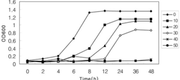
KCTC 3065의 생육곡선Lactic acid
를 배지에 농도별로 첨가하여 농도에 따른 S.mutans의 성장패턴을 조사하였다
. Fig. 1
에서 보듯이 배지에lactic acid
의함량을증가시킬수록균주의생육이저해되는 것을 알 수있으며
40 mM
이상의lactic acid
를 첨가한배지에서는 흡광도의증가가 거의 없었다
. Lactic acid
를 첨가하지않았을 때의 최대 흡광도는
1.35
였고, 10 mM
의lactic acid
첨가배지에서는
1.15
로 정상조건에 비해 생육이감소하였다. 30 mM
의lactic acid
첨가배지에서는최대흡광도가0.86
으로 정상조건에비해 생육이현저하게감소하였으며, 40 mM
에서는최대흡광도가
0.11
로 시간의경과에따른흡광도의증가가 거 의 없었다.
또한lactic acid
를첨가하지않은 정상배지에서는 약8
시간이 경과하여 정지기에도달하는반면, lactic acid
의 농도가증가되는배지일수록정지기에도달하는데시간이오래 소요되어, 10 mM
의lactic acid
첨가배지에서는약12
시간후에 정지기에 도달하였으며
, 20 mM
과30 mM
의lactic acid
첨가배지에서는약
24
시간 후에정지기에도달하여정상조건에 비해 정지기에 도달하는데3
배의 시간이 소요되었다.
균의 성장이가장 활발한 지수증식기일 때
lactic acid
를 첨가한 결과(Fig. 2), 10 mM
의lactic acid
를 첨가한 배지에서는 최대흡광도가
1.30
으로30 mM
의lactic acid
를 첨가한Fig. 1.
Growth Curve. Cells were grown in BHI media(pH 7.2) containing 0 mM, 10 mM, 20 mM, 30 mM, 40 mM and 50 mM of lactic acid for 48hrs at 37oC.Journal of Dental Hygiene Science Vol. 7
,No. 1
,pp. 21~24 (2007) 23
배지에서는
0.95
로 나타났으며40 mM
에서는 최대흡광도가0.84
로시간의경과에따른흡광도의 변화가거의없었다.
또한
10 mM
의lactic acid
를첨가한 배지에서는약8
시간이경과하여 정지기 상태로 접어들었으며
, 20 mM
과30 mM
의lactic acid
첨가배지에서는약10
시간후에정지기에도달하는 것을 관찰할 수 있었다.
이러한결과로 배지에첨가 된
lactic acid
의함량에 비례하여 S. mutans의성장이완만해지는것을 알수 있었으며유
도기일 때
lactic acid
를 첨가한 경우와 지수증식기일 때lactic acid
를 첨가한경우의OD
값을비교해보면 지수증식기에
lactic acid
를 첨가한 경우가 유도기에 비해균의 성장이더 활발한것을 관찰 할 수 있었는데
,
이는 지수증식기까지 성장하는 동안대사과정을 통하여생성되어지는산에 대하여S. mutans가 특유의 산 적응기작에 의하여 적응한 결과로
사료되어진다
.
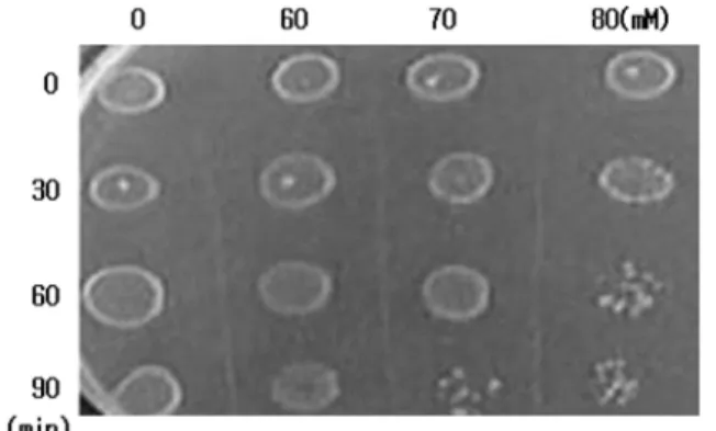
2. 산에 대한 균주의 Survival Test
지수증식기의 균주를 취하여
0, 60, 70
그리고80 mM
농도의
lactic acid
를 처리한 후 각각0, 30, 60, 90 min
후에
colony
의 생성을 관찰하여 균주의 생존유무를 살펴보았다(Fig. 3).
70 mM
의 농도로lactic acid
를 처리하고90
분후부터는colony
의 밀도가 급격히낮아지며생존에 영향을받는 것을관찰할 수있었으며
, lactic acid
의농도가80 mM
이상에서는 생존이 거의 불가능한 것을 관찰할 수 있었다.
이러한 결과로 S. mutans는
80 mM
이상의 고농도의 산에노출되는 경우 생육이 불가능한 것을 알 수 있었다
.
3. Lactic acid 처리 시 세포막 지방산 조성 분석 일반적으로세포막의지방산들은 포화지방산과불포화지방산 으로혼합되어있으며
,
불포화지방산은이중결합의수에따라그 포화도가 조금씩차이를 보인다
.
S. mutansKCTC 3065
의 지방산 조성을 분석한 결과
, C
16:0과C
18:1이 전체의 약70%
를 차지하는것을 알 수 있었으며, acid stress
동안에는C
14:0, C
16:0, C
16:1, 그리고C
18:1에 변화를 나타내었다.
불포화지방산인
C
18:1은30.92%
에서33.89%
로 증가하였으며포화지방산인
C
14:0과C
16:0은 각각5.02%
에서2.62%
로, 39.16%
에서33.69%
로 감소하는 것을볼 수 있었다(Fig. 4).
일반적으로stress
하에서 막지방산들은 막의손상을 받아포화지방산이감소하고불포화지방산의비가증가하며이로인하여막의유 동성이증가한다고알려져있다15,16)
.
위결과에서도stress
가가해지는시간에따라 불포화도가증가하고포화도는감소하는 경향을 볼 수 있었다
.
그러나 불포화지방산인C
16:1의 경우7.21%
에서4.38%
로 감소하는상이한 결과를나타내앞으로의연구에서이에대한원인규명이필요할것으로사료되어진다
.
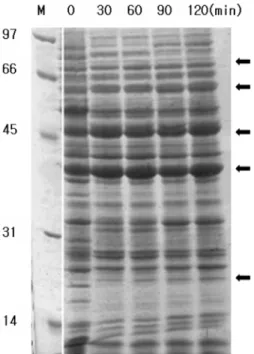
4. Lactic acid 처리 시 단백질 패턴 분석
S. mutans를
BHI
액체배지에서 지수증식기까지 배양시켜20 mM
의lactic acid
를처리하고2
시간동안시간대별로 균체 를 모아단백질을회수하여 단백질패턴을관찰하였다. SDS- PAGE
전기영동을 통하여 확인한 결과, acid stress
동안 발현 이 증가된 단백질은 약70, 60, 45, 40,
그리고23 kDa
의 분자량을 가지는단백질들로band
가 증가하는것을 가시적으 로 확인할 수 있었다(Fig. 5).
이들단백질은
acid stress
에관여하는단백질들로추정되며,
다른 논문에서 발표된 많은
stress proteins
들과도 연관성이큰 겻으로 판단된다17,18)
.
이미 알려진acid stress protein
중에는
Hsp90, Hsp70, Hsp60
등이보고되고있으며acid shock protein
은heat shock stress, oxidative stress
등과 같은stress response
와도 서로 관련이 있다고 보고 되고 있다19,20).
S.mutans가다른 구강내미생물들과는 달리내산성 가질수있
는 요인으로스트레스단백질의 발현은매우중요하며 따라서 앞으로의연구에서는스트레스단백질을동정하고유전자발현 형태및관련대사를 밝혀내는연구가 계속적으로행해져야할 것으로 사료되어 진다
.
Fig. 2.
Growth Curve. Lactic acid was added in the exponential phase of growth with different concentrations of 0 mM, 10 mM, 20 mM, 30 mM, 40 mM and 50 mM for 24hrs at 37 oC.Fig. 3.
Survival test against lactic acid. Cells growing in the exponential phase were treated with 60, 70 and 80 mM of lactic acid for 90min at 37oC.Fig. 4.
Fatty acid composition. Exponentially grown cells were treated with 20 mmol/L lactic acid. Fatty acid composition was measured at 0, 30, 60, 90 and 120 min.24
Analysis of Acid StressResponse inStreptococcus Mutans
KCTC 3065요 약
본 실험에서는 S. mutans
KCTC 3065
을 이용하여lactic acid
를 첨가하였을때 일어나는스트레스 반응을살펴보고자 하였으며 다음과 같은 결론을 얻었다.
1.
배지에lactic acid
를농도별로 첨가하여 농도에따른생 육저해현상을조사한결과,
배 지에 첨가된lactic acid
의함량에비례하여 S. mutans의 성장이완만해지며
,
지 수증식기일때lactic acid
를첨가 하였을경우유도기일때
lactic acid
를 첨가한 경우보다 균의 성장이 활발한것을 알 수 있었다
.
2.
대수증식기의균주를 취하여0, 60, 70, 80 mM
농도의lactic acid
를 처리한후colony
의 생성으로균주의생존 유무를관찰한 결과, 70 mM
의 농도로lactic acid
를 처 리하고90
분후부터는colony
의 밀도가급격히 낮아지며생존에 영향을 받는 것을 관찰할 수 있었으며
, lactic
acid
의 농도가80 mM
이상에서는 생존이 거의불가능한 것으로 나타났다
.
3. Acid stress
동안에 지방산 조성의 변화를 관찰한 결과, C
18:1은30.92%
에서33.89%
로증가하였으며C
14:0은5.02%
에서
2.62%
로C
16:0은39.16%
에서33.69%
로감소하였다. 4. Acid stress
동안에 단백질 패턴의 변화를 관찰한 결과,
약
70 kDa, 60 kDa, 45 kDa, 40 kDa
그리고23 kDa
의 단 백질발현이 증가되는 것을 볼 수 있었다.
참고문헌
1. Houte JV: Bacterial specificity in the etiology of dental caries.
Int den J 30: 305-308, 1980.
2. Marsh PD: Microbiological aspects of the chemical control of plaque and gingivitis. J Dent Res 71(7): 1431-1438, 1999.
3. Inoue M, Koga T: Properties of glucans produced by Streptococcus mutans. Infect Immunity 25: 922-929, 1979.
4. Bende GR, Sutton SCV, Marpuis RE: Acid tolerande, proton permeabilities and membrane ATPase of oral streptococci. Infect Immun 53: 331-338, 1986.
5. Li YH, Lau PCY, Tang N, Svensater G, Ellen RP, Cvitkovitc DG: Novel two-component regulatory system involved in biofilm formation and acid resistance in Streptococcus mutans. J Bacteriol 184: 6333-6342, 2002.
6. Muroi H, Kubo I: Combination effects of anti-bacterial compounds in green tea flavor against Streptococcus mutans. J Agric Food Chem 4: 1102-1105, 1993.
7. Luck TA: Review of its action on Mutans sterptococci and dental plaque-its clinical significance. Int Dent J 45: 77-92, 1999.
8. Belli WA, Marquis RE: Adaptation of Streptococcus mutans and Enterococcus hirae to acid stress in continuous culture. Appl Environ Microbiol 57: 1134-1138, 1991.
9. Goldberg I, Eschar L: Stability of lactic acid bacteria to freezing as related to heir fatty acid composition. Appl Environ Microbiol 33: 489-496, 1997.
10. Guggenheim B: Extracelluar polysaccharides and microbial plaque.
Int Dent J 20: 657-678, 1970.
11. Hanada N, Takehara T: (1-3)-alpha-D-glucan synthase from Strepto- coccus mutans AHT(serotype) does not synthesise glucan withdout primer. Carbohydr Res 168(1): 120-124, 1987.
12. Hwang JK, Kim HJ, Shim JS, Pyun YR: Bactericidal activity of chitosan on S treptococcus mutans . Kor J Food Sci Technol 31:
522-526, 1999.
13. Chun JY, Ryu IH, Lee SU, Lee KS: Isolation and Identification of novel alkalophilc Bacillus alkalophishaggy JY-827 with anticaries micobe Streptoccus mutans . Kor J Appl Microbiol Biotechnlo 28:
243-250, 2000.
14. Han YK, Song SS, Lee IS: Acid tolerance response of Streptoccus mutans at anaerobic condition. J Dent Hygine Sci 1: 7-11, 2001.
15. Bardi LC, Marzona M: Saccharomyces cerevisiae cell fatty acid composition and release during fermentation without aeration and in absence of exogenous lipids. Int J Food Microbio 47: 133- 140, 1999.
16. Benchekroun K, Bonaly R: Physiological properties and plasma membrane composition of Saccharomyces cerevisiae grown in sequential batch culture and in presence of surfactant. Appl Microbiol Biotechnol 36: 673-678, 1992.
17. Van HJ, Lopman J, Kent RO: The final pH of bacteria comprising the predonimant flora of sound and carious human root and enamel surfaces. J Dent Res 75: 1008-1014, 1996.
18. Hahn K, Faustoferri RC, Quivey JR: Induction of an AP endonu- clease activity in Streptococcus mutans during growth at low pH.
Mol Microbiol 31: 1489-1498 1999.
19. Suzuki T, Tagami J, Hanada N: Role of F1F0-ATPase in the growth of S treptococcus mutans GS5. J Appi Microbiol 88: 555- 562, 2000.
20. Svensater G, Sjogreen B, Hamilton I: Multiple stress responses in Streptococcus mutans and the induction of general and stress- specific proteins. Microbiology 146: 107-117, 2000.
(Received February 12, 2007; Accepted March 21, 2007)
Fig. 5.
Protein display using SDS-PAGE Gel Electrophoresis.Protein standards were : rabbit muscle phosphorylase B(97 kDa);
bovin serum albuin(66 kDa); ovalbumin(45 kDa); bovin carbonic anhydrase(31 kDa); hen egg lysozyme(14 kDa)