• 검색 결과가 없습니다.

Virulence genes of Streptococcus mutans and dental caries

N/A
N/A
Protected

Academic year: 2021

Share "Virulence genes of Streptococcus mutans and dental caries"

Copied!
6
0
0

로드 중.... (전체 텍스트 보기)

전체 글

(1)

Dental Biofilm and Streptococcus mutans

Dental caries and periodontal disease are typical oral diseas- es that are prevalent worldwide, and still represent the primary cause of tooth loss, so effective management measures are required [1,2]. Oral bacteria form dental plaque, which is a kind of biofilm formed on the supragingival and the subgingival sur- faces of tooth and the root surface of tooth [3]. Microorganisms in the dental plaque metabolize carbohydrates to organic acids, thereby destroying the hard tissues of teeth and causing dental caries. Periodontitis results from an inflammatory response of

the periodontal tissues to oral bacteria [4-7].

S. mutans is an oral bacterium representing mutans strep- tococci. It is one of important bacteria for formation of supra- gingival dental plaque which is a dental biofilm formed on the supragingival tooth surface, and of dental caries [2,8].

Acquired Pellicle Formation and Bacterial Capture

Formation of the dental plaque occurs via complex multi- stage processes. The first step is the acquired pellicle forma- Int J Oral Biol 44:31-36, 2019

pISSN: 1226-7155 • eISSN: 2287-6618 https://doi.org/10.11620/IJOB.2019.44.2.31

Virulence genes of Streptococcus mutans and dental caries

Yong-Ouk You*

Department of Oral Biochemistry, School of Dentistry, Wonkwang University, Iksan 54538, Republic of Korea

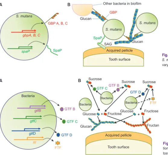
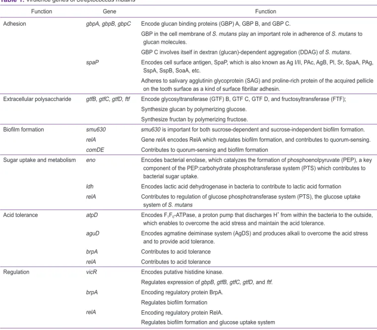
Streptococcus mutans is one of the important bacteria that forms dental biofilm and cause dental caries. Virulence genes in S. mutans can be classified into the genes involved in bacterial adhesion, extracellular polysaccharide formation, biofilm formation, sugar uptake and metabolism, acid tolerance, and regulation. The genes involved in bacterial adhesion are gbps (gbpA, gbpB, and gbpC) and spaP. The gbp genes encode glucan-binding protein (GBP) A, GBP B, and GBP C. The spaP gene encodes cell surface antigen, SpaP. The genes involved in extracellular polysaccharide formation are gtfs (gtfB, gtfC, and gtfD) and ftf, which encode glycosyltransferase (GTF) B, GTF C, and GTF D and fructosyltransferase, respectively. The genes involved in biofilm formation are smu630, relA, and comDE. The smu630 gene is important for biofilm formation. The relA and comDE genes contribute to quorum- sensing and biofilm formation. The genes involved in sugar uptake and metabolism are eno, ldh, and relA. The eno gene encodes bacterial enolase, which catalyzes the formation of phosphoenolpyruvate. The ldh gene encodes lactic acid dehydrogenase. The relA gene contributes to the regulation of the glucose phosphotransferase system. The genes related to acid tolerance are atpD, aguD, brpA, and relA. The atpD gene encodes F 1 F 0 -ATPase, a proton pump that discharges H + from within the bacterium to the outside. The aguD gene encodes agmatine deiminase system and produces alkali to overcome acid stress. The genes involved in regulation are vicR, brpA, and relA.

Keywords: Streptococcus mutans, Dental caries, Virulence factor, Dental biofilm

Received May 14, 2019; Revised June 6, 2019; Accepted June 9, 2019

*Correspondence to: Yong-Ouk You, E-mail: [email protected] https://orcid.org/0000-0002-7754-3033 Copyright © The Korean Academy of Oral Biology

CC

This is an open-access article distributed under the terms of the Creative Commons Attribution Non-Commercial License (http://creativecommons.org/licenses/by- nc/4.0/), which permits unrestricted non-commercial use, distribution, and reproduction in any medium, provided the original work is properly cited.

Invited Review IJOB

(2)

tion (Fig. 1A). The enamel surface surrounded by the hydration layer has negative charge because it has a high concentration of phosphate group [2]. Cations such as calcium ions bind to the negative charge of the enamel surface, eventually chang- ing the enamel surface to positive charge. In saliva, there are acidic proteins such as phosphoproteins and sulfate glycopro- teins, which are negatively charged. Acidic proteins bind to the enamel surface via calcium ions to form acquired pellicle [2].

Calcium ion acts as a bridge between the negative charge on the enamel surface and the negative charge of acid proteins.

Oral bacteria are captured on the acquired pellicle of tooth sur- face (Fig. 1B). Bacterial capture in the early stage is reversible attachment which is caused by ionic bond or van der Waals interaction [2].

Bacterial Adhesion

The captured oral bacteria form irreversible adhesions to the tooth surface covered by the acquired pellicle (Fig. 1B).

Calcium bridge, hydrophobic interaction, polymer bridge, or covalent bond between the tooth surface covered with the ac- quired pellicle and bacterial surface are the main mechanisms of irreversible adhesion [2], and also cell surface adhesins of S. mutans, such as glucan-binding proteins (GBPs) and SpaP, are important for bacterial adhesion to tooth surface. S. mu- tans carry gbps genes (gbpA, gbpB, and gbpC) related with the adhesion. The gbpA, gbpB, and gbpC genes encode GBP

A, B, and C, which are known to play an important role in the adhesion of S. mutans to glucan molecules, a kind of extracel- lular polysaccharide of plaque matrix (Fig. 2). In particular, the GBP C is involved in dextran (glucan)-dependent aggregation (DDAG) of oral bacteria [9,10]. S. mutans also carry the spaP gene, which encodes the cell surface antigen, SpaP. The SpaP is also known as Ag I/II, PAc, AgB, Pl, Sr, SpaA, PAg, SspA, SspB and SoaA, which adheres to salivary agglutinin glycopro- tein (SAG) and proline-rich protein of the acquired pellicle on the tooth surface as a kind of surface fibrillar adhesin (Fig. 2) [11-14]. The irreversible adhesion is followed by oral bacterial growth, division and then colonization.

Extracellular Polysaccharide Formation

Matrix of dental plaque is formed after colonization of the oral microorganisms (Fig. 1C). The plaque matrix can be formed in the absence of food. The plaque matrix is classified into protein matrix and extracellular polysaccharide matrix. S. mutans has gtfB, gtfC, gtfD, and ftf genes each of which encode glycos- yltransferases (GTFs) B, C, D, and fructosyltransferase (FTF), respectively [2,12,13]. After the protein matrix of the dental plaque is formed, S. mutans decomposes sucrose in the oral cavity into glucose and fructose using bacterial invertase, and then synthesizes glucan by polymerizing glucose using GTF.

FTF synthesizes polysaccharides such as fructan by polymer- izing fructose. The extracellular polysaccharides thicken and

Glycoprotein

Salivary flow

Acquired pellicle

Tooth surface

Bacteria

Acquired pellicle

Tooth surface

Acquired pellicle

Tooth surface

Acquired pellicle

Tooth surface

Protein matrix Fructan Glucan

A

C

B

D

Sucrose

Fig. 1. Dental biofilm formation. (A) Acquired

pellicle formation. (B) Bacterial adhesion. (C)

Protein matrix formation. (D) Extracellular

polysaccharide formation.

(3)

harden the dental plaque and lower the oxygen permeability, and play a decisive role in dental plaque maturation (Figs. 1D, 3).

Biofilm Formation

The dental plaque formed in oral cavity is a mixed population of biofilms made by various bacteria combining themselves.

Oral streptococci species such as Streptococcus sanguinis, S. mutans, Streptococcus gordonii, Streptococcus mitis, and Streptococcus oralis play the important role in the formation of supragingival plaque and dental caries. Actinomyces species such as Actinomyces viscosus are closely related to the devel- opment of dental plaque and dental caries on the root surface [2,15].

These bacteria combine with the protein matrix formed by the oral bacteria and the extracellular polysaccharides such as glucan and fructan to form the dental plaque on the tooth sur- face. The gene relA carried by S. mutans encodes RelA, which is known to regulate the formation of biofilm and to contribute to quorum-sensing [16]. Among the genes carried by the

S. mutans, smu630 is reported to be important for sucrose- dependent and sucrose-independent dental biofilm formation [12]. The gene brpA in S. mutans is also known to regulate biofilm formation [17]. The gene comD encodes a histidine ki- nase receptor and the gene comE encodes a cognate response regulator of the competence-stimulating peptide, which are part of the quorum-sensing cascade of S. mutans [17].

Sugar Uptake and Metabolism

Oral bacteria uptake sugars that are consumed in food and metabolize them with glucose, then use glucose to get the necessary energy via glycolysis and fermentation, and produce organic acids as metabolic products (Fig. 4) [2,7]. The relA gene of S. mutans encodes RelA, which is known to contribute to the regulation of phosphoenolpyruvate:carbohydrate phos- photransferase system (PTS), the glucose uptake system [16].

The gene eno of S. mutans encodes the bacterial enolase, which is a major component of the PTS in the bacteria, and is known to contribute to bacterial sugar uptake (Fig. 4) [18]. In addition, the gene ldh of S. mutans encodes lactic acid dehy- gbpA, B, C

spaP SpaP

GBP A, B, C

Other bacteria in biofilm

GBP Glucan

SAG SpaP

Glucan S. mutans

Acquired pellicle Tooth surface

A B

S. mutans S. mutans

Fig. 2. Glucan-binding protein (GBP) and SpaP.

S. mutans, Streptococcus mutans; SAG, sali- vary agglutinin glycoprotein.

Bacteria

GTF B

GTF C

GTF D

FTF ftf

gtfD gtfC

gtfB

Bacteria

Bacteria

Bacteria GTF C GTF B

GTF D

FTF Sucrose Sucrose

Sucrose

Glucose Fructose Glucose Fructose

Fructan Acquired pellicle

Tooth surface Glucan

A B

Fig. 3. Extracellular polysaccharide forma-

tion by glycosyltransferases (GTFs) and fruc-

tosyltransferase (FTF).

(4)

drogenase, which contributes to lactic acid formation.

Acid Tolerance

Oral bacteria metabolize the carbohydrates in the food to produce organic acids. Then the surrounding environment be- comes acidic because the proton concentration is increased.

S. mutans can survive and grow in the acidic environment because of its several genes enabling to overcome acidic en- vironment. The gene atpD in S. mutans encodes the F 1 F 0 - ATPase. F 1 F 0 -ATPase is a proton pump that discharges H + from within the bacteria to the outside, to overcome acid stress and maintain acid tolerance (Fig. 5) [18,19]. Moreover, F 1 F 0 -ATPase has another function as an ATP synthase [19]. The inside of bacterial cells maintains neutral pH, but when the pH is low outside the cell, proton gradient is formed on the boundary of the cell membrane. Proton gradient causes a proton mo- tive force in which H + tries to enter from outside the cell. F 1 F 0 - ATPase uses proton motive force to synthesize ATP required

by bacteria. F-ATPase can play a dual role in obtaining acid tol- erance by releasing protons from inside of cells, and also pro- ducing ATP for the growth and survival of bacteria. In addition, aguD encodes the agmatine deiminase system (AgDS), which produces alkali, enabling to overcome acid stress and maintain acid tolerance [18]. The gene brpA and relA also contribute to acid tolerance.

Regulation

The gene vicR encodes putative histidine kinase, which regulates expression of gbpB, gtfB, gtfC, gtfD, and ftf [18].

The gene brpA encoding regulatory protein BrpA, which regu- lates biofilm formation [20]. The gene relA encoding regulatory protein RelA, which is guanosine tetra (penta)-phosphate syn- thetase, and regulates biofilm formation and glucose uptake system [16].

eno ldh

PEP-PTS

PEP

Enolase LDH

Dietary carbohydrates Salivary amylase

Glucose PTS

Glucose Glycolysis 2PG

PEP LDH

Lactic acid Pyruvate

Lactic acid

Acquired pellicle Damage

Tooth surface

A B

S. mutans Enolase

Fig. 4. Sugar transports, glycolysis, and acid production in Streptococcus mutans.

PEP, phosphoenolpyruvate; PTS, phos- photransferase; LDH, lactate dehydrogenase;

2PG, 2-phosphoglycerate.

aguD

atpD

AgDS

Dietary carbohydrates

Lactic acid

Glycolysis H +

H + H +

H + H + H +

pH

pH ATP

H + H +

H +

Agmatine Agmatine AgDS

A B

S. mutans

ADP

Fig. 5. Mechanism of acid tolerence in Strep- tococcus mutans.

AgDS, agmatine deiminase system.

Data from the article of Lemos and Burne (Mi-

crobiology 2008;154 (Pt 11):3247-55) [19].

(5)

Conclusions

Virulence genes in S. mutans can be classified into genes involved in bacterial adhesion, extracellular polysaccharide formation, biofilm formation, sugar uptake and metabolism, acid tolerance, and regulation (Table 1). The genes related with bacterial adhesion are gbps (gbpA, gbpB, and gbpC) and spaP.

The genes related with extracellular polysaccharide formation are gtfs (gtfB, gtfC, and gtfD) and ftf. The genes involved in biofilm formation are smu630, relA and comDE. The genes in- volved in sugar uptake and metabolism are eno, ldh, and relA.

The genes related with acid tolerance are atpD, aguD, brpA,

and relA. The genes involved in the regulation are vicR, brpA, and relA.

Acknowledgements

This paper was supported by Wonkwang University in 2017.

Conflicts of Interest

No potential conflict of interest relevant to this article was reported.

Table 1. Virulence genes of Streptococcus mutans

Function Gene Function

Adhesion gbpA, gbpB, gbpC Encode glucan binding proteins (GBP) A, GBP B, and GBP C.

GBP in the cell membrane of S. mutans play an important role in adherence of S. mutans to glucan molecules.

GBP C involves itself in dextran (glucan)-dependent aggregation (DDAG) of S. mutans.

spaP Encodes cell surface antigen, SpaP, which is also known as Ag I/II, PAc, AgB, Pl, Sr, SpaA, PAg, SspA, SspB, SoaA, etc.

Adheres to salivary agglutinin glycoprotein (SAG) and proline-rich protein of the acquired pellicle on the tooth surface as a kind of surface fibrillar adhesin.

Extracellular polysaccharide gtfB, gtfC, gtfD, ftf Encode glycosyltransferase (GTF) B, GTF C, GTF D, and fructosyltransferase (FTF);

Synthesize glucan by polymerizing glucose.

Synthesize fructan by polymerizing fructose.

Biofilm formation smu630 smu630 is important for both sucrose-dependent and sucrose-independent biofilm formation.

relA Gene relA encodes RelA which regulates biofilm formation, and contributes to quorum-sensing.

comDE Contributes to quorum-sensing and biofilm formation

Sugar uptake and metabolism eno Encodes bacterial enolase, which catalyzes the formation of phosphoenolpyruvate (PEP), a key component of the PEP:carbohydrate phosphotransferase system (PTS) which contributes to bacterial sugar uptake.

ldh Encodes lactic acid dehydrogenase in bacteria to contribute to lactic acid formation relA Contributes to regulation of glucose phosphotransferase system (PTS), the glucose uptake

system of S. mutans

Acid tolerance atpD Encodes F 1 F 0 -ATPase, a proton pump that discharges H + from within the bacteria to the outside, which enables to overcome the acid stress and maintain the acid tolerance.

aguD Encodes agmatine deiminase system (AgDS) and produces alkali to overcome the acid stress and to provide acid tolerance.

brpA Contributes to acid tolerance relA Contributes to acid tolerance

Regulation vicR Encodes putative histidine kinase.

Regulates expression of gbpB, gtfB, gtfC, gtfD, and ftf.

brpA Encoding regulatory protein BrpA.

Regulates biofilm formation relA Encoding regulatory protein RelA.

Regulates biofilm formation and glucose uptake system

(6)

References

1. National Health Insurance Service, Health Insurance Review

& Assessment Service. 2017 National health insurance sta- tistical yearbook. Wonju: National Health Insurance Service, Health Insurance Review & Assessment Service; 2018.

2. Min BM, Kang JS, Ko SY, Kook JK, Kim JH, Ryoo HM, ParK KK, Park BJ, Park SH, Park JE, You YO, Lee G, Lee TH, Lee HJ, Jang YJ, Chung WY, Cho JY, Gee EJ, Choi JY. Oral bio- chemistry. Seoul: DaehanNarae Publishing Inc.; 2007.

3. Kim EH, Kang SY, Park BI, Kim YH, Lee YR, Hoe JH, Choi NY, Ra JY, An SY, You YO. Chamaecyparis obtusa sup- presses virulence genes in Streptococcus mutans. Evid Based Complement Alternat Med 2016;2016:2396404. doi:

10.1155/2016/2396404.

4. Lee KH, Kim BS, Keum KS, Yu HH, Kim YH, Chang BS, Ra JY, Moon HD, Seo BR, Choi NY, You YO. Essential oil of Curcuma longa inhibits Streptococcus mutans biofilm for- mation. J Food Sci 2011;76:H226-30. doi: 10.1111/j.1750- 3841.2011.02427.x.

5. Lasserre JF, Brecx MC, Toma S. Oral microbes, biofilms and their role in periodontal and peri-implant diseases. Materials (Basel) 2018;11:E1802. doi: 10.3390/ma11101802.

6. Lee DH, Seo BR, Kim HY, Gum GC, Yu HH, You HK, Kang TH, You YO. Inhibitory effect of Aralia continentalis on the cario- genic properties of Streptococcus mutans. J Ethnopharmacol 2011;137:979-84. doi: 10.1016/j.jep.2011.07.015.

7. Yang Y, Park BI, Hwang EH, You YO. Composition analy- sis and inhibitory effect of Sterculia lychnophora against biofilm formation by Streptococcus mutans. Evid Based Complement Alternat Med 2016;2016:8163150. doi:

10.1155/2016/8163150.

8. Banas JA, Drake DR. Are the mutans streptococci still consid- ered relevant to understanding the microbial etiology of dental caries? BMC Oral Health 2018;18:129. doi: 10.1186/s12903- 018-0595-2.

9. Kim BS, Park SJ, Kim MK, Kim YH, Lee SB, Lee KH, Choi NY, Lee YR, Lee YE, You YO. Inhibitory effects of Chrysanthemum boreale essential oil on biofilm formation and virulence factor expression of Streptococcus mutans. Evid Based Complement Alternat Med 2015;2015:616309. doi: 10.1155/2015/616309.

10. Sato Y, Yamamoto Y, Kizaki H. Xylitol-induced elevated ex-

pression of the gbpC gene in a population of Streptococcus mutans cells. Eur J Oral Sci 2000;108:538-45. doi: 10.1034/

j.1600-0722.2000.00928.x.

11. Jeong SI, Kim BS, Keum KS, Lee KH, Kang SY, Park BI, Lee YR, You YO. Kaurenoic acid from Aralia continentalis inhibits biofilm formation of Streptococcus mutans. Evid Based Complement Alternat Med 2013;2013:160592. doi:

10.1155/2013/160592.

12. Shemesh M, Tam A, Steinberg D. Expression of biofilm- associated genes of Streptococcus mutans in response to glucose and sucrose. J Med Microbiol 2007;56(Pt 11):1528- 35. doi: 10.1099/jmm.0.47146-0.

13. Jenkinson HF, Lamont RJ. Streptococcal adhesion and colonization. Crit Rev Oral Biol Med 1997;8:175-200. doi:

10.1177/10454411970080020601.

14. Napimoga MH, H öfling JF, Klein MI, Kamiya RU, Gonçalves RB. Tansmission, diversity and virulence factors of Srepto- coccus mutans genotypes. J Oral Sci 2005;47:59-64. doi:

10.2334/josnusd.47.59.

15. Larsen T, Fiehn NE. Dental biofilm infections-an update. AP- MIS 2017;125:376-84. doi: 10.1111/apm.12688.

16. Lemos JA, Brown TA Jr, Burne RA. Effects of relA on key virulence properties of planktonic and biofilm populations of Streptococcus mutans. Infect Immun 2004;72:1431-40. doi:

10.1128/iai.72.3.1431-1440.2004.

17. Steinberg D, Moreinos D, Featherstone J, Shemesh M, Feuerstein O. Genetic and physiological effects of noncoher- ent visible light combined with hydrogen peroxide on Strep- tococcus mutans in biofilm. Antimicrob Agents Chemother 2008;52:2626-31. doi: 10.1128/AAC.01666-07.

18. Xu X, Zhou XD, Wu CD. The tea catechin epigallocatechin gallate suppresses cariogenic virulence factors of Streptococ- cus mutans. Antimicrob Agents Chemother 2011;55:1229- 36. doi: 10.1128/AAC.01016-10.

19. Lemos JA, Burne RA. A model of efficiency: stress toler- ance by Streptococcus mutans. Microbiology 2008;154(Pt 11):3247-55. doi: 10.1099/mic.0.2008/023770-0.

20. Wen ZT, Baker HV, Burne RA. Influence of brpA on criti-

cal virulence attributes of Streptococcus mutans. J Bacteriol

2006;188:2983-92. doi: 10.1128/JB.188.8.2983-2992.2006.

수치

Fig. 1.   Dental biofilm formation. (A) Acquired  pellicle formation. (B) Bacterial adhesion
Fig. 2.  Glucan-binding protein (GBP) and SpaP.
Fig. 4.   Sugar transports, glycolysis, and  acid production in Streptococcus mutans.
Table 1.  Virulence genes of Streptococcus mutans

참조

관련 문서

High level of nitrogen fertilizer application increased the nitrogen contents in all the parts of citrus trees.. But there was no change in sugar and

Frequency of conidial germination (A) and appressorium formation (B) of Colletotrichum orbiculare and callose formation of plant cells (C) at the

The Study on the Changes of Bacterial Inorganic Phosphate Metabolism in Interactions between Nitric Oxide and Salmonlla enterica serovar Typhimurium..

:Acidulated phosphate fluoride treatment and formation of caries-like lesions in enamel: effect of application time.. Schmidt HF : The fluoride varnish

Biochemical markers of bone turnover can be classified according to the process that underlie in markers of bone formation, products of the osteoblast

In terms of the mould design, the effects of runner system design and the mould temperature on filling characteristics, the weldline formation, the

Table 8.. tors for particle formation in gas phase and scale-up of the reactor system. They have developed models for droplet and particle dynam- ics and applied to the

After the vehicle has become a large-formation and high-speed, loud noise from an express highway is coming to be higher and the number of large-scale