․Correspondence to : Kang-san Kim, Sinyong-dong, Iksan-si, Jeollabuk-do, Korea
Dept. of Internal Medicine, College of Oriental Medicine, Won-Kwang University TEL: 063-859-2803
E-mail: [email protected]
Effect of Lindera obtusiloba extract on cancer metastasis
Hyuk Yun1, Yong-jae Lee3, Hyun-won Seo3, Kyoung-jae Park3, Ha-neul Ko1 Dong-seok Cha3, Jin Kwon4, Hoon Jeon3, Kang-san Kim1,2
1Dept. of Internal Medicine, College of Oriental Medicine, Won-Kwang University
2Research Center of Traditional Korean Medicine,3College of Pharmacy, Woo-Suk University,
4Dept. of Prosthetics and Orthotics, Korea National College of Rehabilitation and Welfare
Effect of Lindera obtusiloba extract on cancer metastasis
Hyuk Yun1, Yong-jae Lee3, Hyun-won Seo3, Kyoung-jae Park3, Ha-neul Ko1 Dong-seok Cha3, Jin Kwon4, Hoon Jeon3, Kang-san Kim1,2
1Dept. of Internal Medicine, College of Oriental Medicine, Won-Kwang University
2Research Center of Traditional Korean Medicine, 3College of Pharmacy, Woo-Suk University,
4Dept. of Prosthetics and Orthotics, Korea National College of Rehabilitation and Welfare
ABSTRACT
Objectives : In the present study, anti-metastatic properties of the methanol extract of L. obtusiloba (MLO) were evaluated.
Methods :To determine the effect of MLO on cancer metastasis, we checked matrix metalloproteinase-2 (MMP-2) and matrix metalloproteinase-9 (MMP-9) activities and expressions in B16F10 melanoma cells. In addition, we performed cell migration assay as well as invasion assay using Matrigel. Finally, we used anin vivo lung metastasis model to confirm the anti-metastatic activity of MLO.
Results :
1. MLO showed potent inhibitory effects on MMP-2 and MMP-9 activities and expressions via down-regulation of activation of NF-κB in B16F10 melanoma cells.
2. Melanoma cell migration and invasion were down-regulated by MLO treatment.
3. Not onlyin vitro model, but MLO also significantly suppressed lung metastasis in vivo.
Conclusions : The present results indicate that MLO has strong inhibitory effect on cancer metastasis. Therefore, L.
obtusiloba could be a valuable anti-metastatic agent.
Key words : Lindera obtusiloba, matrix metalloproteinase (MMP), cancer metastasis
Ⅰ. Introduction Cancer metastasis is the principal cause of mortality among cancer patients and to date, there is a few available therapeutic options. Thus, it is critical to develop effective antimetastatic agents along to low toxicity. Metastasis of cancer cells is generally described as a cascade of several events including primary tumor dissociation, migration,
invasion, adhesion and proliferation at a target site1. Throughout metastatic process, rate-limiting step is the breakdown of connective tissue barriers such as extracellular matrix (ECM) and basement membrane (BM)2, and therefore, the degradation of ECM and BM is crucial events in the cascade of metastasis.
Matrix metalloproteinases (MMPs) are family of zinc-dependent endopeptidases and play a crucial role in proteolysis of ECM and BM which are essential in preventing invasion, metastasis as physiological barrier3. Although other MMPs are also involved in metastatic process, the two gelatinases, MMP-2 and MMP-9, are recognized as key enzymes for tumor invasion and metastasis4. They are abundantly expressed in various cancer cells and play a crucial role in tumor invasion and metastasis5. Therefore, inhibitors of MMP-2 or MMP-9 would be an attractive therapeutic target against tumor invasion and metastasis.
Lindera obtusilobaBlume. (Lauraceae) is widely distributed in Korea and China. In Korea, the stem of L. obtusiloba has been used as a traditional medicine with beneficial effects for the treatment of inflammation, chronic liver diseases and improvement of blood circulation6. Various phytosterols7, obtusilactone derivatives8and Lignans9 were analyzed from L. obtusiloba, and some of them have been found to possess anti-allergic, antifibrotic, antiplatelet and neuroprotective activities10-12. However, until now, studies on the anti-metastatic activity of L. obtusiloba are not reported. Therefore, this study was undertaken to validate the anti-metastatic activity ofL. obtusiloba in the highly metastatic murine melanoma B16F10 cell line. To test the possibility whether L.
obtusiloba can alter the cancer metastasis process,
we checked MMP-2 and MMP-9 activities and expressions as well as migration and invastion in B16F10 cells. We also determined its inhibitory effects on the lung metastasis in vivo.
Ⅱ. Materials and Methods
1. Plant materials
The plant materials were purchased from Hainyakupsa (Jeonbuk, South Korea) in June 2010. A voucher specimen (WME068) has been deposited at the Department of Oriental Pharmacy, College of Pharmacy, Woosuk University.
2. Sample preparation
An extract was obtained twice from the dried sample (1000 g) with 12,000 ml of MeOH under sonification for 2 hours (h). The resultant methanolic extract was concentrated into 36.5 g (Yield : 3.65%) using a rotary evaporator. The sample was lyophilized and then stored at -20 ℃ until use.
3. Animals
Male C57BL/6 mice (5 weeks old) weighing 16-20 g were supplied by Damul Science (Dajeon, Korea).
All animals were housed at 22±1 ℃ with a 12 h light/dark cycle and fed a standard pellet diet with tap water ad libitum.
4. Cell culture
B16F10 murine melanoma cells were obtained from the Korean cell line bank (KCLB, Korea) and cultured in DMEM containing 10% heat- inactivated FBS supplemented with penicillin (100 U/ml), streptomycin (100 Ag/ml), and sodium bicarbonate (2.2 g/l) at 37 ℃ in a 5% CO2 and humidified air atmosphere.
5. Gelatin digestion assay
Agarose solution (1%) was prepared in collagenase buffer (50 mM Tris-HCl, 10 mM CaCl2, 0.15 M NaCl, 7.8 pH) with 0.15% porcine gelatin and allowed to solidify in wells of 6-well plate (3 ml/well) for 1 h at room temperature. Different concentrations of MLO (1 µl) was incubated with 10 µl of bacterial collagenase-1 (0.1 mg/ml) in 89 µl of collagenase buffer for 1 h. The reaction products (5 µl) were loaded onto paper disks placed on gelatin-agarose gel and incubated for 18 h at 37 ℃. The degree of gelatin digestion in agarose gel was visualized by Coomassie Blue staining after removal of the paper disks. Following destaining, the area of light translucent zone over blue background was determined to estimate gelatinase activity.
6. Determination of cancer cell proliferation To evaluate the cytotoxicity activities of MLO, a MTT colorimetric assay was performed. Cells were seeded in 24-well plates at a density of 2.5× 10⁵ cells per well and treated with various concentration of MLO for 24 h. Then the cells were washed with PBS and incubated with 50 µl MTT (5 mg/ml) for 3 h. The viable cell number is directly proportional to the production of formazan following solublization with DMSO, which can be measured spectrophotometrically at 570 nm.
7. Gelatin zymography
Activities of MMP-2 and MMP-9 were determined by gelatin zymography as described previously13. B16f10 cells were incubated in the presence or absence of MLO for 24 h in FBS-free medium.
The conditioned medium was concentrated using centrifugal filter divices (Millipore, MA, USA).
Then the concentrated supernatant was activated with trypsin solution (75 µg/ml trypsin in 0.1 M Tris-HCl, 10 mM CaCl2 buffer, pH 8.0) and resuspended in a 2× sample buffer (125 mM Tris- HCl, 3% SDS, 40% glycerol, 0.02% bromophenol blue, pH 6.8) without boiling and electrophoresed under non-reducing conditions on 10% polyacrylamide gels containing 0.2% gelatin. After electrophoresis, the gels were washed twice with wash solution (50 mM Tris-HCl, 2.5% Triton X-100, pH 7.5) and incubated 18 h at 37 ℃ in a developing buffer containing 10 mM CaCl2, 50 mM Tris-HCl, and 150 mM NaCl. The gels were stained with 0.25%
Coomassie BlueR-250 in 30% methanol and 10%
acetic acid, and de-stained in the same solution without the Coomassie Blue dye. Gelatinolytic bands were observed as clear zones against the blue background.
8. Wound healing assay
For cell motility determination, wound healing assays were performed as described previously14 with minor modifications. A single linear wound was created with a sterile micropipette tip in confluent cultures of murine melanoma cells and human fibrosarcomas, then washed gently with PBS to remove cellular debris. The cells were exposed to various concentrations of MLO (62.5, 125 µg/ml) and 0.1% DMSO as the solvent control.
The wound closure was monitored and photographed at 0 and 24 h using inverted microscope and camera (Nikon, Japan).
9. Matrigel invasion assay
B16F10 cells (1×10⁵ cells) were added into the upper compartment of BioCoat Matrigel invasion chambers (BD biosciences, USA) and cultured in
serum-free DMEM in the precense of various concentrations of MLO. The 8 µm filter pores were precoated with Matrigel basement membrane matrix. The lower chambers were filled with DMEM contatining 10% FBS as an chemoattractant. After 22 h of incubation the noninvading cells were removed from the upper surface of the membrane.
Then the lower surface of membrane were fixed and stained with methanol and 0.5% crystal violet respectively. The membranes were photographed and the invading cells were direct counted from 6 random fields of each filter at the microscope.
10. Preparation of nuclear extracts
Nuclear extracts were prepared essentially according to described previously15. Briefly, the cells were allowed to swell by adding lysis buffer (10 mM HEPES pH 7.9, 10 mM KCl, 0.1 mM EDTA, 1 mM dithiothreitol and 0.5 mM phenylmethylsulfonylfluoride). Pellets containing crude nuclei were resuspended in extraction buffer (20 mM HEPES pH 7.9, 400 mM NaCl, 1 mM EDTA, 1 mM dithiothreitol and 1 mM phenylmethylsulfonyl fluoride) and incubated for 30 minutes on ice. The samples were centrifuged at 12,000 rpm for 10 minutes to obtain the supernatant containing nuclear extracts. Extracts were stored at -70 ℃ until use.
11. Western blot analysis
Whole cell lysates were made by boiling B16F10 cells in sample buffer (62.5 mM Tris-HCl pH 6.8, 2% sodium dodecyl sulfate (SDS), 20% glycerol and 10% 2-mercaptoethanol). Proteins in the cell lysates were then separated by 10% SDS-polyacrylamide gel electrophoresis and transferred to nitrocellulose paper. The membrane was then blocked with 5%
skim milk for 2 h at room temperature and then incubated with anti-MMP-2, anti-MMP-9, anti- NF-κB (SantaCruz, CA, USA) for 3. After washing in with PBS containing 0.05% tween 20 three times, the blot was incubated with secondary antibody (anti-rabbit) for 2 hour and the antibody specific proteins were visualized by the enhanced chemiluminesence detection system according to the recommended procedure (Millipore Corporation. MA, USA).
12. Evaluation of lung metastasis in vivo
C57BL/6 male mice, with an age of 6-week-old, were injected with 5×10⁵ B16F10 melanoma cells (0.2 ml/mouse) in FBS free DMEM subcutaneously.
On the next day, mice were randomly divided into two groups (n=4 for each group). Mice were administrated MLO (500 mg/day/kg of body weight, respectively). After 14 days, animals were sacrificed, and the metastatic nodules on the surface of lungs were pictured counted using a microscopy.
13. Densitometric and Statistical analysis The values are expressed as the mean ± S.D. or mean ± S.E.M depend on the experiments. Data between groups were analyzed by student's unpaired 2-tailed t-test and p-values less than 0.01 were considered significant. Intensity of the bands obtained from western blotting and zymogram studies were estimated with ImageQuantTL (GE Healthcare, Sweden) and the values were expressed as mean ± standard error.
Ⅲ. Results
1. Effects of MLO on bacterial collagenase-1 activity
For the purpose of brief and easy screening of MMP
inhibitors from various sample, we carried out gelatin digestion assay using bacterial collagenase-1 as reported previously16. First, inhibitory effect of MLO on bacterial collagenase-1 was examined.
The 0.1% DMSO-treated control showed significant gelatinolytic activity and it was clearly reduced by MLO treatment. As can be seen Fig. 1, the clear zone by bacterial collagenase activity was decreased following addition of MLO, compared to control group. Thus it was strongly believed that MLO may be one of the effective MMP inhibitor and based on these results, further studies were undertaken.
Fig. 1. Effect of MLO on the bacterial collagenase-1 assessed by gelatin digestion assay.
Reaction mixture, containing bacterial collagenase-1, was incubated with various concentrations of MLO. Then, 5 µl of reaction products were loaded onto paper disks placed on agarose gel with 0.15%
gelatin. The remaining bacterial collagenase-1 activity was assessed by the gelatin digested clear zone visualized by Coomassie Blue staining.
2. Effects of MLO on cell viability
Prior to investigating the anti-metastatic action of MLO in B16F10 cells, we first performed cytotoxicity test using MTT assay. B16F10 cells were pre-treated with various concentrations of MLO for 24 h. As shown in Fig. 2, the addition of MLO altered the cell viability significantly in a dose dependent manner. MLO showed about a 37.54% decreases in cell viability at the maximal concentration. However, the cell viability curve showed that MLO did not affect the growth of the cells at the experimental treatment concentration.
Fig. 2. Effect of MLO on the viability of B16F10 cells.
Cell viability was evaluated by MTT colorimetric assay as described in the method. The results are expressed as means ± S.D. of three independent experiments duplicate in each run. **p<0.001 compared to control group.
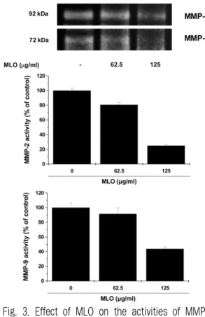
3. Effect of MLO on MMPs activity
The effect of MLO on the secreted MMP-2 and MMP-9 activity was examined using the zymographic method in B16F10 cells. Trypsin activated culture supernatant of untreated control cells showed digested clear areas at 72 and 92 kDa which indicate MMP-2 and MMP-9 respectively (Data not shown). However, treatment of 10 mM EDTA to the zymogram development solution did not exhibit any clear band and this result confirm that the gelatinolytic bands were created due to metalloproteinase (Data not shown). As shown in Fig. 3, a notable reduction in band intensity of both MMP-2 and MMP-9 by the treatement of MLO was observed in a concentration dependent manner. These results show that MLO inhibits the enzymatic activity of MMP-2 and MMP-9 secreted from B16F10 cells.
Fig. 3. Effect of MLO on the activities of MMPs in B16F10 cells.
B16F10 cells were treated with MLO for 24 h and then subjected to gelatin zymography. Determined activities of MMP-2 and MMP-9 were subsequently quantified by densitometric analysis with that of control being 100% as shown just below the gel data.
4. Effect of MLO on MMPs expression
In order to determine whether MLO could attenuate protein levels of MMP-2 and MMP-9 in B16F10 cells, we performed Western blotting. As shown in Fig. 4, non-treated B16F10 melanoma cells exhibited potent expression of MMP-2 and MMP-9. Compared to control, MMP-2 and MMP-9 expression were potentially blocked in the presence of MLO. The down-regulated protein levels were most noticeable at the maximum concentration in
both MMP-2 and MMP-9, demonstrating that MLO could play a crucial role in the inhibition of MMP-2 and MMP-9 expression.
Fig. 4. Effects of MLO on the expression of MMPs in B16F10 cells.
The protein extracts were prepared; samples were analyzed for MMP-9 expression by western blotting as described in the method. Determined activities of MMP-9 were subsequently quantified by densitometric analysis with that of non-treated control being 100% as shown just below the gel data.
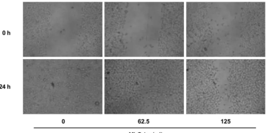
5. Effect of MLO on cell migration
Cell migration (motility) is a critical process of invasion allowing primary tumors to metastasize.
To investigate the inhibitory effect of MLO on B16F10 murine melanoma cells migration, a wound
-healing assay was performed. The treatment of B16F10 cells with increasing concentrations of MLO led to a concentration-dependent decrease in wound healing cell migration (Fig. 5). As shown
in Fig. 30 these cells do not show growth inhibition under treatment concentration. Thus, cell migration inhibitory effect of MLO was not the result of cytotoxicity.
Fig. 5. Effect of MLO on the B16F10 cell migration.
B16F10 cells were plated in a 12-well plate at a density of 2.5×10⁵ cells/well with DMEM and supplemented of 10% FBS. Confluent monolayers were scratched using a yellow tip and then incubated in serum-free medium with or without various concentrations of MLO. Before and 24 h after wounding, the cells were photographed under an invert microscope.
6. Effect of MLO on cell invasion
It is well known that the B16F10 cells have an strong invading properties through Matrigel. In this study, we investigate the inhibitory effect of MLO on cell invasion using BD BioCoat Matrigel chamber. Treatment MLO for 22 h demonstrated that excellent suppression on the melanoma cell invasion in a dose dependent manner (Fig. 6). In the 62.5 µg/ml and 125 µg/ml of MLO treated group showed 38.96% and 75.77% inhibition of cell invasion compared with that of non-treated groups.
These result indicated that MLO could prevent
the spread of melanoma cells. Fig. 6. Effects of MLO on the B16F10 cell invasion.
B16F10 cells were treated with various concentrations MLO for 22 h using Matrigel-coated Transwell.
Data shown are representatives of three independent experiments. **p<0.001 compared to control group.
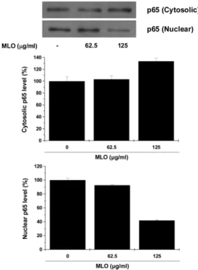
7. Effect of MLO on NF-κB translocation
Because the transcriptional factor NF-κB has an important role in tumor progression and invasion we also investigated the effect of MLO on the activation of NF-κB. To evaluate the effect of MLO on the activation of NF-κB, Western blotting was performed using cytosolic and nuclear extracts of B16F10 melanoma cells. As shown in Fig. 7, in resting conditions, the level of the NF-κB p65 subunit was constitutively enriched in nuclei of B16F10 melanoma cells. Incubation with MLO for 18 h induced significant decrease in the levels of NF-κ B in the nucleus. On the contrary, NF-κB expression in cytosol was increased by MLO treatment in a dose dependent manner. These findings strongly suggest that MLO inhibited the transcriptional activity of NF-κB in B16F10 melanoma cells.
Fig. 7. Effects of MLO on the NF-κB translocation in B16F10 cells.
Cells were incubated with MLO for 18 h and the nuclear extracts were prepared; samples were analyzed by western blotting as described in the method and quantified by densitometry.
8. Anti-metastatic effect of MLO in vivo
Since the above results demonstrated that MLO had potent inhibitory actions in metastasis in vitro, in order to confirm the anti-metastatic effects of MLO, we performed in vivo metastasis study. The lung metastasis induced by an intravenous injection of B16F10 melanoma cells using C57BL/6 mice.
The mice treated with MLO (500 mg/kg/day of body weight) decreased the metastatic lung colonies by 12.18% (p<0.01) compared with that of the untreated control group (Fig. 8). In acute toxicity test, no significant toxicity was observed at this dose level (Data not shown). Thus, it could be concluded that MLO has anti-metastatic activity in both in vitro and in vivo.
Fig. 8. Effects of MLO on the lung metastasis.
The lungs were pictured and observed for any metastasis on the 14th day after injecting B16F10 melanoma cells. The values represented as means
± S.E.M. *p<0.01 compared to control group.
Ⅳ. Discussion
Metastasis, a major problem for cancer patients, is the spread of cancer cells from the primary neoplasm to secondary sites. It may occur only when the cancer cells complete complex multi-step process perfectly. Hence, any disturbance of these steps could be an attractive therapeutic target for cancer metastasis.
Since tumor cells must cross type Ⅳ collagen -rich basement membrane of vessel walls17 before entering blood vessels, degradation of extracellular matrix is a one of the critical stage for the successful metastasis. It is well known that the proteolysis of basement membrane is predominantly achieved by several matrix metalloproteinases (MMPs) such as MMP-2 and MMP-9 which play a crucial role in type Ⅳ collagen degradation, a major component of basement membrane. Therefore, enhanced level of MMP-2 and MMP-9 in many malignant tumor cells including melanoma have been shown to be associated with the progression and invasion of tumors18-20.
Despite recent advances in developing new medicine, there is still a need for effective anti- metastatic agent. In this regard, new drugs originated from medicinal plants have received a lot of attention. In traditional Korean medicine, pathogenesis of cancer metastasis can be explained as deficient and stagnation of Qi results in the blood stagnation formation. Since L. obtusiloba can enhance blood circulating as well as eliminate blood stagnation, we checked the possibility whether this plant has anti-metastatic activities.
In the present study, in order to test the possible effect of methanol extract of L. obtusiloba (MLO) on MMP-2 and MMP-9 inhibition, the
bacterial collagenase-1 gelatin digestion assay was performed and MLO exhibited significant inhibitory effect on bacterial collagenase-1 activity. Based on these results, further anti-metastatic studies were undertaken. Zymogram results from this study demonstrated that MLO inhibited enzymatic activities in both of MMP-2 and MMP-9 in a dose dependent manner. Furthermore, MLO also down-regulated protein levels of MMP-2 and MMP-9. These results suggest that the decreased enzymatic activities of proteases by MLO were due to the suppression of protein expression.
In the cell proliferation assay, MLO exhibited significant anti-proliferative action in B16F10 melanoma cells. Several previous reports suggest that butanolides21 and lignans 9 from L. obtusiloba has potent antitumor activity by exhibiting cytotoxicity against cultured human tumor cell lines. However, the treatment concentration of MLO in zymogram analysis did not show any cytotoxicity representing the MMP inhibitory properties of MLO were not due to its cytotoxicity.
Previous reports have demonstrated that the activation of NF-κB in tumor cells may contribute to the expression of related invasion and metastasis genes including MMP-2 and MMP-922. Moreover, overexpressed nuclear translocation of NF-κB proteins were observed in malignant cancers including colorectal cancer, breast cancer and malignant melanoma23-25. Therefore, we examined whether MLO altered the translocation of NF-κB into the nucleus in B16F10 melanoma cells and MLO showed dose dependent attenuation on the NF-κB level in nucleus indicating down-regulation of metastatic gene expressions. The present results corresponded well with several reports revealed that ursolic acid reduced the levels of NF-κB26,27.
The cancer cells must first migrate from the primary tumor as part of the invasive process in order to spread distant sites28. Invasive cells are defined as several characteristics including altered adherence to the primary tumor, enhanced motility and increased proteolytic degradation of ECM components. Thus, inhibition of cell invasion and migration also could be a useful anti-metastatic strategy. Here in this work, we evaluated the cell invasion using Matrigel invasion assay. The results showed that MLO attenuated invasion of B16F10 cells significantly in a dose dependent manner. In the wound healing assay, the cell migration of B16F10 cells was significantly inhibited in the presence of MLO. Recently, it has become evident that the gelatinases participate not only degradation of ECM matrix, but also stimulate the cell invasion and migration. As mentioned above, MMP-2 and MMP-9 were down-regulated by MLO, and therefore, these inhibitory effects may give a reasonable explanation for the limited invasiveness and motility
of tumor cells by MLO.
In addition to anti-metastasis activity in vitro, in vivo anti-metastatic effect of MLO was also conducted. In the present study, we found that MLO could suppress the formation of metastatic tumor nodules in the lung of C57BL/6 mice which were injected with B16F10 melanoma cells through the tail vein. These results suggest that MLO exert an anti-metastatic influence not only in vitro level but in vivo metastasis.
In summary, it was obviously demonstrated that MLO had potent inhibitory effects on MMP-2 and MMP-9 enzyme activities and expressions via down-regulation of NF-κB activation in B16F10 melanoma cells. The reduced cell invasion and migration were found in the precense of MLO.
MLO also reduces lung metastasis induced by B16F10 melanoma cells. Based on these results, MLO would have a great promise for use in the treatment of cancer as an anti-metastatic agent.
생강나무 추출물의 암전이 억제효과
윤 혁1, 이용재3, 서현원3, 박경재3, 고하늘1, 차동석3, 권 진4, 전 훈3, 김강산1,2
1원광대학교 한의과대학 내과학교실,2한국전통의학연구소
3우석대학교 약학대학, 4한국재활복지대학 의료보장구과
초 록목 적 :본 연구에서는 생강나무 메탄올 추출물이 암전이 억제에 미치는 영향을 조사하고자 하였다.
방 법 :생강나무 추출물의 암전이 억제능을 확인하기 위해서 B16F10 흑색종 세포를 이용하여 금속단백분해효소의 활성
및 발현을 측정하였으며, 암세포의 이동능이나 침윤능도 조사하였다. 폐전이 동물모델에서 생강나무 추출물이 미치는 영향을 조사하여 활성을 최종적으로 확인하였다.
결 과 :
1. 생강나무 추출물은 B16F10 흑색종 세포에서 뚜렷한 금속단백분해효소의 효소활성 및 발현 억제효과를 보였으며 이는 NF-κB의 활성 억제에서 기인한 것임을 확인하였다.
2. 흑색종 세포의 이동이나 침윤 역시 생강나무 추출물 투여에 의해 현저히 감소하였다.
3. 폐전이 동물 모델에서도 생강나무 추출물에 의해 폐로 전이되 집락의 수가 감소하였다.
결 론 :이상의 결과로 생강나무 추출물은 뛰어난 암전이 억제효과가 있는 것을 확인할 수 있었으며, 전이성 암치료에 있
어서 유용하게 사용될 수 있을 것으로 사료된다.
중심단어 : 생강나무, 금속단백분해효소, 암전이
Acknowledgement
This paper was supported by the Won-kwang university in 2010.
References
1. Arvelo F, Cotte, C. Metalloproteinases in tumor progression. Review Invest Clin 2006;47(2) :185-205.
2. Baek WK, Park JW, Lim JH, Suh SI, Suh MH, Gabrielson E, et al. Molecular cloning and characterization of the human budding uninhibited by benomyl (BUB3) promoter.
Gene 2002;295(1):117-23.
3. Gao Z, Sasaoka T, Fujimori T, Oya T, Ishii Y, Sabit H, et al. Deletion of the PDGFR-beta gene affects key fibroblast functions important
for wound healing. J Biol Chem 2005;280(10) :9375-89.
4. Hrabec E, Strek M, Nowak D, Greger J, Suwalski M, Hrabec Z. Activity of type IV collagenases (MMP-2 and MMP-9) in primary pulmonary carcinomas: a quantitative analysis.
J Cancer Res Clin Oncol 2002;128(4):197-204.
5. Jiang J, Grieb B, Thyagarajan A, Sliva D.
Ganoderic acids suppress growth and invasive behavior of breast cancer cells by modulating AP-1 and NF-κB signaling. Int J Mol Med 2008;21(5):577-84.
6. Johnsen M, Lund LR, Romer J, Almholt K, Dano K. Cancer invasion and tissue remodeling:
common themes in proteolytic matrix degradation.
Curr Opin Cell Biol 1998;10(5):667-71.
7. Kohn EC, Liotta LA. Molecular insights into cancer invasion: strategies for prevention and intervention. Cancer Res 1995;55(9):1856-62.
8. Komae H, Hayashi H. Phytosterols of the trunks of Lindera obtusiloba. Phytochemistry 1972;11:1182.
9. Kim MM, Ta QV, Mendis E, Rajapakse N, Jung WK, Byun HG, et al. Phlorotannins in Ecklonia cava extract inhibit matrix metalloproteinase activity. Life Sci 2006;79(15):1436-43.
10. Kim SH, Son JH, Lee SH. Inhibitory effects of water extract of Lindera obtusiloba on the mast cell-mediated allergic inflammation. Kor J Pharmacogn2009;40(3):233-7.
11. Kwon HC, Baek NI, Choi SU, Lee KR. New cytotoxic butanolides from Lindera obtusiloba BLUME. Chem Pharm Bull 2000;48(5):614-6.
12. Kwon HC, Choi SU, Lee JO, Bae KH, Zee OP, Lee KR. Two new lignans from Lindera obtusiloba blume. Arch Pharm Res 1999;22(4) :417-22.
13. Lee KY, Kim SH, Jeong EJ, Park JH, Kim SH, Kim YC, et al. New secoisolariciresinol derivatives from Lindera obtusiloba stems and their neuroprotective activities. Planta Med 2010;76(3):294-7.
14. Liabakk NB, Talbot I, Smith RA, Wilkinson K, Balkwill F. Matrix metalloprotease 2 (MMP-2) and matrix metalloprotease 9 (MMP-9) type IV collagenases in colorectal cancer.Cancer Res 1996;56(15):190-6.
15. Lind DS, Hochwald SN, Malaty J, Rekkas S, Heby P, Mishra G. Nuclear factor-κB is unregulated in colorectal cancer. Surgery 2001;
130(2):363-9.
16. McCawley LJ, Matrisian LM. Matrix metalloproteinases:
multifunctional contributors to tumor progression.
Mol Med Today 2000;6(4):149-56.
17. Nakshatri H, Bhat-Nakshatri P, Martin DA, Goulet RJ, Sledge GW. Constitutive activation
of NF-κB during progression of breast cancer to hormone independent growth. Mol Cell Biol 1997;17(7):3629-39.
18. Niwa M, Iguchi M, Yamamura S. Three new obtusilactones from Lindera obtusiloba Blume.
Chemistry Letters 1975;4:655-8.
19. Oppenheimer SB. Cellular basis of cancer metastasis: A review of fundamentals and neadvances. Acta Histochem2006;108(5):327-34.
20. Zhao W, Liu H, Xu S, Entschladen F, Niggemann B, Zänker KS, et al. Migration and metalloproteinases determine the invasive potential of mouse melanoma cells, but not melanin and telomerase.
Cancer Lett2001;162:S49-S55.
21. Ruehl M, Erben U, Kim K, Freise C, Dagdelen T, Eisele S, et al. Extracts of Lindera obtusiloba induce antifibrotic effects in hepatic stellate cells via suppression of a TGF-beta -mediated profibrotic gene expression pattern.
J Nutr Biochem 2009;20(8):597-606.
22. Scorilas A, Karameris A, Arnogiannaki N, Ardavanis A, Bassilopoulos P, Trangas T, et al.
Overexpression of matrix-metalloproteinase-9 in human breast cancer: a potential favourable indicator in node-negative patients.Br J Cancer 2001;84(11):1488-96.
23. Shishodia S, Majumdar S, Banerjee S, Aggarwal BB. Ursolic acid inhibits nuclear factor-kappaB activation induced by carcinogenic agents through suppression of IkappaBalpha kinase and p65 phosphorylation: correlation with down-regulation of cyclooxygenase 2, matrix metalloproteinase 9, and cyclin D1. Cancer Res 2003;63(15):4375-83.
24. Tryggvason K, Hoyhtya M, Salo T. Proteolytic degradation of extracellularmatrix in tumor invasion. Biochimica et Biophysica Acta 1987;
907(3):191-217.
25. Yang J, Richmond A. Constitutive IkappaB kinase activity correlates with nuclear factor- kappaB activation in human melanoma cells.
Cancer Res 2001;61(12):4901-9.
26. Yeh CT, Wu CH, Yen GC. Ursolic acid, a naturally occurring triterpenoid, suppresses migration and invasion of human breast cancer cells by modulating c-Jun N-terminal kinase, Akt and mammalian target of rapamycin
signaling. Mol Nutr Food Res 2010;54(9):1285-95.
27. Yook C. Lindera obtusiloba. Medical plants of Korea. Seoul: Jinmyeong Publishing Co.; 1989, p. 184.
28. Yoon SO, Park SJ, Yun CH, Chung AS. Roles of matrix metalloproteinases in tumor metastasis and angiogenesis. J Biochem Mol Biol 2003;
36(1):128-37.