51
and Safety
Available online at http://www.foodhygiene.or.kr
http://dx.doi.org/10.13103/JFHS.2016.31.1.51
흰민들레, 고들빼기, 씀바귀 추출물의 암세포 증식 억제 효과
신성아·이해님·추강식·김형진·박병권·김병수·정지윤*
공주대학교 특수동물학과
Induction of Apoptosis in Human Cancer Cells with Extracts of Taraxacum coreanum, Youngia sonchifolia and Ixeris dentate
Seong-Ah Shin, Hae-Nim Lee, Gang-Sik Choo, Hyeong-Jin Kim, Byung-Kwon Park, Byeong-Soo Kim, and Ji-Youn Jung*
Department of Companion and Laboratory Animal Science, Kongju National University (Received December 24, 2015/Revised January 7, 2016/Accepted January 13, 2016)
ABSTRACT - This research examined the effects of Compositae extract on the inhibition of proliferation and apoptosis in human breast and human gastric cancer cells. Compositae extracts which is used in the experiment are Taraxacum coreanum (TC), Youngia sonchifolia (YS) and Ixeris dentata (ID). The proliferation of SK-BR-3, MDA- MB-231 and AGS cells were investigated by MTT assay. ID and YS extracts inhibited proliferation of SK-BR-3, MDA-MB-231 and AGS cells in a dose-dependent manner, but TC have barely affected. In addition, the most effec- tive extract was ID. To assess the apoptosis of ID extract, the nuclei of human cancer cells were stained with DAPI solution respectively. Chromatin condensation, indicated apoptosis, was increased in a dose-dependent manner. We investigated change of ID extract-induced apoptosis proteins on human cancer cells by western blot analysis. The level of Bcl-2 decreased, whereas the level of Bax, cleaved-PARP increased in dose-dependent manner compared with non-treatment. Also Bax/Bcl-2 ratio, which is used in clinical indicator of apoptosis, was increased at ID extract treat- ment group compared with non-treatment. Moreover the Bax/Bcl-2 ratio of MDA-MB-231 cell was significantly increased as against SK-BR-3, AGS cells. These results indicated that ID extract have anti-proliferation effect better than YS or TC, and induced apoptosis in human breast cancer MDA-MB-231 cell better than human breast cancer SK- BR-3 cell, human gastric cancer. Even if further research is needed, ID can be developed as a chemopreventive or therapeutic agent of breast cancer.
Key words : Taraxacum coreanum, Youngia sonchifolia, Ixeris dentata, apoptosis, anticancer
암의 발생과 사망률이 증가함에 따라 이를 예방하거나 치료할 수 있는 물질에 대한 관심이 고조되고 있으며, 기 존에 사용되고 있는 약물의 부작용을 극복하기 위해 천연 물로부터 항암성분을 찾기 위한 연구가 꾸준히 진행 되고 있다1,2).
매년 사망률이 증가하고 있는 유방암은 여성에서 발생 하는 대표적인 질환으로 자궁암, 위암 다음으로 많은 비 중을 차지하고 있는 암으로 보고 되고 있다. 유방암은 외 과적 절제술, 방사선치료 및 항암제 투여 등으로 암 환자 의 생존율을 향상시키고는 있지만, 기존 항암제에 대한 부
작용이 심하여 지속적으로 투여 할 수 없기 때문에 부작 용이 적은 새로운 항암제의 개발이 필요한 실정이다3,4,5). 위암은 전 세계적으로 가장 발생 빈도가 높은 암의 하나 로 미국과 유럽에 비해 특히 우리나라를 포함한 동아시아 에서 발병률이 상대적으로 높게 나타난다. 위암의 치료법 으로는 항암제투여, 위 절제술, 방사선요법 등이 있지만, 조기에 발견되지 못할 경우 완치가 거의 불가능하다. 또 한, 화학요법에 이용되는 다양한 종류의 항암제들은 개개 인에 따라 감수성이 다르게 나타나고 독성에 의한 부작용 이 있기 때문에 이를 줄일 수 있는 새로운 천연물 유래 항암제에 대한 연구가 필요하다6,7,8).
개체가 발달하는 과정에서 형태를 이루고, 세포 수를 일 정하게 유지하며, 손상된 세포를 제거하는데 중요한 역할 을 하는 apoptosis는 종양의 생성을 억제하는 기능을 가진 다. Apoptosis는 내부와 외부경로로 나뉘는데 이들은 서로
*Correspondence to: Ji-Youn Jung, Department of Companion and Laboratory Animal Science, Kongju National University, Yesan 32439, Korea
Tel: 82-41-330-1526, Fax: 82-41-330-1529 E-mail: [email protected]
연관되어 있으며, 내외부 자극에 의해 DNA 손상을 받은 세포에서 유도된다. 이후 다양한 세부과정을 거치지만 궁 극적으로는 caspase의 활성화에 의해 세포가 사멸한다.
Apoptosis 과정에서 Bcl-2, Bcl-w, Bcl-xL 등은 apoptosis 억제인자이며, Bax, Mtd/Bok, Bak 등은 apoptosis를 유도 하는 활성인자이다9,10). 따라서 이러한 apoptosis를 유도하 는 식품 또는 식물의 성분들은 향후 천연항암제, 예방제 로 개발할 수 있는 가능성이 매우 크다.
국화과(Compositae) 식물은 세계에서 가장 널리 분포하 고 있으며, 우리나라에는 흰민들레, 고들빼기, 씀바귀, 좁 밥나물, 구절초 등 300여 종이 자생 또는 귀화되어있는 것 으로 알려져 있다11,12,13). 이러한 국화과 식물은 전통식품의 기능성 소재로도 사용되며, 한방에서는 해열, 소염, 혈압 저하, 두통 등에 유효하고14) 바이러스억제효과, 항균활성 및 항암활성 등에 효과가 있다고 알려져 있다11).
흰민들레(Taraxacum coreanum, TC)는 포공영, 금잔로 및 지정이라고도 불리며, 우리나라 전국의 산야, 길가 밭 둑 등에서 흔히 볼 수 있다. 민들레의 뿌리, 잎, 꽃 등 식 물의 전체는 동양에서 오랫동안 약용으로 사용되어 왔다.
민들레에는 특히 taraxacin과 inulin이 풍부하며, luteolin과 quercetin 등 플라보노이드 유도체를 함유하고 있다13,15). 이 러한 성분들로 항염증16), 항산화, 항균 활성17), 항당뇨18), 항암, 면역 관련 활성, 위장 보호 효과19) 등 다양한 효능 을 가지는 것으로 보고되어 있다.
고들빼기(Youngia sonchifolia, YS)는 약사초, 고채라고도 하며, 우리나라에서는 주로 중부 이남에 분포하고, 산기슭, 들, 밭두둑, 길가에서 흔히 볼 수 있다13). 고들빼기의 주성 분은 inulin으로, 이 외에도 flavonoid, amino acids, 식물 sterol 같은 유효성분을 함유하고 있으며20,21), 항염, 간 보 호22), 항산화23), 항종양24)효과가 있다고 보고되어 있다. 하 지만, 고들빼기의 생리활성에 대한 연구는 부족한 편이다.
씀바귀(Ixeris dentata, ID)의 뿌리를 포함한 식물의 전체 는 식용 또는 약용으로 사용되어 왔으며, 건위나 소염제 로서 소화불량, 식용 증진 등에 사용되는 것으로 알려져 있다13). 씀바귀의 주요 구성물질은 aliphatics, triterpenoids 및 sesquiterpene glicoside이고, 비타민 A, C, E와 카로티 노이드 및 flavonoids 등 다양한 생리활성 물질을 포함하 고 있다25,26,27). 또한 항돌연변이28,29), 항고지혈30), 항염증31), 항알레르기32), 항산화33,34) 및 항증식35) 작용 등 다양한 생 리적 활성이 보고되어 있다.
따라서 본 연구에서는 국화과 식물 중 흰민들레(TC), 고 들빼기(YS), 씀바귀(ID) 추출물이 유방암세포인 SK-BR-3, MDA-MB-231과 위암세포인 AGS의 세포 증식에 미치는 영향을 조사하고, 가장 효과 있는 추출물 한 가지를 선택 하여, 그 추출물이 apoptosis 기전 발현에 의한 효과인지 를 확인하였다.
Materials and Methods
세포 및 실험 재료
본 연구에 사용된 유방암 세포 SK-BR-3, MDA-MB-231 과 위암 세포 AGS는 한국 세포주 은행(KCLB, Korea Cell Line Bank, Seoul, Korea)에서 구입하였다. 세포배양에 사 용된 RPMI-1640은 Welgene (Gyeonsan, Korea)에서 구입하 였고, fetal bovine serum과 streptomycin/penicillin은 Gibco BRL (Grand Island, NY, USA)에서 구입하였다. 본 연구 에 사용한 일반적인 시약은 Sigma Chemical Co. (St.Louis, MO, USA)에서 구입하였다. 1차 항체 anti-Bax, anti-Bcl-2, anti-PARP, anti-β-actin와 2차 항체 Anti-rabbit IgG는 Cell signaling Technology (Danvers, MA, USA)에서 구입하였다.
시료 추출
본 실험에 사용한 흰민들레, 고들빼기, 씀바귀 추출물은 건조(음지, 실온 5일)한 후 분쇄하고 45oC에서 시료와 methyl alcohol을 15분 동안 sonication 후 2시간 정치를 1일 10회 반복하여 총 3일간 추출하였다. Rotary Evaporator를 사용 하여 45oC에서 감압하여 농축시킨 후 저온에 보관한 시료 를 한국식물추출물은행에서 분양 받아 실험에 사용하였다.
Cell culture
유방암세포 SK-BR-3, MDA-MB-231과 위암세포 AGS 는 5% fetal bovine serum, 1% streptomycin/penicillin을 첨 가한 RPMI-1640 배지를 사용하여 37oC, 5% CO2가 유지 되는 incubator에서 배양하였다. 175T flask에 세포 밀도가 80-90% 정도 찼을 때, phosphate buffered saline solution (PBS, pH 7.4)로 세포의 단층을 washing 한 후 trypsin- EDTA를 처리하여 계대배양 하였고, 배지는 2-3일 주기로 refresh 하였다.
MTT assay
SK-BR-3, MDA-MB-231, AGS 세포를 96 well plate에 2× 10⁴cells/mL로 분주한 후, 24시간 동안 배양시킨 다음 각각의 세포에 TC, YS, ID 추출물을 0, 50, 100, 200 μg/
mL의 농도로 처리하였다. 24시간 후 각 세포가 배양된 96 well plate에 MTT[3-(4,5-dimethylthiazol-2-yl)-2,5-diphenyl- tetrazolium bromide] reagent (mg/mL)을 40 μL 씩 처리한 후 1시간 30분 동안 37oC, 5% CO2가 유지되는 incubator 에서 배양하였다. MTT reagent을 제거한 후 dimethylsulfoxid (DMSO)를 100 μL/well로 첨가하여 well에 형성된 formazan 을 모두 녹인 후 ELISA-reader (Bio-Rad Laboratories Inc, Hercules, CA, USA)로 595 nm에서 흡광도를 측정하였다.
DAPI staining
SK-BR-3, MDA-MB-231, AGS 세포를 60 mm dish에 1
× 105cells/mL로 분주한 후, 24시간 동안 안정화 하였다.
ID 추출물을 0, 25, 50 μg/mL의 농도로 처리하여 24시간 동안 incubated 한 다음 PBS로 두 번 washing 한 후 4%
paraformaldehyde reagent로 15분간 고정하였다. 다시 한 번 washing 한 후 PBS에 10배로 희석한 DAPI reagent를 2 mL 씩 처리하여 형광현미경(Zeiss fluorescence microscope, Thornwood, NY, USA)으로 400배 시야에서 관찰하였다.
Western blot analysis
175T flask에 SK-BR-3, MDA-MB-231, AGS 세포를 37oC, 5% CO2가 유지되는 incubator에서 배양하였다. ID 추출물 을 0, 25, 50 μM 농도로 처리하고 24시간 동안 배양하여 trypsin-EDTA를 첨가한 후 세포를 부유시켜 원심분리(1,200 rpm, 5 min, 4oC)하였다. PBS로 세포를 washing하고 원심 분리하여 cell pellet에 cell lysis buffer (Invitrogen, Carlsbad, CA, USA)를 첨가한 후 4oC 에서 20분간 반응 시켰다. Lysate는 13,000 rpm에서 5분 동안 원심분리하여 상등액을 취해 cell lysate로 사용하였다. 추출한 단백질의 농도는 Bradford protein assay를 이용해 측정하였다. 단백 질을 12% sodium dodecyl sulfate polyacrylamide gel electrophoresis (SDS-PAGE)로 크기별로 분리한 후 nitro- cellulose membrane (Bio-Rad, Hercules, CA)에 이동시켰 다. Membrane은 5% skim milk로 2시간 동안 blocking 한 후 anti-Bax, anti-Bcl-2, anti-PARP, anti-β-actin의 1차 항체 를 각각 첨가하여 4oC 에서 overnight 하였다. 그 후 2차 항체 anti-rabbit IgG를 첨가하여 2시간 반응시켰다. 각 protein band는 ECL detection reagents (Pierce, Rockford, IL, USA)를 이용하여 실험결과를 확인하였다. 밴드의 밀 도는 imaging program인 Image J Launcher (provided by NCBI)를 이용하여 측정하였다.
통계학적 분석
모든 실험결과는 평균치와 표준편차를 사용하여 나타내 고 각 군간 비교는 one-way ANOVA에 이은 t-test 분석을 실시하였다. 대조군과 비교하여 p값이 0.05 미만일 때를 통계학적으로 유의성이 있다고 판정하였다.
Results and Discussion
TC, YS, ID 추출물이 인체 암세포의 생존에 미치는 영향 국화과 식물인 TC, YS, ID 추출물이 유방암세포인 SK- BR-3, MDA-MB-231과 위암세포인 AGS 성장에 미치는 영향을 확인하기 위해 MTT assay를 수행하였다. TC, YS, ID 추출물을 0, 50, 100, 200 μg/mL의 농도로 24시간 처 리하여 각각 암세포의 생존율을 확인한 결과, TC에서는 90-100%로 생존에 거의 영향을 주지 않았지만(Fig. 1A), YS와 ID 추출물은 대조군과 비교하였을 때 생존율이 농
도 의존적으로 감소하는 것을 확인하였다. YS 추출물을 처리한 SK-BR-3, MDA-MB-231, AGS 세포의 생존율은 50μg/mL에서 89%, 93%, 78%, 100 μg/mL에서 91%, 89%, 68%, 200μg/mL에서 83%, 69%, 58%로 억제되었고 (Fig. 1B), ID 추출물을 처리한 SK-BR-3, MDA-MB-231, AGS 세포의 생존율은 50 μg/mL에서 71%, 60%, 58%, 100 μg/mL에서 64%, 55%, 45%, 200 μg/mL에서 59%, 45%, 30%로 억제되었다(Fig. 1C). 특히 ID 추출물의 경우 50 μg/
mL에서부터 암세포의 생존율이 유의적으로 억제된 것을 확인할 수 있었다.
TC, YS, ID 추출물 중에서 저농도인 50 μg/mL에서 유 Fig. 1. Effect of TC, YS and ID extracts on the cell viability of human cancer cells. The results are showed cell viability inhibi- tory effect of TC extract(A), YS extract(B) and ID extract(C) on cancer cells. To study effect of TC, YS and ID extracts on SK- BR-3, MDA-MB-231 and AGS cells growth, 2× 10⁴cells were plated in 96 well plate and after 24 h, cells were incubated in medium with absence or presence of various concentrations of extracts respectively. Cell viability was measured by the MTT assay. Datas are shown as means ± SD of three independent experiments performed in triplicate. Significance was determined by Dunnett's t-test with *P < 0.05 considered as statistically signifi- cant compared with non-treated controls. TC: Taraxacum core- anum, YS: Youngia sonchifolia, ID: Ixeris dentate.
의적으로 암세포 생존 억제율을 보인 ID 추출물을 이용하 여 이후 실험을 진행하였다. ID 추출물의 농도 설정을 위 해 이전 실험보다 더 낮은 농도(0, 6.25, 12.5, 25, 50 μg/
mL)에서 MTT assay를 수행하여 SK-BR-3, MDA-MB-231, AGS 세포의 생존율을 확인하였다(Fig. 2). ID 추출물의 각 농도에 따른 생존율은 유방암 세포인 SK-BR-3에서는 83%, 82%, 78%, 77%, MDA-MB-231은 84%, 79%, 72%, 63%
였고, 위암세포인 AGS는 81%, 79%, 67%, 50%로 농도 의존적으로 감소하는 경향을 보였다. ID 추출물은 6.25, 12.5μg/mL의 농도에서는 생존율에 큰 차이를 보이지 않 았지만, 25, 50 μg/mL에서는 유방암세포인 SK-BR-3보다 유방암세포인 MDA-MB-231과 위암세포인 AGS에서 생존 억제율이 다소 높음을 확인할 수 있었다.
추출용매와 추출 부위에 차이는 보이지만, Lee19)등의 연 구에 의하면 페암세포 Calu-6, 대장암세포 HCT-116, 위암 세포 SNU-601에 ethanol로 추출한 TC의 잎과 뿌리 추출 물을 0, 200, 400, 600, 800 μg/mL 농도로 48시간 처리하 였을 때, TC의 잎에 비해 뿌리 추출물에서 농도 의존적으 로 세포 생존율이 감소하는 것을 확인하였으며, Calu-6, HCT-116, SNU-601 세포에 각각 약 522, 532, 614 μg/mL 에서 50%의 억제율을 보였다. Chon20)등의 연구에 의하면 폐암 세포 CALU-601에 개화기 때 수확하여 methanol로 추출한 부위별 YS 추출물을 처리하였을 때, 부위별로 차 이는 있으나 농도 의존적으로 감소하는 결과를 보였으며 뿌리, 잎, 꽃, 줄기 순으로 약 196, 298, 483, 751 mg kg−1 에서 50%의 억제율을 보였다. 또한, Kim29)의 연구에 의 하면 골육암 세포인 MG-63에 ID methanol 추출물을 0,
50, 100μg/plate로 처리하였을 때, 농도 의존적으로 세포 의 생존율이 감소한 것을 확인하였으며 대조군과 비교하 여 농도 별로 각각 54%, 76% 억제율을 보였다.
Lee19)등의 연구결과와 달리 본 연구에서는 TC 추출물에 의한 암세포 생존율에 변화가 거의 없는 것을 확인하였고, 반면, YS와 ID추출물의 경우 Chon20)등과 Kim29)의 연구결 과와 동일하게 농도 의존적으로 암세포 생존율이 억제되 는 것을 확인하였다. 또한, YS 추출물에 비해 ID 추출물이 암세포생존율에 가장 강한 생존 억제 효과를 보였으며, 이 러한 결과는 유방암과 위암세포의 농도 의존적인 세포사 멸이 ID 추출물의 영향을 받은 것으로 사료된다.
ID 추출물이 인체암세포의 핵 형태에 미치는 영향 Apoptosis는 인간의 질병에 있어 다양한 생물학적 과정 에 관여하며, 세포 수축, 염색질 분해와 핵 응축 등의 일 반적인 특징을 가진다. DAPI stain은 apoptosis에 의해 유 도된 핵 응축으로 인한 핵분열을 가시화 시켜주는 하나의 실험기법으로, apoptosis가 유도된 세포들은 정상세포에 비 해 약간 작은 형태로 관찰된다36).
MTT assay 결과를 통해 세가지 추출물 중 암세포들에 가장 유의적인 억제율을 보였던 ID 추출물을 이용하여 각 세포의 생존율 억제가 apoptosis에 의해 유도된 결과인지 확인하기 위해 DAPI staining을 실시하였다. SK-BR-3, MDA-MB-231, AGS 세포에 ID 추출물을 0, 25, 50 μg/mL 의 농도로 24시간 동안 처리한 후 DAPI 염색을 하여 형 광현미경으로 관찰하였다(Fig. 3A). 그 결과 MTT assay와 동일하게 유방암세포 SK-BR-3보다 MDA-MB-231, 위암 세포 AGS에서 더 많은 세포질의 응축과 apoptotic body 를 확인할 수 있었다.
Apoptosis가 유도된 세포를 정량적으로 분석하기 위해 DAPI 형광에 반응한 세포를 counting한 결과, ID 추출물 (25, 50μg/mL) 처리에 의해 모든 세포에서 Apoptotic 세 포가 유의적으로 증가된 것을 확인하였다(Fig. 3B-D). 이 러한 연구결과를 통해 ID 추출물 처리에 의한 암세포의 증식억제효과가 apoptosis에 의한 것임을 확인하였다.
ID 추출물이 Bcl-2 family 단백질 발현에 미치는 영향 Bcl-2 family 단백질들은 미토콘드리아의 막 외부에 통 로를 만들거나 차단하여 apoptosis를 조절한다9). 이 중 pro- apoptosis 인자인 Bax는 세포질에서 미토콘드리아로 이동 하여, cytochrome C의 분비 활성을 높여 세포사멸을 유도 시키는 반면, anti-apoptosis 인자인 Bcl-2는 미토콘드리아 로 이동하는 Bax를 억제함으로써 세포사멸을 억제시키는 작용을 한다37,38). DAPI staining 실험 결과, SK-BR-3, MDA- MB-231, AGS 세포에서 대조군과 비교하였을 때, ID 추 출물을 처리한 군에서 apoptosis 양성세포의 증가가 관찰 되었으므로, western blot을 통해 ID 추출물이 apoptosis를 Fig. 2. Effect of ID extract of the cell viability on SK-BR-3,
MDA-MB-231 and AGS cells. The results are showed cell viabil- ity inhibitory effect of ID extract on cancer cells. For studies effect of ID extracts on AGS, MDA-MB-231 and SK-BR-3 cells growth, 2× 10⁴cells were plated in 96 well plate and after 24 h cells were incubated in medium with absence or presence of ID extracts(6.25, 12.5, 25 and 50μg/mL) respectively. Cell viability was measured by the MTT assay. Datas are shown as means ± SD of three independent experiments performed in triplicate. Signifi- cance was determined by Dunnett's t-test with *P < 0.05 consid- ered as statistically significant compared with non-treated controls.
ID: Ixeris dentate.
조절에 관여하는 Bcl-2 family 단백질의 발현 양상에 어떤 영향을 미치는지 확인하였다. 그 결과, pro-apoptotic 인자 인 Bax 단백질의 발현은 ID 추출물(25, 50 μg/mL) 처리에 의해 SK-BR-3, MDA-MB-231, AGS 세포에서 농도의존적 으로 증가하였다(Fig. 4A). 또한, anti-apoptotic 인자인 Bcl- 2 단백질은 SK-BR-3 세포에서는 발현량의 차이가 거의 없었지만, MDA-MB-231, AGS 세포에서는 농도의존적으 로 감소하는 경향을 확인 할 수 있었다(Fig. 4B).
세포의 생존과 사멸의 결정은 Bcl-2 family간의 균형에 의존하며, 개별적인 단백질 발현량을 확인하는 것보다 세 포사멸의 주요 임상지표로 이용되기도 하는 Bax/Bcl-2 ratio 을 확인하는 것이 더 중요하다고 볼 수 있다39). 본 실험에 서 ID 추출물 처리에 의한 β-actin에 대한 상대적인 Bax/
Bcl-2 ratio는 유방암세포 SK-BR-3와 위암세포 AGS에 비 해 유방암세포 MDA-MB-231에서 유의적으로 증가하였다 (Fig. 4C).
따라서, ID 추출물에 의한 유방암세포 SK-BR-3, MDA- MB-231과 위암세포 AGS의 증식 억제 효과가 apoptosis에 의한 것임을 Bcl-2 family 단백질의 발현 변화로 확인하였 고, 이러한 결과는 세포에 따라 증식억제와 apoptosis 유
도에서 차이를 보였지만, 유방암에서 더 효과가 있을 것 으로 사료된다.
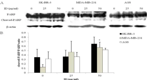
ID 추출물이 cleaved- PARP 단백질 발현에 미치는 영향 Poly-ADP ribose polymerase (PARP)는 세포핵 내에 존 재하며, 손상된 DNA를 수선하여 세포의 생존을 유지하는 데 있어 중요한 역할을 담당한다40). 내외부적 자극에 의해 단백질 분해효소인 caspase의 활성화가 이루어지고, 이들 은 apoptosis의 최종단계라 할 수 있는 PARP를 분절하여 apoptosis를 유도한다41). 따라서 cleaved-PARP의 발현량이 증가한다는 것은 apoptosis의 대표적인 특징이라고 볼 수 있다.
이러한 PARP 단백질의 발현 양상을 확인한 결과, SK- BR-3, MDA-MB-231, AGS 세포에 ID 추출물을 25, 50 μg/
mL 처리한 군에서 PARP의 분절이 증가하였다(Fig. 5A).
또한 각 세포의 cleaved-PARP/PARP ratio를 비교한 결과, β-actin에 대한 상대적인 ratio가 MDA-MB-231 세포에 ID 추출물을 50 μg/mL 처리한 군에서 유의적으로 증가한 것 을 확인하였다(Fig. 5B).
결과적으로 ID 추출물은 유방암세포 SK-BR-3, MDA- Fig. 3. The effect of ID extract-induced apoptosis on SK-BR-3, MDA-MB-231 and AGS cells. A: SK-BR-3, MDA-MB-231, AGS cells were treated with absence or presence of ID extract (25 and 50μg/mL) for 24 h and apoptotic cells were stained with DAPI. Chromatin condensation, representing apoptotic cell death (arrow), was examined using a fluorescence microscope (× 400). B-D: The graphs show quantification of DNA fragmentation and nuclear condensation in each cancer cells. Each bar represents the mean ± SD calculated from three independent experiments in triplicate. Significance was determined by Dunnett's t-test with *P < 0.05 considered as statistically significant compared with non-treated controls. ID: Ixeris dentate.
Fig. 4. The effect of ID extract on Bcl-2 family protein in SK-BR-3, MDA-MB-231 and AGS cells. A-B: Cells were treated with/without ID extract (25 and 50μg/ml) for 24 h and cell harvested to measure protein levels of Bax and Bcl-2 by western blotting. The blots were also probed with anti-β-actin antibodies to confirm equal sample loading. C: The Bax/Bcl-2 ratio was calculated from the Bax and Bcl-2 over β-actin ratios. Each bar represents the mean ± SD calculated from three independent experiments in triplicate. Significance was deter- mined by Dunnett's t-test with *P < 0.05 considered as statistically significant compared with non-treated controls. ID: Ixeris dentate.
Fig. 5. The effect of ID extract on PARP protein in SK-BR-3, MDA-MB-231 and AGS cells. A: Cells were treated with/without ID extract (25 and 50μg/ml) for 24 h and cell harvested to measure protein levels of PARP by western blotting. The blots were also probed with anti-β- actin antibodies to confirm equal sample loading. B: The cleaved-PARP/PARP ratio was calculated from the cleaved-PARP and total-PARP over β-actin ratios. Each bar represents the mean ± SD calculated from three independent experiments in duplicate. Significance was deter- mined by Dunnett's t-test with *P < 0.05 considered as statistically significant compared with non-treated controls. ID: Ixeris dentate.
MB-231과 위암세포 AGS에서 Bax 및 Bcl-2 단백질의 발 현 수준을 변화시키고 PARP을 분절하여 apoptosis를 유도 하는 것으로 여겨지며, 위암보다 유방암에서 항암제로의 잠재력이 있을 것으로 사료된다.
Acknowledgment
이 논문은 2013년도 정부(교육과학기술부)의 재원으로 한국연구재단의 지원을 받아 수행된 기초연구사업임(NRF- 2013R1A1A4A01012315).
국문요약
본 연구는 인체 유방암세포 SK-BR-3, MDA-MB-231과 위암세포 AGS를 대상으로 국화과 식물 중 흰민들레 (Taraxacum coreanum, TC), 고들빼기(Youngia sonchifolia, YS), 씀바귀(Ixeris dentate, ID) 추출물에 의한 증식 억제 효과를 비교 한 후, 가장 억제효율이 높은 추출물을 선택 하여 apoptosis 유도 효과를 조사하였다. 암세포의 증식 억 제효과는 TC 추출물에 의한 영향은 약한 반면, YS와 ID 추출물에 의한 영향은 농도 의존적으로 증가하는 것을 확 인하였다. 세 가지 추출물 중 가장 효과가 뛰어난 ID 추 출물을 이용하여 추가적으로 농도 설정을 진행한 다음 이 후 실험을 진행하였다. ID 추출물에 의한 apoptosis 양성 세포를 확인하기 위해 DAPI assay를 진행한 결과, ID 추 출물을 처치한 군에서 apoptotic body와 세포질 응축을 확 인하였다. MTT assay와 DAPI staining의 결과를 바탕으로 ID 추출물이 SK-BR-3, MDA-MB-231, AGS 세포에서 apoptosis와 관련한 단백질 발현 양상에 미치는 영향을 확 인하기 위해 western blot을 실시하였다. ID 추출물에 의 해 SK-BR-3, MDA-MB-231, AGS 세포에서 pro-apoptosis 인 Bax 단백질 발현은 농도 의존적으로 증가하였고, anti- apoptosis인 Bcl-2 단백질 발현은 SK-BR-3 세포에서는 발 현의 변화가 거의 없었지만, MDA-MB-231과 AGS 세포 에서는 감소하였다. 세포사멸의 주요임상지표인 Bax/Bcl- 2 ratio는 MDA-MB-231세포에서 가장 유의적으로 증가하 였다. 또한, 손상된 DNA를 복구하는 단백질인 PARP의 발 현은 감소하면서, cleaved-PARP이 증가하였다. 이러한 결 과들을 종합하였을 때, YS와 ID 추출물은 TC 추출물에 비해 암세포의 생존을 효과적으로 억제하였고, 특히 ID 추 출물은 낮은 농도에서도 암세포의 생존 억제효과가 우수 한 것을 확인하였다. 또한, ID 추출물에 의한 유방암세포 SK-BR-3, MDA-MB-231와 위암세포 AGS의 생존율 억제 는 apoptosis 유도를 통해 이루어 지는 것으로 사료되며, 그 중 유방암세포 MDA-MB-231에서 apoptosis 유도 효과 가 뛰어난 것을 미루어 보아, 유방암의 종류와 그 특징에 따른 차이를 보이지만 ID는 향후 유방암 예방 및 치료제
로 개발 가능성을 제시하며, 추후 지속적인 연구를 통하 여 in vivo에서의 ID 추출물의 항암효과에 대해 심도 있 는 연구가 이루어 져야 할 것으로 사료된다.
References
1. Moon Y.O., Park E.C., Shin H.R., Woo Y.J., Jung K.W., LEE J.H., Kong H.J., Hwang S.S., Lee J.K., Kong I.S., Hahm M.I.: Regional variation in accessing regional hospitals for cancer patients. Korean J. Epidemiol, 28, 152-161 (2006).
2. Nam S.H. and Yang M.S.: Isolation of cytotoxic substances from Chrysanthemum boreale M., J. Korean Soc. Appl. Biol.
Chem, 38, 273-277 (1995).
3. Kim J.E, Park J.H, Kang B.W, Seo M.J, Choi Y.H, Lim H.S, Seo K.I, Kim J.I, Joo W.H, Lee B.K, Jeong Y.K.: Anticancer Activity of Ethanol Extract from Peel of Citrus junos and Pon- cirus trifoliata on MCF-7 Breast Cancer Cells. J. Life Sci, 18, 1435-1441 (2008).
4. Dorssers, L.C.J., van der Flier, S., Brinkman, A., van Agth- oven, T., Veldscholte, J., Berns, E.M.J.J., Klijn, J.G.M., Beex, L.V.A.M., Foekens, J.A.: Tamoxifen resistance in breast can- cer. Drugs, 61, 1721-1733 (2001).
5. Kang J.O.: Special Editions : Radiation Therapy of Breast Cancer. Kyung Hee Medical, 21, 37-42 (2005).
6. Zhang L., Hou Y.H., Wu K., Zhai J.S., Lin N.: Proteomic analysis reveals molecular biological details in varioliform gastritis without Helicobacter pylori infection. World J. Gas- troentero, 16, 3664-3673 (2010).
7. Roth, A. and Ajani, J.: Docetaxel-based chemotherapy in the treatment of gastric cancer. Annals of oncology, 14, ii41-4 (2003).
8. Rivera, F., Vega-Villegas, M.E., López-Brea M.F.: Chemo- therapy of advanced gastric cancer. Cancer Treat. Rev, 33, 315-324 (2007).
9. Pecorino, L.: Molecular biology of cancer: mechanisms, targets, and therapeutics. Oxford university press, 157-182 (2012).
10. Lhee H.Y., Lee K.B., Kim H., Chae S.W., Park M.H.: Apop- tosis in Autosomal Dominant Polycystic Kidney Disease.
Korean J Nephro, 24, 366-374 (2005).
11. An I.J., Kwon J.K., Lee J.S., Park H.S., Kim D.C., Choi B.J., Lee K.M., Park Y.J., Jung J.Y.,: induction of Apoptosis in Human Cancer Cells with Compositae Extracts. J. Korean Soc. Food Sci. Nutr, 41, 584-590 (2012).
12. Woo, J.H., Shin S.L., Lee C.H.: Antioxidant effects of etha- nol extracts from flower species of compositae plant. J.
Korean Soc. Food Sci. Nutr, 39, 159-164 (2010).
13. Lee, T.B.: Coloured Flora of Korea, 388-402 (2003).
14. Lee M.K., Moon H.C., Lee J.H., Kim J.A., Yu C.Y., Lee H.Y.: Screening of immune enhancing activities in medicinal herbs, Compositae. Korean J. Medicinal Crop Sci, 10, 51-57 (2002).
15. Kang, M.J. and Kim K.S.: Current trends of research and biological activities of dandelion. Food industry and nutri- tion, 6, 60-67 (2001).
16. Lee M.H., Kang H., Lee K.J., Yang G.S., Ham I.H., Bu Y.M., Kim H.C., Choi H.Y.: The aerial part of Taraxacum core- anum extract has an anti-inflammatory effect on peritoneal macrophages in vitro and increases survival in a mouse model of septic shock. J. Ethnopharmacol, 146, 1-8 (2013).
17. Im D.Y. and Lee K.I.: Antioxidative and Antibacterial Activ- ity and Tyrosinase Inhibitory Activity of the Extract and Fractions from Taraxacum coreanum Nakai. Korean J.
Medicinal Crop Sci, 19, 238-245 (2011).
18. Schütz K., Carle R., Schieber A.: Taraxacum—a review on its phytochemical and pharmacological profile. J. Ethnop- harmacol, 107, 313-323 (2006).
19. Lee H.H. and Lee S.Y.: Cytotoxic and antioxidant effects of Taraxacum coreanum Nakai. and T. officinale WEB. Extracts.
Korean J. Medicinal Crop Sci, 16, 79-85 (2008).
20. Chon S.U. and Kang J.G.: Phenolics Level and Antioxidant Activity of Methanol Extracts from Different Plant Parts in Youngia sonchifolia. Korean J. Medicinal Crop Sci, 58, 20- 27 (2013).
21. Park S.S.: Original Articles; Studies on the Constituents and Their Biological Activities of Ixeris Sonchifolia Hance (1).
BMB Rep, 10, 241-252 (1977).
22. Bae S.J., Kim N.H., Koh J.B., Roh S.B., Jung B.M.: Effects of Godulbaegi (Ixeris sonchifolia H.) diet on enzyme activi- ties of CCl4 induced hepatotoxicity in rats. Korean J. Nutr, 30, 19-24 (1997).
23. Kim, M.J., Kim S.H, Lee C.I., Park H.S., Son J.C., Huh W., Kim P.N., Lee D.Y.: Component Analysis and Antioxidant Effects of Youngia sonchifolia Max. J. Food Hyg. Saf, 25, 354- 359 (2010).
24. Shin S.C.: Exploitation of the biologically active components in Youngia sonchifolia Max. J. Korean Agric. Chem. Soc, 36, 134-137 (1993).
25. Ahn E.M., Bang M.H., Song M.C., Park M.H., Kim H.Y., Kwon B.M., Baek N.I.: Cytotoxic and ACAT-inhibitory ses- quiterpene lactones from the root of Ixeris dentata forma albiflora. Arch. Pharmacal Res, 29, 937-941 (2006).
26. Cha M.R., Choi Y.H., Choi C.W., Yoo D.S., Choi S.U., Kim Y.H., Ryu S.Y.: New guaiane sesquiterpene lactones from Ixeris dentata. Planta Med, 77, 380-382 (2011).
27. Chung H.S., Jeong H.J., Han M.J., Park S.T., Seong K.K., Baek S.H., Jeong D.M., Kim M.J., Kim H.M.: Nitric oxide and tumor necrosis factor-α production by Ixeris dentata in mouse peritoneal macrophages. J. Ethnopharmacol, 82, 217- 222 (2002).
28. Kim M.J., Kim J.S., Kang W.H., Jeong D.M.: Effect on anti- mutagenic and cancer cell growth inhibition of Ixeris dentata Nakai. Korean J. Medicinal Crop Sci, 10, 139-143 (2002).
29. Kim, S.H.: Inhibitory effects of Ixeris dentata on the mutage-
nicity of aflatoxin B1, N-methyl-N'-nitro-N-nitrosoguani- dine and the growth of MG-63 human osteosarcoma cells. J.
Korean Soc. Food Sci. Nutr, 24, 305-312 (1995).
30. Lim S.S. and Lee J.H.: Effect of Aster scaber and Ixeris den- tata on contractility and vasodilation of cardiovascula and endothelial cell in hyperlipidemic rat. J. Korean Soc. Food Sci. Nutr, 26, 300-307 (1997).
31. Lee E.: Effects of Ixeris dentata extract on the production of pro-inflammatory cytokines in the LPS stimulated rat and Raw 264.7 cells. Korean J. Plant Res, 24, 604-612 (2011).
32. Park E.K., Sing J.H., Trinh H.T., Bae E.A., Yun H.K., Hong S.S., Kim D.H.: Lactic acid bacterial fermentation increases the antiallergic effects of Ixeris dentata. J. Microbiol. Bio- technol, 18, 308-313 (2008).
33. Kim M.J., Kim J.S., Jeong D.M., Ham S.S., Yu C.Y.: Effect of antioxidant, antimutagenicity and anticancer of root extract from Ixeris dentata Nakai. Korean J. Medicinal Crop Sci, 10, 222-229 (2002).
34. Yoon B.R., Cho B.J., Lee H.K., Kim D.J., Rhee S.K., Hong H.D., Kim K.T., Cho C.W., Choi H.S., Lee B.Y., Lee O.H.:
Antioxidant and anti-adipogenic effects of ethanolic extracts from Tartary and Common Buckwheats. Korean J. Food Pre- serv, 19, 123-130 (2012).
35. Heo B.G., Chon S.U., Park Y.J., Bae J.H., Park S.M., Park Y.S., Jang H.G., Gorinstein S.: Antiproliferative activity of Korean wild vegetables on different human tumor cell lines.
Plant Foods Hum. Nutr, 64, 257-263 (2009).
36. Doonan F. and Cotter T.G.: Morphological assessment of apoptosis. Methods, 44, 200-204 (2008).
37. Oltval Z.N., Milliman C.L., Korsmeyer S.J.: Bcl-2 het- erodimerizes in vivo with a conserved homolog, Bax, that accelerates programed cell death. Cell, 74, 609-619 (1993).
38. Yang J., Liu X., Bhalla K., Kim C.N., Ibrado A.M., Cai J., Peng TI., Jones D.P., Wang X.: Prevention of apoptosis by Bcl-2: Release of cytochrome c from mitochondria blocked.
Science, 275, 1129-1132 (1997).
39. Reed J.C.: Bcl-2 and the regulation of programmed cell death. J. Cell Biol, 124, 1-6 (1994).
40. Oliver, F.J., de la Rubia, G., Rolli, V., Ruiz-Ruiz, M.C., de Murcia, G., Ménissier-de Murcia, J.: Importance of poly (ADP-ribose) polymerase and its cleavage in apoptosis Lesson from an uncleavable mutant. J. Biol. Chem, 273, 33533-33539 (1998).
41. Kothakota, S., Azuma, T., Reinhard, C., Klippel, A., Tang, J., Chu, K., McGarry, J.T., Kirschner, M.W., Koths, K., Kwiat- kowski, D.J., Williams, L.T.: Caspase-3-generated fragment of gelsolin: effector of morphological change in apoptosis.
Science, 278, 294-298 (1997).