573 REVIEW
DOI 10.4070 / kcj.2008.38.11.573
Print ISSN 1738-5520 / On-line ISSN 1738-5555 Copyright ⓒ 2008 The Korean Society of Cardiology
Transesophageal Echocardiographic Evaluation of Atherosclerosis
Masami Nishino, MD and Jun Tanouchi, MD
Division of Cardiology, Osaka Rosai Hospital, Osaka, Japan ABSTRACT
Transesophageal echocardiography (TEE) is a promising method for evaluating thoracic aortic atherosclerosis and coronary atherosclerosis. The highest impact of TEE as a clinical tool is in searching for cardiac embolic sou- rces in patients with stroke and atrial fibrillation and in conducting detailed evaluations in patients with valvular disease, especially those with mitral valvular disease. However, it is also clinically useful in the evaluation of tho- racic aortic atherosclerosis and coronary atherosclerosis. TEE is capable of evaluating thoracic aortic atherosis (in- tima-media complex thickness) and sclerosis (stiffness parameter β) simultaneously. In addition, TEE can evaluate coronary atherosclerosis by non-invasively revealing narrowing or occlusion of the coronary arteries and providing information about coronary flow reserve. TEE imaging has improved with the advent of harmonic imaging, multi- plane probes, contrast agents, and three-dimensional TEE. Future technology, including integrated backscatter (IBS), tissue Doppler, and strain imaging, will lead to further improvements in TEE. Thoracic aortic atherosclerosis and coronary atherosclerosis assessment should be performed in any patient undergoing TEE. (Korean Circ J 2008;38:573-582) KEY WORDS: Transesophageal echocardiography; Atherosclerosis; Aorta, thoracic; Coronary artery disease.
Introduction
Previous reports have shown that thoracic aortic athe- rosclerosis correlates with systemic embolism and vascular disease and is a marker of coronary artery disease.1-3) Tra- nsesophageal echocardiography (TEE) provides high re- solution imaging of the thoracic aorta and is a reliable tool for evaluating the degree of thoracic aortic athero- sclerosis.4)5)
Blankenhorn et al.6) reported that atherosclerosis con- sists of two components-atherosis and sclerosis-and that future studies should be directed toward evaluating both.
TEE is capable of evaluating atherosis and sclerosis si- multaneously.
Therefore, it is a very useful tool for evaluating tho- racic aortic atherosclerosis.4) Moreover, TEE can provi- de direct visualization through Doppler technique,7) co- ronary arterial flow reserve,8)9) and coronary sinus flow reserve.10-12)
This review focuses on TEE evaluation of thoracic ao- rtic atherosclerosis and coronary atherosclerosis and de- tails some of the data we have collected.
Evaluation of Thoracic Aortic Atherosclerosis
TEE has made relatively noninvasive, clear visualiza- tion of the aortic arch and descending aorta possible.
Therefore, this procedure can be used to select patients at high risk for stroke. Some prospective studies have demonstrated TEE finding of large atheromas (4-5 mm) with mobile attachments are strongly correlated with fu- ture embolic disease.13)14) In addition to looking for em- bolic sources like thoracic atheromas, it is clinically im- portant to evaluate atherosis and sclerosis of the thoracic aorta using TEE. In general, atherosis is evaluated based on intima-media complex thickness (IMT), and sclerosis is evaluated based on arterial stiffness. Atherosis (incre- ased IMT) and sclerosis (increased arterial stiffness) are correlated with generalized atherosclerosis,15) and both are strong predictors of ischemic stroke.16) These parame- ters are usually evaluated in the carotid artery,15)16) but they can also be evaluated in the aorta.
Measurement of thoracic aortic atherosis
Of the various noninvasive imaging methods available, carotid IMT measurement obtained with B-mode ultra- sound is the most widely used method and is recom- mended by the American Heart Association for evalua- tion of risk.17) This index was proposed by Pignoli et al.,18) and the authors of the current study applied it to
Correspondence: Masami Nishino, MD, Division of Cardiology, Osaka Rosai Hospital, 1179-3 Nagasone-cho, Sakai-city, Osaka 591-8025, Japan
Tel: 81-72-252-3561, Fax: 81-72-250-5492 E-mail: [email protected]
574·TEE Evaluation of Atherosclerosis
the aorta using TEE.4)19) We then performed pathological examination to determine if IMT as determined by TEE can accurately demonstrate pathological IMT in autopsy cases.4) According to the pathological findings, we beli- eve that IMT can be applied to aortic atherosclerosis by means of TEE.
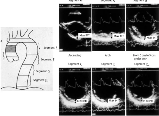
In our previous study, we determined maximum IMTs in 6 segments, according to Pignoli’s method (Fig. 1).18) The mean maximum IMT among the 6 segments was used as an index of aortic atherosis. We discovered that dyslipidemia and age were significantly and independe- ntly correlated with atherosis, but age was not.4) In addi- tion, Tomochika et al.20) reported that, in patients with familial hypercholesterolemia, cholesterol-lowering thera- pies could lead to regression of aortic atherosis, using a similar method. This finding can be partially connected to several recent intravascular ultrasound studies showing the effects of cholesterol-lowering therapies on coronary artery plaque regression.21-23) Furthermore, maximum IMT as evaluated by TEE has contributed to the search for new lipid serum markers of aortic atherosis. The authors found that lipoprotein (a) is a significant inde- pendent risk factor for aortic atherosclerosis.19) There is a close homology between apolipoprotein (a) and plas- minogen,24) so both may play a role in thrombosis and atherosclerosis. One decade after our report detailed ab- ove, Peltier et al. supported our data in a study using a similar TEE method in a larger population.25) However,
because lipoprotein (a)-lowering medications have not been developed, and because the correlation between coronary atherosclerosis and lipoprotein (a) is contro- versial,26-28) medical intervention to decrease lipoprotein (a) has not progressed in the meantime. The other se- rum marker of aortic atherosclerosis that was found using this method was fibrinogen,29) but it was not applied to therapeutic strategies for aortic atherosclerosis.
Measurement of thoracic aortic sclerosis
Sclerosis was clinically measured using three ultraso- und methods: stiffness parameter β,30) distensibility,31) and strain parameters.32) The formulae for these parame- ters are as follows:
Stiffness parameter β=ln (Ps-Pd)×Dd/(Ds-Dd) Distensibility =2×(Ds-Dd)/(Ps-Pd)×Dd Strain=(Ds-Dd)×100/Dd
Ds, aortic dimension at systole; Dd, aortic dimension at diastole
Ps, systolic arterial pressure; Dd, diastolic arterial pressure.
For the assessment of aortic sclerosis, the authors prin- cipally used stiffness parameter β. The formulae for the other parameters are straightforward, but the stiffness parameter β is supported by several basic science and engineering studies30)33) and has already been applied in many clinical studies.4)20)34) Some reports30)33) have ad-
Fig. 1. The thoracic aorta was divided into six segments as depicted in the illustration on the left (length of one segment 5 cm). The maxi- mum (Max) intima-media complex thickness (IMT) was measured in each segment, and the mean value for the maximum IMT among the 6 segments was used as an index of aortic atherosis.
Segment ③
Segment ④
Segment ⑤ Segment ⑥ Segment ②
Segment ①
Segment ① Segment ② Segment ③
Segment ④ Segment ⑤ Segment ⑥ Ascending Arch From 0 cm to 5 cm
under arch
From 5 cm to From 10 cm to From 15 cm to 10 cm under arch 15 cm under arch 20 cm under arch
Max IMT Max IMT Max IMT
Max IMT Max IMT
Max IMT
Masami Nishino, et al.·575
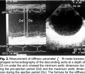
dressed the mechanical behavior of human arterial walls by noting changes in external radii due to distending pres- sure. These reports have demonstrated that the stiffness parameter β is the slope of the exponential function between the logarithm of the relative arterial pressure and the distension ratio of the artery. This parameter characterizes the full deformation behavior of the vas- cular wall and is independent of the intramural pressure within the physiological range. Therefore, it indicates ao- rtic sclerosis itself. In the interest of measuring the stiff- ness parameter β, instantaneous dimensional variables of the aorta (descending aorta) were evaluated. The di- mensional changes of the sites with intimal echo were simultaneously recorded during cardiac cycles using M- mode TEE, while a transverse 2D echogram of the des- cending aorta was recorded at a depth of 15 cm under the arch. Blood pressure was measured at the same time.
The minimum aortic dimension during the preejection period (Dd), maximum aortic dimension during the ejection period (Ds), and systolic distension (Ds-Dd) measurements were determined in millimeters. The stif- fness parameter β was calculated using the above-men- tioned formula (Fig. 2).
Many reports on aortic atherosclerosis have depicted only the grade of aortic atheroma. However, measure- ment of both the aortic atherosis and sclerosis parameters would improve the characterization of atherosclerosis in the aorta and serve as a more sensitive marker of athe- rosclerosis progression and regression in clinical trials, compared to atheroma (IMT) measurement alone. Arte- rial stiffness is related to structural and anatomic changes such as increased collagen to elastin ratio and qualitative deficiency of wall elements. Excluding age, aortic athe- rosis is significantly correlated with dyslipidemia and diabetes mellitus,4)34) while aortic sclerosis is correlated with hypertension.4) Atherosis and sclerosis seem to repre-
sent different atherosclerotic vessel wall properties.4)16)20) Nevertheless, the correlation between the two is weak, but significant.4) Atherosis and sclerosis are two inde- pendent processes.15)16) Therefore, TEE is one of the best methods for evaluating aortic atherosclerosis because it can demonstrate aortic atherosis and sclerosis simultaneously.
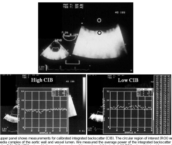
Possible new index of aortic atherosclerosis The measurement of integrated backscatter (IBS) is based on the use of unprocessed radiofrequency signals to derive quantitative ultrasonic indices with which to differentiate normal and pathological myocardial struc- tures.35) Quantitative ultrasonic tissue characterization of the biophysical composition of plaque and vessel wall has been performed with high-frequency, high-resolution acoustic microscopy in vitro,36) but such tools are available only in an experimental setting. Acoustic densitometry is a clinically applicable ultrasonic backscatter imaging technology that provides an integrated on-line capability to measure, display, and analyze the average acoustic ima- ge intensity within a region of interest (ROI). Thus, with acoustic densitometry, IBS offers a clinically promising method for assessing myocardial contractile performance independent of wall motion37)38) or quantitative evalua- tion of the vessel wall plaque composition: fibrosis, cal- cification, or lipid deposition.39)40) The latter approach can provide for a new evaluation of thoracic aortic athe- rosclerosis. Wickline et al.36) have shown that IBS can be applied to aortic atherosclerosis in vivo. However, it is not known if these tools can be applied in humans. The authors, therefore, tried to use this imaging technique to evaluate thoracic aortic atherosclerosis by means of TEE.41) We placed circular ROIs in the intima-media complex of the aortic wall and vessel lumen and meas- ured the average power of the IBS signal contained with the ROIs. We expressed the relative IBS values of the intima-media complex of the aortic wall as the difference from the IBS values obtained from a reference ROI pla- ced in the vessel lumen {CIB: calibrated IBS value (dB)}
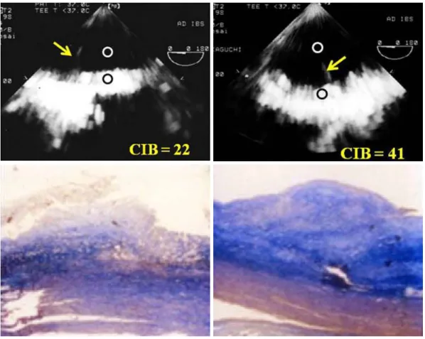
(Fig. 3). As a baseline measurement, the authors com- pared ultrasonic tissue characterization data for excised human aorta specimens with histological findings: fibro- fatty or fibrosis tissue specimen. In a trunk filled with a saline solution, we evaluated 24 sections of aorta in which TEE examinations were delimited by a metallic pin: this marker remained fixed in the specimens so the same segment that was imaged by TEE could be eval- uated pathologically (Fig. 4). Interestingly, the CIB of the fibrofatty change group (n=14) was significantly lower than that of the fibrous change group (n=10) (23.2±3.2 vs. 31.0±5.7, p<0.001). There were no significant differ- ences in the IMT values for the two groups. Furthermore, we noted a significant, though weak, relation between CIB and stiffness parameter β (y=0.065x+1.47, r=
0.50, p=0.003). The correlation between CIB and IMT
Fig. 2. Measurement of stiffness parameter β. M-mode transeso- phageal echocardiography of the descending aorta at a depth of 15 cm under the arch showed the minimum aortic dimension dur- ing the pre-ejection period (Dd) and the maximum aortic dimen- sion during the ejection period (Ds). The formula for the stiffness parameter β is shown in the text. APW: aortic pressure wave, PAPW: pulmonary artery pressure wave, ECG: electrocardiogram.
576·TEE Evaluation of Atherosclerosis
was not significant. The above-mentioned phenomenon was partially attributable to the fact that IMT indicated a morphological change of aortic atherosclerosis, and sti- ffness parameter β was shown to be a functional change (including distribution of elastin and collagen of the wall elements). We believe CIB can serve as a new index of aortic atherosclerosis, obtained by TEE. However, IBS has not been established as a standard reference. The authors have used the IBS signal from the vessel lumen as a reference object, while other investigators have used left ventricular cavity,41) myocardium,42) pericardium,43) and adventitia39) as references. The established standard reference tissue for quantitative ultrasonic tissue charac- terization with IBS analysis is indispensable when this index is used as a ‘noninvasive ultrasonic biopsy’ in the clinical setting.
Tissue Doppler imaging and strain imaging can be used to evaluate thoracic aortic stiffness. Vitarelli et al.44)45) assessed the aortic elastic properties using tissue Doppler imaging and strain rate imaging in patients with Marfan syndrome and those with a history of coarctoplasty. They found that both groups of patients had abnormal thora- cic aortic elastic properties. These new technologies com- bined with TEE may spread in the field of thoracic aortic atherosclerosis research.
Evaluation of Coronary Atherosclerosis
In 1988, Zwicky et al.46) became the first to report that the coronary arteries could be visualized using color- coded TEE. Since that time, improvements in TEE tec- hnology-including multiplane TEE,47) digital imaging,48) and contrast agents49)50) -have made coronary artery vi- sualization clearer, especially with respect to stenosis (atherosclerosis).9)
Visualization of coronary artery stenosis or occlusion
The TEE approach, in which the proximity of the transducer to the coronary artery serves as an advantage, may be useful in evaluating or detecting coronary artery stenosis of the left main coronary artery (LMCA) and prox- imal left anterior descending coronary artery (LAD).7) A combination of Doppler color flow mapping improves the detection for LMCA and LAD proximal stenosis on TEE.7) In addition, the development of multiplane TEE,47) contrast agents,49)50) second harmonic imaging techni- ques,51) and three-dimensional TEE52) has increased the accuracy and efficacy of coronary artery imaging on TEE.
Coronary flow velocity can be measured by pulsed Dop-
Fig. 3. The upper panel shows measurements for calibrated integrated backscatter (CIB). The circular region of interest (ROI) was placed in the intima-media complex of the aortic wall and vessel lumen. We measured the average power of the integrated backscatter (IBS) signal contained in the ROI. CIB={IBS value of intima-media complex of the aortic wall (black circle)}-{(IBS values obtained from a reference ROI placed within the vessel lumen (white circle}. Lower panels show representative cases with high and low CIB. The CIB of the case on the left is 36 (CIB=38-2), and that of the case on the right is 17 (CIB=18-1).
Masami Nishino, et al.·577
pler technique, and TEE can demonstrate the proximal segments of the LAD clearly.9) It has already been shown that transthoracic echocardiography with a relatively low velocity range setting (approximately 25 cm/sec) can vi- sualize distal LAD flow.53) Investigators have reported that LAD distal flow can be detected by TTE in 94%
of patients,53) but TTE mainly visualizes distal LAD flow.
Some investigators have shown that the left circumflex coronary artery (LCX)54) and right coronary artery (RCA)55) can be evaluated by transthoracic echocardiography with contrast agents. However, the detection of coronary flow is not satisfactory in the clinical setting. TEE is obviously superior to TTE in the evaluation of the coronary arteries, mainly due to image quality. However, TEE is semi-in- vasive. If a small intravenous injection of sedative agents is used, almost all patients can accept this procedure wi- thout significant discomfort. Accordingly, in the authors’
laboratory, 43 patients underwent TEE with a 5-MHz mul- tiplane transesophageal probe after sedation with a small intravenous injection of propofol. The LAD, LCX, and RCA were evaluated in each patient before and after in- jection of Levovist (300 mg/mL in 2 mL). Detection of color flow in the LAD, LCX, and RCA was 100%. Co- ronary flow detection by pulsed Doppler technique was
100%, 77%, and 82% for the LAD, LCX, and RCA, respectively. Therefore, contrast-enhanced multiplane TEE can be used to evaluate three major coronary arteries in the clinical setting. Moreover, detection of total coronary artery occlusion can be performed using echocardiogra- phy. It has been reported that patients with total coronary occlusion have a high incidence of cardiac events56) and may benefit from mechanical revascularization.57) Invasive coronary angiography is a definitive diagnostic tool for detecting total coronary occlusion. However, it has been reported that total LAD occlusion can be detected on TTE.58) These reports have shown that the sensitivity and specificity of TTE in identifying LAD occlusion are al- most 100%.58)59) However, it is very difficult to apply this TTE technique to the LCX or RCA. It has been re- ported that only the distal RCA can be evaluated using this technique,60) but due to image quality, anatomy, and the small echo-windows seen in the intercostal area, this technique has many limitations in the evaluation of the LCX and RCA. Thus, the authors evaluated whether this technique could be applied to three major coronary arteries (LAD, LCX, and RCA) using contrast-enha- nced multiplane TEE. Seven patients with total proxi- mal coronary occlusion (2 with LAD occlusion, 3 with
Fibrofatty change Fibrous change
Fig. 4. Pathological examination. In a trunk filled with a saline solution, the aorta in which TEE examinations were performed to measure calibrated integrated backscatter (CIB) were delimited by a metallic pin (pins are shown in the upper panels). This marker remained fixed in the specimens (Masson trichrome stain) so the same segment that was imaged by TEE could be evaluated pathologically. The left panel case shows low CIB (22). There was fibrofatty change on pathologic examination. The right case shows high CIB (41) and fibrotic change.
578·TEE Evaluation of Atherosclerosis
LCX occlusion, and 2 with RCA occlusion) were eva- luated using multiplane TEE. All patients were eva- luated by contrast-enhanced TEE. In addition to re- trograde flow detection, contrast-enhanced TEE can directly visualize the proximal occlusion site in the co- ronary artery. TEE can provide clear visualization of almost all of the proximal coronary vessel wall (LAD, LCX, and RCA), so abrupt disappearance of color Dop- pler flow in a coronary artery suggests total occlusion of the proximal coronary artery. A representative case of total RCA occlusion is shown in Fig. 5. TEE revealed almost no color Doppler flow just proximal to the seg- ment of the RCA considered to be the site of occlusion.
Retrograde flow in the RCA suggested functional oc- clusion of this area. Another representative case with total LCX occlusion is shown in Fig. 6. The abrupt dis- appearance of color Doppler flow in the LCX was con- sidered to represent the total occlusion; this correspo- nded with the coronary angiogram results. Contrast- enhanced multiplane TEE can detect proximal LAD, LCX, and RCA functional occlusion in the clinical setting.
Coronary flow reserve
Several reports have shown that CFR measured by
TEE is useful in the assessment of significant LAD ste- nosis.8)61) CFR is usually expressed as the ratio of coro- nary flow under maximal vasodilation to coronary flow under resting conditions. In the measurement of CFR, vasodilating agents, dipyridamole,61) adenosine,8) and adenosine triphosphate62) have been used. Coronary flow velocities are measured before and immediately after peak vasodilation activity is achieved, and the CFR is expressed as the ratio of the peak diastolic flow velocity during maximal vasodilation to the basal diastolic velo- city. Reported TEE cut-off values to predict ischemia span 2.18) to 2.3.61) CFR is now relatively easily measured by TTE, which more clinicians are now using. According to several studies concerning significant LAD stenosis on TTE, the suitable cut-off value of CFR is considered to be 2.0.53)55)63) TTE is superior to TEE in the evaluation of LAD ischemia with CFR, because the former is a com- pletely noninvasive method. However, it is difficult to measure the CFR of the LCX or RCA using TTE, and very few reports have been published concerning the RCA.64)65)
CFR is influenced not only by hemodynamically sig- nificant stenosis of the coronary arteries (stenosis >70%), but also by other physiological variables, including myo- cardial hypertrophy,66) microvascular disease,67) aortic
Fig. 5. Left upper panel: contrast-enhanced transesophageal echocardiography (TEE) revealed no color Doppler flow (black arrow) just proximal to the right coronary artery (RCA), while the wall of the coronary artery was visualized clearly. Left lower panel: small amounts of retrograde flow (white arrows) were found in the proximal RCA on contrast-enhanced pulsed Doppler TEE. The findings in the left panels suggest functional occlusion of the proximal RCA area. Right panels: coronary angiography showed abrupt proximal occlusion (black arrow) of the RCA (upper panel) and relatively poor retrograde RCA flow (black arrows) consisting of collateral flow from the left coronary artery (lower panel).
Masami Nishino, et al.·579
Diabetic patient Control Baseline Baseline
Hyperemia Hyperemia
CFR:1.4 CFR:2.2
Fig. 7. The upper panel shows a sample point of coronary sinus (CS) flow (arrow). The lower panels show representative cases (a diabetic patient and a control patient) for measuring coronary flow reserve (CFR) of CS flow. Adenosine triphosphate was used to induce hyperemia in this study. RA: right atrium, S: maximum antegrade systolic flow velocity, D: maximum antegrade diastolic flow velocity.
Fig. 6. A: contrast-enhanced transesophageal echocardiography (TEE) showed abrupt disappearance of color Doppler flow (black arrow) in the left circumflex coronary artery (LCX). B: coronary angiogram showed the location of total occlusion (black arrow), which corresponded with the results of contrast-enhanced TEE.
A B
580·TEE Evaluation of Atherosclerosis
stenosis,68) and left bundle branch block.69) Youn et al.70) reported that CFR using TTE is useful for the detection of microvasculature-induced ischemia; other investigators have reported on the clinical impact of CFR by TTE in the setting of cardiomyopathy, a kind of microvascular disease.71-73) However, CFR using TTE has mainly been focused on the LAD. Simultaneous measurement of the CFR of the LAD, LCX, and RCA requires considerable time, and this technique is not suitable in clinical settings.
On the other hand, the authors12) of the current study and some other investigators10)11) have described nonin- vasive assessment of CFR using transesophageal Doppler evaluation of coronary sinus flow. Calculations based on coronary sinus flow may better represent the CFR of the entire coronary artery system, compared to CFRs of the LAD, LCX, or RCA. Therefore, the authors believe the CFR of coronary sinus flow is superior in evaluating microvascular disease. In order to obtain coronary sinus flow recordings, the TEE probe was advanced to the gas- tric level, and with the transducer at a dorsal angulation, the probe was cautiously withdrawn until a modified four-chamber view was achieved with visualization of the coronary sinus near its ostium into the right atrium.
The transducer position was then optimized to obtain an angle of <30° between the Doppler beam and the long axis of the coronary sinus and to achieve continuous visualization of the vessel throughout the cardiac cycle.
Flow recordings were performed after placement of a sample volume into the coronary sinus no more than 1.5 cm from its ostium (Fig. 7).
Compared to TTE, TEE makes it much easier to detect and measure coronary sinus flow. The authors investigated whether the CFR of coronary sinus flow, which can be measured by TEE (especially contrast-en- hanced TEE), was useful in evaluating diabetic micro- vascular dysfunction. We found that, using 1.7 of CFR as a cut-off value, diabetic microvascular dysfunction could be detected with 82% sensitivity and 83% speci- ficity.12) Zehetgruber et al.10) has also reported on the clinical usefulness of the CFR of coronary sinus flow in other microvascular diseases. Therefore, in order to evaluate microvascular disease, the CFR of coronary sinus flow using TEE procedures may be a clinically useful index.
Conclusion
The major clinical role for TEE is the detection of car- diac emboli in patients with stroke and atrial fibrillation, or detailed evaluation of valvular disease. However, TEE has many advantages in the evaluation of thoracic aortic atherosclerosis and coronary atherosclerosis. Clinical TEE measurements should include detailed evaluation of ath- erosclerosis, with special attention directed to the thora- cic aorta and coronary arteries.
REFERENCES
1) Fazio GP, Redberg RF, Winslow T, Schiller NB. Transesopha- geal echocardiographically detected atherosclerotic aortic plaque is a marker for coronary artery disease. J Am Coll Cardiol 1993;
21:144-50.
2) Di Tullio MR, Homma S, Jin Z, Sacco RL. Aortic atherosclerosis, hypercoagulability, and stroke the APRIS (Aortic Plaque and Risk of Ischemic Stroke) study. J Am Coll Cardiol 2008;52:855-61.
3) Frogoudaki A, Barbetseas J, Aggeli C, et al. Thoracic aorta athe- rosclerosis burden index predicts coronary artery disease in pa- tients undergoing transesophageal echocardiography. Atheros- clerosis 2008;197:232-6.
4) Nishino M, Masugata H, Yamada Y, Abe H, Hori M, Kamada T.
Evaluation of thoracic aortic atherosclerosis by transesophageal echocardiography. Am Heart J 1994;127:336-44.
5) Khatibzadeh M, Mitusch R, Stierle U, Gromoll B, Sheikhzadeh A. Aortic atherosclerotic plaques as a source of systemic embolism.
J Am Coll Cardiol 1996;27:664-9.
6) Blankenhorn DH, Kramsch DM. Reversal of atherosis and scle- rosis: the two components of atherosclerosis. Circulation 1989;
79:1-7.
7) Yamagishi M, Yasu T, Ohara K, Kuro M, Miyatake K. Detection of coronary blood flow associated with left main coronary artery stenosis by transesophageal Doppler color flow echocardiogra- phy. J Am Coll Cardiol 1991;17:87-93.
8) Redberg RF, Sobol Y, Chou TM, et al. Adenosine-induced co- ronary vasodilation during transesophageal Doppler echocardio- graphy: rapid and safe measurement of coronary flow reserve ratio can predict significant left anterior descending coronary stenosis. Circulation 1995;92:190-6.
9) Youn HJ, Foster E. Transesophageal echocardiography (TEE) in the evaluation of the coronary arteries. Cardiol Clin 2000;18:
833-48.
10) Zehetgruber M, Mundigler G, Christ G, et al. Estimation of coro- nary flow reserve by transesophageal coronary sinus Doppler measurements in patients with syndrome X and patients with significant left coronary artery disease. J Am Coll Cardiol 1995;
25:1039-45.
11) Vrublevsky AV, Boshchenko AA, Karpov RS. Reduced coronary flow reserve in the coronary sinus is a predictor of hemodynami- cally significant stenoses of the left coronary artery territory. Eur J Echocardiogr 2004;5:294-303.
12) Nishino M, Hoshida S, Egami Y, et al. Coronary flow reserve by contrast enhanced transesophageal coronary sinus Doppler meas- urements can evaluate diabetic microvascular dysfunction. Circ J 2006;70:1415-20.
13) Tunick PA, Kronzon I. Atheromas of the thoracic aorta: clinical and therapeutic update. J Am Coll Cardiol 2000;35:545-54.
14) Kronzon I, Tunick PA. Aortic atherosclerotic disease and stroke.
Circulation 2006;114:63-75.
15) van Popele NM, Grobbee DE, Bots ML, et al. Association between arterial stiffness and atherosclerosis: the Rotterdam Study. Stroke 2001;32:454-60.
16) Harloff A, Strecker C, Reinhard M, et al. Combined measurement of carotid stiffness and intima-media thickness improves predic- tion of complex aortic plaques in patients with ischemic stroke.
Stroke 2006;37:2708-12.
17) Smith SC Jr, Allen J, Blair SN, et al. AHA/ACC guidelines for secondary prevention for patients with coronary and other ather- osclerotic vascular disease: 2006 update: endorsed by the Na- tional Heart, Lung, and Blood Institute. Circulation 2006;113:
2363-72.
18) Pignoli P, Tremoli E, Poli A, Oreste P, Paoletti R. Intimal plus
Masami Nishino, et al.·581
medial thickness of the arterial wall: a direct measurement with ultrasound imaging. Circulation 1986;74:1399-406.
19) Nishino M, Ito T, Yasuno M, et al. Serum lipoprotein (a) as a risk factor for thoracic aortic atherosclerosis in subjects aged > 40 years. Am J Cardiol 1993;72:227-9.
20) Tomochika Y, Okuda F, Tanaka N, et al. Improvement of atheros- clerosis and stiffness of the thoracic descending aorta with cho- lesterol-lowering therapies in familial hypercholesterolemia. Arte- rioscler Thromb Vasc Biol 1996;16:955-62.
21) Matsuzaki M, Hiramori K, Imaizumi T, et al. Intravascular ultra- sound evaluation of coronary plaque regression by low density lipoprotein-apheresis in familial hypercholesterolemia: the Low Density Lipoprotein-Apheresis Coronary Morphology and Reserve Trial (LACMART). J Am Coll Cardiol 2002;40:220-7.
22) Okazaki S, Yokoyama T, Miyauchi K, et al. Early statin treat- ment in patients with acute coronary syndrome: demonstration of the beneficial effect on atherosclerotic lesions by serial volu- metric intravascular ultrasound analysis during half a year after coronary event: the ESTABLISH Study. Circulation 2004;110:1061-8.
23) Takayama T, Hiro T, Yamagishi M, et al. Rationale and design for a study using intravascular ultrasound to evaluate effects of rosuvastatin on coronary artery atheroma in Japanese subjects:
COSMOS study (Coronary Atherosclerosis Study Measuring Ef- fects of Rosuvastatin Using Intravascular Ultrasound in Japanese Subjects). Circ J 2007;71:271-5.
24) McLean JW, Tomlinson JE, Kuang WJ, et al. cDNA sequence of human apolipoprotein(a) is homologous to plasminogen. Nature 1987;330:132-7.
25) Peltier M, Iannetta Peltier MC, Sarano ME, Lesbre JP, Colas JL, Tribouilloy CM. Elevated serum lipoprotein(a) level is an inde- pendent marker of severity of thoracic aortic atherosclerosis.
Chest 2002;121:1589-94.
26) Cantin B, Gagnon F, Moorjani S, et al. Is lipoprotein(a) an inde- pendent risk factor for ischemic heart disease in men?: The Quebec Cardiovascular Study. J Am Coll Cardiol 1998;31:519-25.
27) Nishino M, Malloy MJ, Naya-Vigne J, Russell J, Kane JP, Red- berg RF. Lack of association of lipoprotein(a) levels with coronary calcium deposits in asymptomatic postmenopausal women. J Am Coll Cardiol 2000;35:314-20.
28) Bennet A, Di Angelantonio E, Erqou S, et al. Lipoprotein(a) levels and risk of future coronary heart disease: large-scale prospective data. Arch Intern Med 2008;168:598-608.
29) Tribouilloy C, Peltier M, Colas L, et al. Fibrinogen is an inde- pendent marker for thoracic aortic atherosclerosis. Am J Cardiol 1998;81:321-6.
30) Hayashi K, Handa H, Nagasawa S, Okumura A, Moritake K.
Stiffness and elastic behavior of human intracranial and extra- cranial arteries. J Biomech 1980;13:175-84.
31) Reneman RS, van Merode T, Hick P, Muytjens AM, Hoeks AP.
Age-related changes in carotid artery wall properties in men.
Ultrasound Med Biol 1986;12:465-71.
32) Lacombe F, Dart A, Dewar E, Jennings G, Cameron J, Laufer E.
Arterial elastic properties in man: a comparison of echo-Doppler indices of aortic stiffness. Eur Heart J 1992;13:1040-5.
33) Hirai T, Sasayama S, Kawasaki T, Yagi S. Stiffness of systemic arteries in patients with myocardial infarction: a noninvasive met- hod to predict severity of coronary atherosclerosis. Circulation 1989;80:78-86.
34) Tomochika Y, Tanaka N, Ono S, et al. Assessment by transeso- phageal echography of atherosclerosis of the descending thora- cic aorta in patients with hypercholesterolemia. Am J Cardiol 1999;83:703-9.
35) Wickline SA, Verdonk ED, Wong AK, Shepard RK, Miller JG.
Structural remodeling of human myocardial tissue after infarction:
quantification with ultrasonic backscatter. Circulation 1992;85:
259-68.
36) Wickline SA, Shepard RK, Daugherty A. Quantitative ultrasonic characterization of lesion composition and remodeling in athero- sclerotic rabbit aorta. Arterioscler Thromb 1993;13:1543-50.
37) Takiuchi S, Ito H, Iwakura K, et al. Ultrasonic tissue characteri- zation predicts myocardial viability in early stage of reperfused acute myocardial infarction. Circulation 1998;97:356-62.
38) Nishino M, Hoshida S, Egami Y, et al. Ultrasonic integrated back- scatter discloses intramyocardial hemorrhage in patients with acute myocardial infarction. Echocardiography 2007;24:52-60.
39) Takiuchi S, Rakugi H, Honda K, et al. Quantitative ultrasonic tis- sue characterization can identify high-risk atherosclerotic altera- tion in human carotid arteries. Circulation 2000;102:766-70.
40) Katakami N, Yamasaki Y, Kosugi K, et al. Tissue characteriza- tion identifies subjects with high risk of cardiovascular diseases.
Diabetes Res Clin Pract 2004;63:93-102.
41) Klein AL, Murray RD, Black IW, et al. Integrated backscatter for quantification of left atrial spontaneous echo contrast. J Am Coll Cardiol 1996;28:222-31.
42) Ito T, Suwa M, Nakamura T, Miyazaki S, Hirota Y, Kawamura K.
Influence of warfarin therapy on left atrial spontaneous echo con- trast in nonvalvular atrial fibrillation. Am J Cardiol 1999;84:
857-9, A8.
43) Suwa M, Ito T, Kobashi A, et al. Myocardial integrated ultrasonic backscatter in patients with dilated cardiomyopathy: prediction of response to beta-blocker therapy. Am Heart J 2000;139:905-12.
44) Vitarelli A, Conde Y, Cimino E, et al. Aortic wall mechanics in the Marfan syndrome assessed by transesophageal tissue Doppler echocardiography. Am J Cardiol 2006;97:571-7.
45) Vitarelli A, Conde Y, Cimino E, et al. Assessment of ascending aorta distensibility after successful coarctation repair by strain Do- ppler echocardiography. J Am Soc Echocardiogr 2008;21:729-36.
46) Zwicky P, Daniel WG, Mugge A, Lichtlen PR. Imaging of coro- nary arteries by color-coded transesophageal Doppler echocar- diography. Am J Cardiol 1988;62:639-40.
47) Pandian NG, Hsu TL, Schwartz SL, et al. Multiplane transesoph- ageal echocardiography: imaging planes, echocardiographic ana- tomy, and clinical experience with a prototype phased array OmniPlane probe. Echocardiography 1992;9:649-66.
48) Memmola C, Iliceto S, Rizzon P. Detection of proximal stenosis of left coronary artery by digital transesophageal echocardiogra- phy: feasibility, sensitivity, and specificity. J Am Soc Echocardiogr 1993;6:149-57.
49) Iliceto S, Caiati C, Aragona P, Verde R, Schlief R, Rizzon P. Im- proved Doppler signal intensity in coronary arteries after intra- venous peripheral injection of a lung-crossing contrast agent (SHU 508A). J Am Coll Cardiol 1994;23:184-90.
50) Redberg RF. Coronary flow by transesophageal Doppler echo- cardiography: do saccharide-based contrast agents sweeten the pot? J Am Coll Cardiol 1994;23:191-3.
51) Mulvagh SL, Foley DA, Aeschbacher BC, Klarich KK, Seward JB. Second harmonic imaging of an intravenously administered echocardiographic contrast agent: visualization of coronary arter- ies and measurement of coronary blood flow. J Am Coll Cardiol 1996;27:1519-25.
52) Fischer TA, Menzel T, Kolsch B, Kolfhaus B, Mohr-Kahaly S.
Quantitative analysis of the left main coronary artery by 3D echo- cardiography: role, possibilities and limitations of noninvasive imaging of the left coronary artery. Z Kardiol 2002;91:33-9.
53) Hozumi T, Yoshida K, Akasaka T, et al. Noninvasive assessment of coronary flow velocity and coronary flow velocity reserve in the left anterior descending coronary artery by Doppler echocardio- graphy: comparison with invasive technique. J Am Coll Cardiol
582·TEE Evaluation of Atherosclerosis
1998;32:1251-9.
54) Fujimoto K, Watanabe H, Hozumi T, et al. New noninvasive diag- nosis of myocardial ischemia of the left circumflex coronary artery using coronary flow reserve measurement by transthoracic Doppler echocardiography: comparison with thallium-201 single photon emission computed tomography. J Cardiol 2004;43:109-16.
55) Youn HJ, Foster E. Demonstration of coronary artery flow using transthoracic Doppler echocardiography. J Am Soc Echocardiogr 2004;17:178-85.
56) Puma JA, Sketch MH Jr, Tcheng JE, et al. The natural history of single-vessel chronic coronary occlusion: a 25-year experience.
Am Heart J 1997;133:393-9.
57) Bell MR, Berger PB, Bresnahan JF, Reeder GS, Bailey KR, Holmes DR Jr. Initial and long-term outcome of 354 patients af- ter coronary balloon angioplasty of total coronary artery occlu- sions. Circulation 1992;85:1003-11.
58) Watanabe N, Akasaka T, Yamaura Y, et al. Noninvasive detection of total occlusion of the left anterior descending coronary artery with transthoracic Doppler echocardiography. J Am Coll Cardiol 2001;38:1328-32.
59) Hirata K, Watanabe H, Hozumi T, et al. Simple detection of occl- uded coronary artery using retrograde flow in septal branch and left anterior descending coronary artery by transthoracic Doppler echocardiography at rest. J Am Soc Echocardiogr 2004;17:108-13.
60) Otsuka R, Watanabe H, Hirata K, et al. A novel technique to detect total occlusion in the right coronary artery using retrograde flow by transthoracic Doppler echocardiography. J Am Soc Ech- ocardiogr 2005;18:704-9.
61) Hutchinson SJ, Shen A, Soldo S, Hla A, Kawanishi DT, Chandra- ratna PA. Transesophageal assessment of coronary flow velocity reserve during “regular” and “high”-dose dipyridamole stress testing. Am J Cardiol 1996;77:1164-8.
62) Sonoda S, Takeuchi M, Nakashima Y, Kuroiwa A. Safety and optimal dose of intracoronary adenosine 5’-triphosphate for the measurement of coronary flow reserve. Am Heart J 1998;135:
621-7.
63) Matsumura Y, Hozumi T, Watanabe H, et al. Cut-off value of co- ronary flow velocity reserve by transthoracic Doppler echocardio- graphy for diagnosis of significant left anterior descending artery stenosis in patients with coronary risk factors. Am J Cardiol 2003;92:1389-93.
64) Lethen H, P Tries H, Kersting S, Lambertz H. Validation of noni- nvasive assessment of coronary flow velocity reserve in the right coronary artery: a comparison of transthoracic echocardiographic results with intracoronary Doppler flow wire measurements. Eur Heart J 2003;24:1567-75.
65) Watanabe H, Hozumi T, Hirata K, et al. Noninvasive coronary flow velocity reserve measurement in the posterior descending coronary artery for detecting coronary stenosis in the right co- ronary artery using contrast-enhanced transthoracic Doppler echocardiography. Echocardiography 2004;21:225-33.
66) Opherk D, Mall G, Zebe H, Schwarz F, Weihe E, Manthey J, et al.
Reduction of coronary reserve: a mechanism for angina pectoris in patients with arterial hypertension and normal coronary ar- teries. Circulation 1984;69:1-7.
67) Cannon RO 3rd, Watson RM, Rosing DR, Epstein SE. Angina caused by reduced vasodilator reserve of the small coronary arte- ries. J Am Coll Cardiol 1983;1:1359-73.
68) Marcus ML, Doty DB, Hiratzka LF, Wright CB, Eastham CL.
Decreased coronary reserve: a mechanism for angina pectoris in patients with aortic stenosis and normal coronary arteries. N Engl J Med 1982;307:1362-6.
69) Youn HJ, Park CS, Cho EJ, et al. Left bundle branch block dis- turbs left anterior descending coronary artery flow: study using transthoracic Doppler echocardiography. J Am Soc Echocardiogr 2005;18:1093-8.
70) Youn HJ, Park CS, Cho EJ, et al. Pattern of exercise-induced ST change is related to coronary flow reserve in patients with chest pain and normal coronary angiogram. Int J Cardiol 2005;101:
299-304.
71) Kim HK, Kim YJ, Sohn DW, Park YB, Choi YS. Transthoracic echocardiographic evaluation of coronary flow reserve in patients with hypertrophic cardiomyopathy. Int J Cardiol 2004;94:167-71.
72) Sugioka K, Hozumi T, Takemoto Y, et al. Relation of early im- provement in coronary flow reserve to late recovery of left ven- tricular function after beta-blocker therapy in patients with idio- pathic dilated cardiomyopathy. Am Heart J 2007;153:1080, e1-6.
73) Meimoun P, Malaquin D, Sayah S, et al. The coronary flow re- serve is transiently impaired in tako-tsubo cardiomyopathy: a prospective study using serial Doppler transthoracic echocardio- graphy. J Am Soc Echocardiogr 2008;21:72-7.