Endocrinol Metab 2020;35:122-131 https://doi.org/10.3803/EnM.2020.35.1.122 pISSN 2093-596X · eISSN 2093-5978
Original Article
Associations of Perirenal Fat Thickness with Renal and Systemic Calcified Atherosclerosis
Bo Kyung Koo1,2, Julie O. Denenberg3, C. Michael Wright3, Michael H. Criqui3, Matthew A. Allison3
1Department of Internal Medicine, Seoul National University College of Medicine, Seoul; 2Department of Internal Medicine, Seoul Metropolitan Government Seoul National University Boramae Medical Center, Seoul, Korea; 3Department of Family Medicine and Public Health, University of California San Diego, La Jolla, CA, USA
Background: We investigated associations between perirenal fat thickness and atherosclerotic calcification in six different vascular beds.
Methods: Using a community-based cohort (n=3,919), perirenal fat thickness was estimated from computed tomography scans. It was classified as Q1 (the lowest quartile) to Q4 (the highest quartile) in each sex. Calcification in the carotid arteries, coronary arter- ies, thoracic aorta, abdominal aorta, iliac arteries, and renal arteries was evaluated.
Results: Perirenal fat thickness was associated with older age (P<0.01) and a higher prevalence of obesity, hypertension, and dys- lipidemia (P<0.01 for all). Perirenal fat thickness was independently associated with renal arterial calcification even after adjust- ment for age, sex, body mass index, hypertension, dyslipidemia, smoking history, and family history of heart diseases in first-degree relatives (odds ratio [OR] per quartile of perirenal fat thickness, 1.25; 95% confidence interval [CI], 1.09 to 1.44). Compared to Q1, the odds of renal arterial calcification in Q4 was about two times higher (OR, 2.05; 95% CI, 1.29 to 3.25). After adjustment for renal arterial calcification and atherosclerotic risk factors, the only other vascular bed where perirenal fat thickness showed a significant association with calcification was the abdominal aorta (OR, 1.11; 95% CI, 1.00 to 1.23; P=0.045).
Conclusion: Perirenal fat thickness was independently associated with vascular calcification in the renal artery and abdominal aorta.
Keywords: Atherosclerosis; Vascular calcification; Obesity; Renal artery; Aorta, abdominal
INTRODUCTION
Visceral fat, intrahepatic fat, and intramuscular fat are involved in lipid and glucose metabolism and contribute to the develop- ment of metabolic abnormalities [1-5]. They also cause lipotox- icity and inflammation [6-8], and consequently, atherosclerosis [1,8-10].
Perirenal fat has the same developmental origins as visceral fat [11]. It is associated not only with waist circumference and metabolic syndrome [12], but also with the risk of hypertension [13] and chronic kidney disease [14]. However, few studies have investigated the association between perirenal fat thick- ness and systemic atherosclerosis. In addition, since renal sinus fat has been reported to exert a local effect on the renin-angio-
Received: 15 October 2019, Revised: 27 November 2019, Accepted: 2 January 2020
Corresponding author: Bo Kyung Koo
Department of Internal Medicine, Seoul Metropolitan Government Seoul National University Boramae Medical Center, Seoul National University College of Medicine, 20 Boramae-ro 5-gil, Dongjak-gu, Seoul 07061, Korea
Tel: +82-2-870-2225, Fax: +82-2-831-2826 E-mail: [email protected]
Copyright © 2020 Korean Endocrine Society
This is an Open Access article distributed under the terms of the Creative Com- mons Attribution Non-Commercial License (https://creativecommons.org/
licenses/by-nc/4.0/) which permits unrestricted non-commercial use, distribu- tion, and reproduction in any medium, provided the original work is properly cited.
pISSN 2093-596X · eISSN 2093-5978
tensin-aldosterone system [15,16], perirenal fat might have dif- ferent associations with atherosclerosis in other arterial beds de- pending on their location.
Perirenal fat thickness was assessed by computed tomography (CT), which has been reported to be a reliable method of mea- suring perirenal fat volume [17]. In the current study, we investi- gated the association between perirenal fat thickness and arterial calcification using a large community-based cohort that was evaluated for the extent of calcified atherosclerosis in six differ- ent vascular beds: the carotid arteries, coronary arteries, thoracic aorta, abdominal aorta, iliac arteries, and renal arteries. In addi- tion, differences in the association between perirenal fat and vas- cular calcification among different arterial beds were evaluated.
METHODS
Study population
From October 1999 to July 2003, 9,763 asymptomatic subjects visited a university-affiliated disease prevention center in San Diego, California and had a CT scan for preventive health screening [10]. Most subjects were self-referred or referred by their primary care provider. The study protocol conformed to the ethical guidelines of the 1975 Declaration of Helsinki and was reviewed and approved by the Institutional Review Board (IRB) of the University of California, San Diego, Human Re- search Protections Program (IRB number: 150603). All subjects provided informed consent.
For the current study, we analyzed subjects who had CT scans of the carotid arteries, coronary arteries, thoracic aorta, abdomi- nal aorta, iliac arteries, and renal arteries (n=4,671). To mini- mize confounders between ectopic fat and systemic atheroscle- rosis, subjects with diabetes mellitus [18] or a history of coro- nary bypass, cardiac or carotid stent, or stroke were excluded (n=577). After additionally excluding subjects with intra-ab- dominal masses on their CT scans (n=98) or with a poor-quality CT scan in terms of perirenal fat on both sides (n=77), 3,919 subjects were included in the analysis.
Clinical and laboratory assessments
Before undergoing the scanning procedure, a questionnaire was used to obtain medical history, current medications, smoking his- tory, and family history of cardiovascular diseases. Height and weight were measured using a stadiometer and standardized scale. From these measurements, body mass index (BMI) was calculated as kilograms divided by meters squared (kg/m2) and obesity was defined as a BMI ≥30 kg/m2 [19]. Systolic and dia-
stolic blood pressure measurements were obtained by a trained technician after the participant rested for 5 minutes. Hypertension was defined as systolic blood pressure >140 mm Hg, diastolic blood pressure >90 mm Hg, or the use of anti-hypertensive med- ications. Casual serum lipid and glucose levels were obtained via a finger-stick using the Cholestech LDX system (Alere, Hay- ward, CA, USA). Dyslipidemia was defined as a ratio of total to high-density lipoprotein cholesterol (HDL-C) of >5, or the use of cholesterol-lowering medication. Diabetes mellitus was identi- fied by the current use of anti-diabetic medications or a casual serum glucose level ≥200 mg/dL. Smoking status was classified as current smokers, ex-smokers, and never-smokers.
Imaging
The determination of quantitative calcium scores in various ar- terial beds has been described previously [10,20-22]. Briefly, all participants underwent imaging with an Imatron C-150 scanner (GE Imatron, San Francisco, CA, USA). Each bed was obtained by a distinct scan of the segment in question using the following slice thicknesses: 3 mm for the coronary bed; 6 mm through the neck, abdomen, and pelvis; and 5 mm for the thorax. Cardiac tomographic imaging was performed with electrocardiogram- gated triggering. The coronary artery calcium score was calcu- lated according to the method described by Agatston et al. [23].
Atherosclerotic calcification was defined as a plaque of ≥2 pix- els (area=0.67 mm2) with a density of ≥130 Hounsfield units.
Data from the left and right sides were combined to give the ex- tent of calcium in the carotid, iliac, and renal arterial beds.
On CT scans, perirenal fat thickness was evaluated as the dis- tance from the kidney to the nearest viscera or muscle, and five perirenal measurements were taken (medial, posterior, lateral, anterolateral, and posterolateral) on a slice passing through the renal vein (Supplemental Fig. S1) [17,24,25]. All five measure- ments were made on both sides for each patient. One reader measured the scans blinded to the demographic data. The repro- ducibility of the measurements was examined using 50 random- ly selected participant scans, for which intra-reader variability was assessed by calculating the intraclass correlation coefficient (ICC) [26]. The ICCs for perirenal fat thickness were 0.984 (95% confidence interval [CI], 0.972 to 0.991; P<0.01) and 0.872 (95% CI, 0.785 to 0.925; P<0.01) for the left and right kidney, respectively. The total perirenal fat thickness was de- fined as the sum of perirenal fat thickness on both sides, and it was classified as Q1 (the lowest quartile) to Q4 (the highest quartile) in each sex, as males have more perirenal fat than fe- males (Supplemental Table S1) [24].
Statistical analysis
Patients’ clinical characteristics according to the quartiles of perirenal fat thickness were compared using analysis of vari- ance or the Kruskal-Wallis test for continuous variables and the chi-square test for categorical variables. To investigate indepen- dent factors determining the presence of calcification, a binary logistic regression model was generated with adjustment for age, sex, hypertension, dyslipidemia, obesity, current smoking status, and family history of heart diseases in first-degree rela- tives. A receiver operating characteristic curve was used to eval- uate the diagnostic performance of the detection of arterial bed calcification, and values of the area under the receiver operating characteristic curve (AUROC) were compared using the boot- strap method (n=1,000). P values <0.05 were considered to in- dicate statistical significance. All statistical analyses were con- ducted using SPSS version 24.0 (IBM Inc., Armonk, NY, USA) and the R 3.5.3. program (http://www.R-project.org; Vienna, Austria).
RESULTS
Clinical characteristics according to perirenal fat thickness on CT
The mean age of the sample was 56±11 years, and 55.6% were male (Table 1). The prevalence of arterial calcification was 53.3%
in the coronary arteries, 26.2% in the carotid arteries, 34.8% in the thoracic aorta, 52.4% in the abdominal aorta, 47.1% in the ili- ac arteries, and 12.5% in the renal arteries (9.2% and 8.3% in the left and right renal arteries, respectively). Among all sub- jects with renal arterial calcification (n=488), 198 (40.6%) had calcification in both renal arteries, 126 (25.8%) in the right kid- ney only, and 164 (33.6%) in the left kidney only. The median total perirenal fat thickness was 13.0 cm (interquartile range [IQR], 8.1 to 18.4) and 5.3 cm (IQR, 3.4 to 8.1) in males and fe- males, respectively (Supplemental Table S1). There was a strong correlation between right and left perirenal fat thickness (Spearman ρ=0.866; P<0.001).
Increasing perirenal fat thickness was associated with older age (mean age 50.4±10.3, 54.7±10.4, 57.4±10.7, and 60.6±
10.2 years in Q1 to Q4, respectively; P<0.01) and more severe obesity (median BMI, 23.9 kg/m2 [IQR, 21.9 to 26.0], 26.0 kg/
m2 [IQR, 23.5 to 28.0], 27.0 kg/m2 [IQR, 25.0 to 29.8], and 29.9 kg/m2 [IQR, 27.0 to 32.3], respectively; P<0.01). The preva- lence of obesity, hypertension, and dyslipidemia also increased as perirenal fat thickness increased (P<0.01) (Table 1). In the crude analysis, a significant association was found between
perirenal fat thickness quartiles and vascular calcification in each arterial bed (P<0.01 in all arterial beds) (Table 1).
Prevalence of arterial calcification according to perirenal fat thickness
In the crude analysis, as perirenal fat thickness increased by quartile, the odds of coronary and carotid calcification increased (odds ratio [OR], 1.386; 95% CI, 1.308 to 1.468; and OR, 1.520; 95% CI, 1.420 to 1.627, respectively) (Table 2). Calcifi- cation in the thoracic aorta, abdominal aorta, iliac arteries, and renal arteries also showed significant associations with perirenal fat thickness (Table 2). After adjustment for age, sex, BMI, hy- pertension, HDL-C, non-HDL-C, smoking history, and family history of heart disease in first-degree relatives, only vascular calcification in the coronary, carotid, and renal arteries were sig- nificantly associated with perirenal fat thickness (model 2 in Ta- ble 2). Specifically, renal arterial calcification was independent- ly associated with perirenal fat in this model (OR per quartile of perirenal fat thickness, 1.251; 95% CI, 1.091 to 1.435) (model 2 in Table 2). Moreover, compared to Q1, the odds of renal arteri- al calcification increased along with perirenal fat thickness, with ORs of 1.399 (95% CI, 0.893 to 2.191; P=0.142), 1.786 (95%
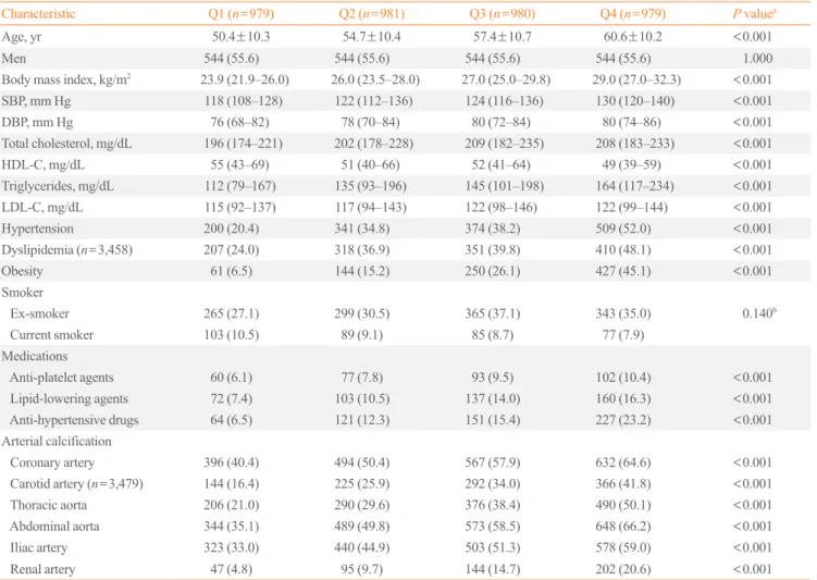
CI, 1.153 to 2.768; P=0.009), and 2.050 (95% CI, 1.294 to 3.250; P=0.002) for Q2 to 4, respectively (Table 2, Fig. 1A, Supplemental Table S2). Similarly, the ORs for carotid artery, thoracic aorta, and abdominal aorta calcification for each in- crease in the perirenal fat thickness quartile were modest, but significant: 1.118 (95% CI, 1.006 to 1.241; P=0.037), 1.141 (95% CI, 1.030 to 1.264; P=0.011), and 1.117 (95% CI, 1.013 to 1.23; P=0.027), respectively (Table 2, Fig. 1A).
A sensitivity analysis according to sex also confirmed the sig- nificance of the association between perirenal fat thickness and renal artery calcification in both sexes (Supplemental Table S3).
A significant association between perirenal fat thickness and ar- terial calcification was not found in both sexes for any arterial bed other than the renal artery (Supplemental Table S3).
The risk of renal arterial calcification on each side was evalu- ated according to the ipsilateral and contralateral perirenal fat thickness. No location-specific differences in the association between renal arterial calcification and perirenal fat thickness were found (Supplemental Table S4), and the AUROC values for the prediction of renal arterial calcification by the ipsilateral and contralateral perirenal fat thickness were similar (Supple- mental Table S5). The AUROC for the prediction of renal artery calcification using total perirenal fat was higher than the AU- ROCs obtained when using either the contralateral fat or ipsilat-
Table 1. Clinical Characteristics of the Study Subjects According to Perirenal Fat Thickness
Characteristic Q1 (n=979) Q2 (n=981) Q3 (n=980) Q4 (n=979) P valuea
Age, yr 50.4±10.3 54.7±10.4 57.4±10.7 60.6±10.2 <0.001
Men 544 (55.6) 544 (55.6) 544 (55.6) 544 (55.6) 1.000
Body mass index, kg/m2 23.9 (21.9–26.0) 26.0 (23.5–28.0) 27.0 (25.0–29.8) 29.0 (27.0–32.3) <0.001
SBP, mm Hg 118 (108–128) 122 (112–136) 124 (116–136) 130 (120–140) <0.001
DBP, mm Hg 76 (68–82) 78 (70–84) 80 (72–84) 80 (74–86) <0.001
Total cholesterol, mg/dL 196 (174–221) 202 (178–228) 209 (182–235) 208 (183–233) <0.001
HDL-C, mg/dL 55 (43–69) 51 (40–66) 52 (41–64) 49 (39–59) <0.001
Triglycerides, mg/dL 112 (79–167) 135 (93–196) 145 (101–198) 164 (117–234) <0.001
LDL-C, mg/dL 115 (92–137) 117 (94–143) 122 (98–146) 122 (99–144) <0.001
Hypertension 200 (20.4) 341 (34.8) 374 (38.2) 509 (52.0) <0.001
Dyslipidemia (n=3,458) 207 (24.0) 318 (36.9) 351 (39.8) 410 (48.1) <0.001
Obesity 61 (6.5) 144 (15.2) 250 (26.1) 427 (45.1) <0.001
Smoker
Ex-smoker 265 (27.1) 299 (30.5) 365 (37.1) 343 (35.0) 0.140b
Current smoker 103 (10.5) 89 (9.1) 85 (8.7) 77 (7.9)
Medications
Anti-platelet agents 60 (6.1) 77 (7.8) 93 (9.5) 102 (10.4) <0.001
Lipid-lowering agents 72 (7.4) 103 (10.5) 137 (14.0) 160 (16.3) <0.001
Anti-hypertensive drugs 64 (6.5) 121 (12.3) 151 (15.4) 227 (23.2) <0.001
Arterial calcification
Coronary artery 396 (40.4) 494 (50.4) 567 (57.9) 632 (64.6) <0.001
Carotid artery (n=3,479) 144 (16.4) 225 (25.9) 292 (34.0) 366 (41.8) <0.001
Thoracic aorta 206 (21.0) 290 (29.6) 376 (38.4) 490 (50.1) <0.001
Abdominal aorta 344 (35.1) 489 (49.8) 573 (58.5) 648 (66.2) <0.001
Iliac artery 323 (33.0) 440 (44.9) 503 (51.3) 578 (59.0) <0.001
Renal artery 47 (4.8) 95 (9.7) 144 (14.7) 202 (20.6) <0.001
Values are expressed as mean±standard deviation, number (%), or median (interquartile range). Hypertension was defined as SBP >140 mm Hg, DBP
>90 mm Hg, or the use of anti-hypertensive medications. Dyslipidemia was defined as a ratio of total to HDL-C >5, or the use of cholesterol-lowering medication.
SBP, systolic blood pressure; DBP, diastolic blood pressure; HDL-C, high-density lipoprotein cholesterol; LDL-C, low-density lipoprotein cholesterol.
aUsing analysis of variance for age, the Kruskal-Wallis test for the other continuous variables, and the chi-square test for categorical variables; bFrom lin- ear-by-linear association.
Table 2. Risk of Arterial Calcification According to Perirenal Fat Thickness (per Quartile)
Variable Unadjusted Age and sex–adjusted Model 1 Model 2
OR (95% CI) P value OR (95% CI) P value OR (95% CI) P value OR (95% CI) P value Coronary artery 1.386 (1.308–1.468) <0.001 1.162 (1.088–1.240) <0.001 1.083 (1.01–1.171) 0.047 1.079 (0.990–1.176) 0.082 Carotid artery 1.520 (1.420–1.627) <0.001 1.162 (1.074–1.258) <0.001 1.115 (1.016–1.224) 0.022 1.118 (1.006–1.241) 0.037 Thoracic aorta 1.548 (1.455–1.647) <0.001 1.096 (1.014–1.184) 0.021 1.156 (1.053–1.268) 0.002 1.141 (1.030–1.264) 0.011 Abdominal aorta 1.522 (1.436–1.614) <0.001 1.077 (1.001–1.159) 0.046 1.136 (1.041–1.240) 0.004 1.117 (1.013–1.233) 0.027 Iliac artery 1.414 (1.335–1.498) <0.001 1.056 (0.985–1.131) 0.125 1.042 (0.959–1.132) 0.327 1.012 (0.924–1.110) 0.793 Renal artery 1.666 (1.519–1.827) <0.001 1.227 (1.104–1.365) <0.001 1.289 (1.137–1.460) <0.001 1.251 (1.091–1.435) 0.001 Model 1: with adjustment for age, sex, and body mass index. Model 2: with adjustment for hypertension, high-density lipoprotein cholesterol (HDL-C), non-HDL-C, smoking history, and family history of heart disease in first-degree relatives in addition to Model 1.
OR, odds ratio; CI, confidence interval.
4 3 2 1 0
2.5 2.0 1.5 1.0 0.5 0
20 156
4 2 0 OR (95% CI)OR (95% CI) OR (95% CI)
Perirenal fat thickness
Perirenal fat thickness
Perirenal fat thickness RACS>0
Entire
RACS=0 Q1 Q2 Q3 Q4
Q1 Q2 Q3 Q4
Q1 Q2 Q3 Q4
Q1 Q2 Q3 Q4
Q1 Q2 Q3 Q4
Q1 Q2 Q3 Q4
Q1 Q2 Q3 Q4
Q1 Q2 Q3 Q4
Q1 Q2 Q3 Q4
Q1 Q2 Q3 Q4
Q1 Q2 Q3 Q4 Q1 Q2 Q3 Q4
Pc=0.037
Q1 Q2 Q3 Q4 Pc=0.011
a a a
Q1 Q2 Q3 Q4 Pc=0.027
Q1 Q2 Q3 Q4 Q1 Q2 Q3 Q4 Pc=0.001
Fig. 1. The risk of arterial calcification according to the quartiles of perirenal fat thickness. Total perirenal fat thickness was defined as the sum of perirenal fat thickness of both sides, and it was classified as Q1 (the lowest quartile) to Q4 (the highest quartile). Odds ratios (ORs) with 95% confidence interval (CIs) of the calcification in each arterial bed according to the perirenal fat thickness are shown (A) in the entire popu- lation, (B) those without renal artery calcification, and (C) in those with renal artery calcification. All statistical values were adjusted for age, sex, body mass index, hypertension, high-density lipoprotein cholesterol (HDL-C), non-HDL-C, smoking history, and family history of heart disease in first-degree relatives. RACS, renal artery calcification score. aP<0.05 compared to Q1; bP<0.005 compared to Q1; cP for trend.
Coronary artery
Coronary artery
Coronary artery Carotid artery
Carotid artery
Carotid artery Thoracic aorta
Thoracic aorta
Thoracic aorta Abdominal aorta
Abdominal aorta
Abdominal aorta Iliac artery
Iliac artery
Iliac artery Renal artery
A
B
C
a b
a a a
Pc=0.045
eral fat, although there was no statistically significant differ- ence. In females, the AUROC of right perirenal fat thickness for the prediction of left renal arterial calcification was significantly lower than that of total perirenal fat thickness (P=0.008); how- ever, it was not significantly different from that of left perirenal fat thickness (P=0.452). Similarly, the AUROC of left perirenal fat thickness for the prediction of right renal arterial calcifica- tion was lower than that of total perirenal fat thickness (P=
0.006) in females (Supplemental Table S5).
Calcification in different arterial beds according to the status of renal arterial calcification
Since renal arterial calcification itself is a well-known risk fac- tor for both hypertension and systemic atherosclerosis [27], and
because a strong association between renal fat thickness and re- nal arterial calcification was found in the current study (Fig.
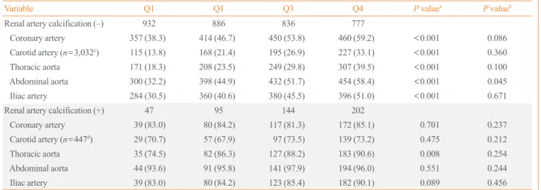
1A), the association between perirenal fat thickness and vascu- lar calcification in the other arterial beds was re-evaluated ac- cording to the presence or absence of renal arterial calcification (Fig. 1B, C). In subjects with renal arterial calcification, the prevalence of vascular calcification in each arterial bed reached
>70% irrespective of perirenal fat thickness (Table 3). Even in Q1, the prevalence of vascular calcification in the coronary ar- teries, carotid arteries, thoracic aorta, and abdominal aorta was 83.0%, 70.7%, 74.5%, and 93.6%, respectively. No significant association was found between the prevalence of arterial calcifi- cation and perirenal fat thickness in those with renal arterial cal- cification after adjustment for confounders (Table 3).
Table 4. Risk of Arterial Calcification According to Perirenal Fat Thickness (per Quartile) in Subjects without Renal Artery Calcification
Variable Unadjusted Age and sex–adjusted Model 1 Model 2
OR (95% CI) P value OR (95% CI) P value OR (95% CI) P value OR (95% CI) P value Coronary artery 1.329 (1.250–1.413) <0.001 1.155 (1.079–1.237) <0.001 1.076 (0.991–1.169) 0.080 1.082 (0.989–1.185) 0.086 Carotid artery 1.437 (1.330–1.554) <0.001 1.146 (1.051–1.250) 0.002 1.059 (0.956–1.174) 0.273 1.056 (0.940–1.186) 0.360 Thoracic aorta 1.426 (1.330–1.528) <0.001 1.050 (0.966–1.141) 0.249 1.105 (1.000–1.222) 0.050 1.098 (0.982–1.227) 0.100 Abdominal aorta 1.424 (1.339–1.516) <0.001 1.065 (0.988–1.147) 0.099 1.122 (1.026–1.228) 0.012 1.109 (1.003–1.228) 0.045 Iliac artery 1.321 (1.242–1.406) <0.001 1.028 (0.957–1.105) 0.449 1.002 (0.918–1.093) 0.972 0.979 (0.889–1.079) 0.671 Model 1: with adjustment for age, sex, and body mass index. Model 2: with adjustment for hypertension, high-density lipoprotein cholesterol (HDL-C), non-HDL-C, smoking history, and family history of heart diseases in the first-degree relatives in addition to Model 1.
OR, odds ratio; CI, confidence interval.
Table 3. Prevalence of Vascular Calcification According to Perirenal Fat Thickness
Variable Q1 Q1 Q3 Q4 P valuea P valueb
Renal artery calcification (–) 932 886 836 777
Coronary artery 357 (38.3) 414 (46.7) 450 (53.8) 460 (59.2) <0.001 0.086
Carotid artery (n=3,032c) 115 (13.8) 168 (21.4) 195 (26.9) 227 (33.1) <0.001 0.360
Thoracic aorta 171 (18.3) 208 (23.5) 249 (29.8) 307 (39.5) <0.001 0.100
Abdominal aorta 300 (32.2) 398 (44.9) 432 (51.7) 454 (58.4) <0.001 0.045
Iliac artery 284 (30.5) 360 (40.6) 380 (45.5) 396 (51.0) <0.001 0.671
Renal artery calcification (+) 47 95 144 202
Coronary artery 39 (83.0) 80 (84.2) 117 (81.3) 172 (85.1) 0.701 0.237
Carotid artery (n=447d) 29 (70.7) 57 (67.9) 97 (73.5) 139 (73.2) 0.475 0.212
Thoracic aorta 35 (74.5) 82 (86.3) 127 (88.2) 183 (90.6) 0.008 0.254
Abdominal aorta 44 (93.6) 91 (95.8) 141 (97.9) 194 (96.0) 0.551 0.244
Iliac artery 39 (83.0) 80 (84.2) 123 (85.4) 182 (90.1) 0.089 0.456
Values are expressed as number(%).
aP for trend, unadjusted; bP for trend with adjustment for age, sex, body mass index, hypertension, high-density lipoprotein cholesterol (HDL-C), non- HDL-C, smoking history, and family history of heart disease in first-degree relatives; cNumber of subjects in each quartile: 836, 785, 726, and 685 in Q1, Q2, Q3, and Q4, respectively; dNumber of subjects in each quartile: 41, 84, 132, and 190 in Q1, Q2, Q3, and Q4, respectively.
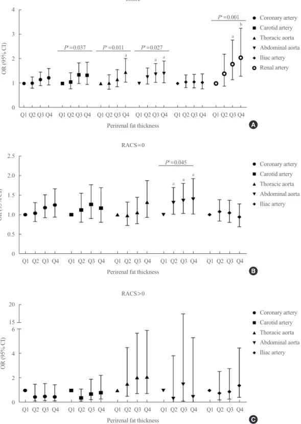
By contrast, in subjects without renal arterial calcification, a significant trend in the prevalence of arterial calcification ac- cording to perirenal fat thickness was found (P<0.001 for all in the crude analysis) (Table 3). Subsequent adjustment for age, sex, BMI, hypertension, HDL-C, non-HDL-C, smoking history, and family history of heart disease in first-degree relatives atten- uated the statistical significance of these trends. Only abdominal aorta calcification remained significant (P for trend=0.045) (Ta- ble 3). For each 1-quartile increase in perirenal fat thickness, the odds of abdominal aorta calcification increased by 10% (OR, 1.109; 95% CI, 1.003 to 1.228; P=0.045) (model 2 in Table 4).
The ORs for abdominal aorta calcification in Q2, Q3, and Q4 compared to Q1 were 1.305 (95% CI, 1.010 to 1.305; P=0.042), 1.357 (95% CI, 1.029 to 1.791; P=0.031), and 1.393 (95% CI, 1.013 to 1.916; P=0.041), respectively (Fig. 1B).
DISCUSSION
The results of the current study suggest that perirenal fat thick- ness is significantly associated with the presence of renal arteri- al calcification. Even after adjustment for atherosclerotic risk factors such as age, obesity, dyslipidemia, hypertension, smok- ing history, and family history, the risk of renal arterial calcifica- tion increased by 25% with every quartile of perirenal fat thick- ness. Perirenal fat thickness was also associated with calcified atherosclerosis in the other vascular beds evaluated (the carotid arteries, coronary arteries, thoracic aorta, abdominal aorta, iliac arteries, and renal arteries); however, after adjustment for renal arterial calcification and atherosclerotic risk factors, it was only associated with abdominal aorta calcification.
Perirenal fat is, like other ectopic fat, an active secretory or- gan that produces adipokines and cytokines [28,29], which in- duce inflammation [6-8], and consequently, atherosclerosis [1,8- 10]. The current findings that perirenal fat thickness was signifi- cantly associated not only with dyslipidemia or hypertension, but also with systemic arterial calcification, and that the signifi- cant association between vascular calcification and perirenal fat thickness was attenuated by adjustment for atherosclerotic risk factors, suggest a systemic effect of perirenal fat on the athero- sclerotic process. Nevertheless, the presence of an independent association between renal artery calcification and perirenal fat thickness in a dose-dependent manner suggests that perirenal fat exerts a local effect on the renal arteries. Furthermore, in fe- males, the prediction of renal artery calcification by contralater- al perirenal fat thickness was less powerful than the use of total perirenal fat thickness. Perirenal fat is known to induce a local
inflammatory response [30] and to regulate renal physiology [30,31]. Nonetheless, no significant difference was found in the effect of perirenal fat thickness between the ipsilateral and con- tralateral renal arteries in the current study. The AUROC for the prediction of renal artery calcification using total perirenal fat was higher than the AUROC values obtained for both the con- tralateral fat and ipsilateral fat, even though the difference did not reach statistical significance. We could not differentiate fat inside the renal fascia from the fat outside the fascia, which may have attenuated the local effects of perirenal fat. Considering that renal sinus fat increases renal hydrostatic pressure and acti- vates the renin-angiotensin-aldosterone system [15,16], fat in- side the renal fascia might have a more direct effect on the renal arteries than perirenal fat outside the fascia. In addition, in the current study, both perirenal fat thickness and renal arterial cal- cification on both sides were strongly correlated with each oth- er, which might have caused the absence of a difference be- tween the ipsilateral and contralateral associations. Further studies to determine the local effects of perirenal fat are needed.
Renal arterial calcification is an independent risk factor for all-cause mortality even in healthy outpatients without any car- diovascular disease [27]. A stratified analysis according to the presence of renal arterial calcification in the current study con- firmed that the presence of renal artery calcification reflects a high risk of systemic atherosclerosis. Specifically, the preva- lence of vascular calcification in the other arterial beds among those with renal artery calcification was more than 70% in each arterial bed, irrespective of the amount of perirenal fat. By con- trast, in those without renal arterial calcification, a significant trend in the prevalence of arterial calcification according to perirenal fat thickness was found in all arterial beds in the crude analysis, which implies that the effects of ectopic fat such as perirenal fat might be different according to the severity of the systemic atherosclerosis burden, and that reduction of ectopic fat might have a more favorable effect on cardiometabolic health in the early stages of atherosclerosis.
Interestingly, in those without renal arterial calcification, the only independent association between vascular calcification and perirenal fat thickness was found for abdominal aorta calcifica- tion, although its OR per quartile was around 1.2. Currently, it is not apparent why perirenal fat thickness was independently as- sociated with calcification in the abdominal aorta, but not in the other vascular beds. The prevalence of calcification in abdomi- nal aorta and coronary arteries was relatively high compared to the other arteries in the current study, which might have strengthened the statistical significance of this finding despite
the similar ORs. In addition, the relatively short anatomical dis- tance between perirenal fat and the abdominal aorta might have contributed to this finding, and this possibility should be ex- plored in another independent study.
A limited number of studies have estimated perirenal fat thickness using CT scans [17,24]. In most previous studies, perirenal fat thickness was assessed using ultrasonography [12- 14]. Although ultrasonography is more convenient, CT scans enable a full-scale measurement [32]. In addition, perirenal fat thickness assessed by CT scans was reported to be a reliable marker of perirenal fat volume [17].
The current study has several limitations. First, as mentioned above, we did not differentiate fat inside the renal fascia from the fat outside the fascia, which might have masked the local ef- fects of perirenal fat. Few studies have compared the effects of fat on both sides. Although both pararenal fat [13] as well as re- nal sinus fat [33,34] have been reported to be independent risk factors for hypertension even after adjustment for visceral fat, the fat inside the renal fascia might exert a more direct effect on the renal arteries than the perirenal fat outside the fascia via the renin-angiotensin-aldosterone system [15,16] and local inflam- mation [8]. Second, we did not measure waist circumference or intra-abdominal visceral fat, although a comparison of the effect of perirenal fat to that of other visceral fat on systemic athero- sclerotic calcification would enable the pathophysiological role of perirenal fat distinct from other visceral fat to be elucidated.
In addition, no information on fasting lipid levels was analyzed in the current study. However, the ratio of total to HDL-C [35,36] and non-HDL-C [37] used in the current study has been reported to be a good predictor of future cardiovascular diseas- es. Lastly, the cross-sectional nature of the study makes it im- possible to identify the causal relationships underlying the asso- ciation between perirenal fat and arterial calcification.
A major strength of the study is that the current findings are based on a large community-based cohort that underwent exten- sive evaluations of systemic calcification, including six arterial beds in each of 3,919 subjects. Furthermore, few previous stud- ies have investigated the association between perirenal fat thick- ness and systemic atherosclerotic calcification.
In the current study, we confirmed that perirenal fat thickness was independently associated with vascular calcification in the renal artery and abdominal aorta. Further epidemiological stud- ies to replicate the current findings are needed in diverse study populations.
CONFLICTS OF INTEREST
No potential conflict of interest relevant to this article was re- ported.
ACKNOWLEDGMENTS
This study was supported by the National Institute of Health (Ro 1 DK080015).
AUTHOR CONTRIBUTIONS
Conception or design: B.K.K., M.A.A. Acquisition, analysis, or interpretation of data: B.K.K., J.O.D., C.M.W., M.H.C., M.A.A.
Drafting the work or revising: B.K.K., C.M.W., M.H.C., M.A.A. Final approval of the manuscript: B.K.K., J.O.D., C.M.W., M.H.C., M.A.A.
ORCID
Bo Kyung Koo https://orcid.org/0000-0002-6489-2656
REFERENCES
1. Byrne CD, Targher G. NAFLD: a multisystem disease. J Hepatol 2015;62(1 Suppl):S47-64.
2. Meshkani R, Adeli K. Hepatic insulin resistance, metabolic syndrome and cardiovascular disease. Clin Biochem 2009;
42:1331-46.
3. Komiya H, Mori Y, Yokose T, Kurokawa N, Horie N, Taji- ma N. Effect of intramuscular fat difference on glucose and insulin reaction in oral glucose tolerance test. J Atheroscler Thromb 2006;13:136-42.
4. Therkelsen KE, Pedley A, Speliotes EK, Massaro JM, Mu- rabito J, Hoffmann U, et al. Intramuscular fat and associa- tions with metabolic risk factors in the Framingham Heart Study. Arterioscler Thromb Vasc Biol 2013;33:863-70.
5. Krssak M, Roden M. The role of lipid accumulation in liver and muscle for insulin resistance and type 2 diabetes melli- tus in humans. Rev Endocr Metab Disord 2004;5:127-34.
6. Tilg H, Hotamisligil GS. Nonalcoholic fatty liver disease:
cytokine-adipokine interplay and regulation of insulin resis- tance. Gastroenterology 2006;131:934-45.
7. Wieckowska A, Papouchado BG, Li Z, Lopez R, Zein NN, Feldstein AE. Increased hepatic and circulating interleukin-6 levels in human nonalcoholic steatohepatitis. Am J Gastro-
enterol 2008;103:1372-9.
8. Lim S, Meigs JB. Links between ectopic fat and vascular disease in humans. Arterioscler Thromb Vasc Biol 2014;34:
1820-6.
9. Targher G, Day CP, Bonora E. Risk of cardiovascular dis- ease in patients with nonalcoholic fatty liver disease. N Engl J Med 2010;363:1341-50.
10. Koo BK, Allison MA, Criqui MH, Denenberg JO, Wright CM. The association between liver fat and systemic calci- fied atherosclerosis. J Vasc Surg 2020;71:204-11.
11. Chau YY, Bandiera R, Serrels A, Martinez-Estrada OM, Qing W, Lee M, et al. Visceral and subcutaneous fat have different origins and evidence supports a mesothelial source.
Nat Cell Biol 2014;16:367-75.
12. Roever L, Resende ES, Veloso FC, Diniz AL, Penha-Silva N, Casella-Filho A, et al. Perirenal fat and association with metabolic risk factors: the Uberlandia Heart Study. Medi- cine (Baltimore) 2015;94:e1105.
13. De Pergola G, Campobasso N, Nardecchia A, Triggiani V, Caccavo D, Gesualdo L, et al. Para- and perirenal ultrasono- graphic fat thickness is associated with 24-hours mean dia- stolic blood pressure levels in overweight and obese sub- jects. BMC Cardiovasc Disord 2015;15:108.
14. Lamacchia O, Nicastro V, Camarchio D, Valente U, Grisorio R, Gesualdo L, et al. Para- and perirenal fat thickness is an independent predictor of chronic kidney disease, increased renal resistance index and hyperuricaemia in type-2 diabetic patients. Nephrol Dial Transplant 2011;26:892-8.
15. Dwyer TM, Mizelle HL, Cockrell K, Buhner P. Renal sinus lipomatosis and body composition in hypertensive, obese rabbits. Int J Obes Relat Metab Disord 1995;19:869-74.
16. Spit KA, Muskiet MH, Tonneijck L, Smits MM, Kramer MH, Joles JA, et al. Renal sinus fat and renal hemodynam- ics: a cross-sectional analysis. MAGMA 2020;33:73-80.
17. Favre G, Grangeon-Chapon C, Raffaelli C, Francois-Chal- min F, Iannelli A, Esnault V. Perirenal fat thickness mea- sured with computed tomography is a reliable estimate of perirenal fat mass. PLoS One 2017;12:e0175561.
18. Shulman GI. Ectopic fat in insulin resistance, dyslipidemia, and cardiometabolic disease. N Engl J Med 2014;371:1131- 41.
19. Executive summary of the clinical guidelines on the identifi- cation, evaluation, and treatment of overweight and obesity in adults. Arch Intern Med 1998;158:1855-67.
20. Allison MA, Criqui MH, Wright CM. Patterns and risk fac- tors for systemic calcified atherosclerosis. Arterioscler
Thromb Vasc Biol 2004;24:331-6.
21. Hughes-Austin JM, Dominguez A 3rd, Allison MA, Wassel CL, Rifkin DE, Morgan CG, et al. Relationship of coronary calcium on standard chest CT scans with mortality. JACC Cardiovasc Imaging 2016;9:152-9.
22. Lin TC, Wright CM, Criqui MH, Allison MA. Superior mesenteric artery calcification is associated with cardiovas- cular risk factors, systemic calcified atherosclerosis, and in- creased mortality. J Vasc Surg 2018;67:1484-90.
23. Agatston AS, Janowitz WR, Hildner FJ, Zusmer NR, Vi- amonte M Jr, Detrano R. Quantification of coronary artery calcium using ultrafast computed tomography. J Am Coll Cardiol 1990;15:827-32.
24. Eisner BH, Zargooshi J, Berger AD, Cooperberg MR, Doyle SM, Sheth S, et al. Gender differences in subcutaneous and perirenal fat distribution. Surg Radiol Anat 2010;32:879-82.
25. Hervochon R, Bobbio A, Guinet C, Mansuet-Lupo A, Rab- bat A, Regnard JF, et al. Body mass index and total psoas area affect outcomes in patients undergoing pneumonecto- my for cancer. Ann Thorac Surg 2017;103:287-95.
26. Portney LG, Watkins MP. Foundations of clinical research:
applications to practice. 2nd ed. Upper Saddle River: Pren- tice Hall Health; 2000.
27. Rifkin DE, Ix JH, Wassel CL, Criqui MH, Allison MA. Re- nal artery calcification and mortality among clinically as- ymptomatic adults. J Am Coll Cardiol 2012;60:1079-85.
28. Wu H, Cheng XW, Hao C, Zhang Z, Yao H, Murohara T, et al. Regulation of apelin and its receptor expression in adi- pose tissues of obesity rats with hypertension and cultured 3T3-L1 adipocytes. Exp Anim 2014;63:257-67.
29. Kwon EY, Shin SK, Cho YY, Jung UJ, Kim E, Park T, et al.
Time-course microarrays reveal early activation of the im- mune transcriptome and adipokine dysregulation leads to fi- brosis in visceral adipose depots during diet-induced obesity.
BMC Genomics 2012;13:450.
30. Hoogduijn MJ, Crop MJ, Peeters AM, van Osch GJ, Balk AH, Ijzermans JN, et al. Human heart, spleen, and perirenal fat-derived mesenchymal stem cells have immunomodula- tory capacities. Stem Cells Dev 2007;16:597-604.
31. Niijima A. Reflex effects from leptin sensors in the white ad- ipose tissue of the epididymis to the efferent activity of the sympathetic and vagus nerve in the rat. Neurosci Lett 1999;
262:125-8.
32. Liu BX, Sun W, Kong XQ. Perirenal fat: a unique fat pad and potential target for cardiovascular disease. Angiology 2019;
70:584-93.
33. Chughtai HL, Morgan TM, Rocco M, Stacey B, Brinkley TE, Ding J, et al. Renal sinus fat and poor blood pressure control in middle-aged and elderly individuals at risk for cardiovascular events. Hypertension 2010;56:901-6.
34. Foster MC, Hwang SJ, Porter SA, Massaro JM, Hoffmann U, Fox CS. Fatty kidney, hypertension, and chronic kidney dis- ease: the Framingham Heart Study. Hypertension 2011;58:
784-90.
35. Lemieux I, Lamarche B, Couillard C, Pascot A, Cantin B, Bergeron J, et al. Total cholesterol/HDL cholesterol ratio vs LDL cholesterol/HDL cholesterol ratio as indices of isch-
emic heart disease risk in men: the Quebec Cardiovascular Study. Arch Intern Med 2001;161:2685-92.
36. Lamarche B, Moorjani S, Lupien PJ, Cantin B, Bernard PM, Dagenais GR, et al. Apolipoprotein A-I and B levels and the risk of ischemic heart disease during a five-year follow-up of men in the Quebec cardiovascular study. Circulation 1996;
94:273-8.
37. Cui Y, Blumenthal RS, Flaws JA, Whiteman MK, Langen- berg P, Bachorik PS, et al. Non-high-density lipoprotein cholesterol level as a predictor of cardiovascular disease mortality. Arch Intern Med 2001;161:1413-9.