509 http://dx.doi.org/10.4196/kjpp.2014.18.6.509
ABBREVIATIONS: RKO cells, RKO Human Colorectal Cancer Cells;
[Ca2+]i, intracellular Ca2+ activity; NCX, Na+-Ca2+ exchanger; IP3, Inositol-1,4,5-triphosphate; SOCI, Store-operated Ca2+ influx; PMCA, plasma membrane Ca2+-ATPase; SERCA, sarcoplasmic/endoplasmic Ca2+-ATPase; ER, endoplasmic reticulum.
Received July 21, 2014, Revised October 13, 2014, Accepted October 13, 2014
Corresponding to: Chang Kook Suh, Department of Physiology and Biophysics, Inha University College of Medicine, 253 Yong-hyun- dong, Nam-gu, Incheon 402-751, Korea. (Tel) 82-32-890-0921, (Fax) 82-32-884-5997, (E-mail) [email protected]
This is an Open Access article distributed under the terms of the Creative Commons Attribution Non-Commercial License (http://
creativecommons.org/licenses/by-nc/3.0) which permits unrestricted non-commercial use, distribution, and reproduction in any medium, provided the original work is properly cited.
Distinct Cellular Calcium Metabolism in Radiation-sensitive RKO Human Colorectal Cancer Cells
Yun Tai Kim1,2, Soo Shin Jo1, Young Jun Park1, Myung Za Lee3, and Chang Kook Suh1
1Department of Physiology and Biophysics, Inha University College of Medicine, Incheon 401-751, 2Research Group of Food Functionality, Korea Food Research Institute, Seongnam 463-746, Division of Food Biotechnology, Korea University of Science and Technology, Daejeon 305-350, 3Department of Radiation Oncology, Hanyang University College of Medicine, Seoul 133-791, Korea
Radiation therapy for variety of human solid tumors utilizes mechanism of cell death after DNA damage caused by radiation. In response to DNA damage, cytochrome c was released from mitochondria by activation of pro-apoptotic Bcl-2 family proteins, and then elicits massive Ca2+ release from the ER that lead to cell death. It was also suggested that irradiation may cause the deregulation of Ca2+
homeostasis and trigger programmed cell death and regulate death specific enzymes. Thus, in this study, we investigated how cellular Ca2+ metabolism in RKO cells, in comparison to radiation-resistant A549 cells, was altered by gamma (γ )-irradiation. In irradiated RKO cells, Ca2+ influx via activation of NCX reverse mode was enhanced and a decline of [Ca2+]i via forward mode was accelerated. The amount of Ca2+ released from the ER in RKO cells by the activation of IP3 receptor was also enhanced by irradiation. An increase in [Ca2+]i via SOCI was enhanced in irradiated RKO cells, while that in A549 cells was depressed. These results suggest that γ-irradiation elicits enhancement of cellular Ca2+
metabolism in radiation-sensitive RKO cells yielding programmed cell death.
Key Words: A549 cells, Inositol-1,4,5-triphosphate receptors, Na+-Ca2+ exchanger, RKO cells, Store- operated Ca2+ influx
INTRODUCTION
Radiation therapy is one of common conventional treat- ment modalities for variety of human solid tumors.
Apoptosis plays an important role in cell death after DNA damage caused by radiation. In response to DNA damage caused by irradiation, p53 (53 kDa protein) activates vari- ous genes [1-3]. Protein products of pro-apoptotic Bcl-2 gene-family members cause cytochrome c released from the mitochondria into the cytosol and released cytochrome c ac- tivates the caspase cascade [4-6]. Mitochondrial apoptotic involvement could depend on signals that originate from other intracellular compartments. Namely, Ca2+ released from the endoplasmic reticulum (ER) could induce and/or play a facilitative role in the apoptotic changes [7-9].
Cell death has always been known to be one of the numer- ous cellular events triggered by an increase in intracellular Ca2+ evoked by physiological or pathological stimuli. The
role of intracellular Ca2+ activity ([Ca2+]i) in apoptosis was appreciated more recently [10]. It has been reported that the expression and/or localization of Bcl-2 can modulate Ca2+
fluxes during the course of cell death [11-13].
Increase in intracellular Ca2+ causing apoptosis can arise from a variety of sources. Mechanisms for increasing [Ca2+]i
include the entry of extracellular Ca2+ via Ca2+ channels (voltage-gated channels, receptor-mediated channels) and the transient receptor potential channel of store-operated Ca2+ influx (SOCI), or the release of stored Ca2+ form intra- cellular stores via IP3 receptors and ryanodine receptors in intracellular Ca2+ stores [14-17]. Once Ca2+ has served its signaling function, [Ca2+]i is lowered to resting levels to maintain intracellular Ca2+ homeostasis. Ca2+ is seques- tered into intracellular stores by pumps such as the sarco- plasmic/endoplasmic Ca2+-ATPase (SERCA) or is extruded to the extracellular environment by transporters such as the Na+-Ca2+ exchanger (NCX) and the plasma membrane Ca2+-ATPase (PMCA) [14,15,17]. Mitochondria also de- crease intracellular Ca2+ via the mitochondrial uniporter located in the inner mitochondrial membrane [18], although their role in regulating intracellular Ca2+ levels appears to be clearing Ca2+ in restricted microdomains such as the microenvironment of IP3 receptor channels [19].
The role of Ca2+ in promoting cell proliferation and cell
Table 1. Effects of γ-irradiation on basal level of R340/380 in RKO cells
Incubation time 30 min 1 hr
30 min 3 hrs 6 hrs 12 hrs 24 hrs 48 hrs
Basal level of R340/380 Control 0.80±0.05 0.80±0.05 0.81±0.05 0.80±0.05 0.81±0.05 0.82±0.05 0.81±0.05 γ-ray irradiated 0.82±0.05 0.80±0.05 0.81±0.05 0.79±0.05 0.83±0.11 0.83±0.05 0.84±0.11 Note that p=0.1379 at 48 hrs after γ-irradiation. n=30.
death has been regarded as signaling checkpoints in cancer, which determine how these processes are remodeled in can- cer [20]. Many studies have reported that a large influx of Ca2+ triggering apoptosis in cancer cells is provided by Ca2+ influx mediated by store-operated Ca2+ entry chan- nels, which suggest a pivotal role of SOCI in apoptosis and cancer progression [21]. Moreover, the anti-apoptotic pro- tein Bcl-2, which is commonly degraded in cancer, appears to modulate IP3-receptor Ca2+ channel activity on the ER Ca2+ stores [10,22,23]. It was also reported that the reduc- tion in ER means that Ca2+ release is insufficient to pro- duce apoptosis [10,24]. All these results suggest that the deregulation of cellular Ca2+ homeostasis caused under non-physiologic condition such as irradiation can elicit cell death and determine the sensitivity of cancer cells to radiotherapy. It was also suggested that ion transports may contribute to the intrinsic radio-resistance and the survival of the tumor cell, by controlling cell cycle, metabolic adapta- tions or DNA repair [25]. In this study, to explore the role of cellular Ca2+ metabolism in sensitivity of tumor cells to radiation, the effects of gamma (γ)-ray irradiation on cel- lular Ca2+ metabolism in radiosensitive RKO human color- ectal cancer cells and A549 human lung cancer cells, one of known radiation-resistant cells, were examined.
METHODS
Cell culture and Irradiation of cell cultures
RKO human colorectal cancer cells and A549 human lung cancer cells were used. The cells were grown in DMEM sup- plemented with 10% fetal bovine serum and 1% pen- icillin/streptomycin. The cells were cultured in 25 cm2 plas- tic tissue culture flasks at 37oC in a humidified 5% CO2/95%
air atmosphere. When the cells were in exponential growth phase at a cell density of 3×10⁶ cells/ 25 cm2 flasks, cells were irradiated with 10 Gy of γ-rays at a dose rate of 5.0 Gy/min with a 137 Cs irradiator (Cis biointernational IBL437C, France).
Measurements of [Ca2+]i
Intracellular free Ca2+ concentration was measured as described previously [26]. Cells were washed with PBS and incubated in 2 ml of buffer (0.05% trypsin and 0.02%
EDTA). The cells were then resuspended in Tyrode solution (in mM: 140 NaCl, 4 KCl, 2 CaCl2, 1 MgCl2, 1 NaH2PO4, 5 HEPES, 5.5 Glucose and pH 7.4) and incubated at 37oC with 3 μM fura-2 AM (Molecular Probe, Eugene, Oregon, USA) for 30 min and transferred to a recording chamber on an epifluorescence inverted microscope (Nikon Diaphot 300, Tokyo, Japan). Experimental solutions were super- fused at a flow rate of 2 ml/min. Fluorescence intensity was
measured using a cooled CCD camera (Photometrics PXL37, Tucson, Arizona, USA) and processed using the Axon Imaging Workbench v.2.2 (Axon Instrument, Foster city, CA, USA). [Ca2+]i was presented as the ratio of flur- escence intensities (R340/380) excited by alternating illumina- tion of 340 nm and 380 nm. Fluorescence intensity through 510 nm wavelength filter was collected using a cooled CCD digital camera (PXL-37, Photometrics, Tucson, AZ, USA).
Experiments were done at 37oC.
Solutions
The composition of Tyrode’s solution was 140 mM NaCl, 2.5 mM CaCl2, 5 mM KCl, 1 mM MgCl2, 1 mM NaH2PO4, 5 mM N-[2-hydroxyethl] piperazine-N’[2-ethanosulfonic acid] (HEPES), and 5.5 mM glucose at pH 7.4. In the 0 mM Na+/2.5 mM Ca2+ solution (Na+-free solution), NaCl was isosmotically replaced by N-methyl-D-glucamine (NMDG). 140 mM Na+/0 mM Ca2+ solution (Ca2+-free sol- ution) was made by omitting CaCl2. To isolate NCX activity from other Ca2+ pathways, 1 μM thapsigargin (ER Ca2+- ATPase inhibitor), 5 mM caffeine (ryanodine receptor in- hibitor), and 250 μM La3+ (plasma membrane Ca2+- ATPase inhibitor) were added to the superfusing solutions.
The 0 Ca2+ solution, which was used to empty the internal Ca2+ stores, also contained 0.1 mM EGTA and 1 μM thapsigargin.
Statistical analysis
All data were expressed as mean±SD. Statistical analysis was performed by independent t-test, with p<0.05 as cri- teria of significance.
RESULTS
Basal level of intracellular Ca2+ activity
Basal levels of intracellular Ca2+ activities in γ-ray irra- diated RKO cells were compared to those in non-irradiated control cells (Table 1). When the cells were incubated for various durations up to 48 hrs after irradiation, basal levels of R340/380 in RKO cells were not fluctuated both in the con- trol and irradiated cells. And no difference in R340/380 was observed between the control and irradiated cells. Even when the basal levels of R340/380 were deviated most, such as in cells incubated for 48 hrs (0.81±0.05 vs, 0.84±0.11), no statistical significance was observed (p=0.14). Based on these findings, further experiments were carried out with cells which were incubated for 48 hrs after γ-ray irradia- tion.
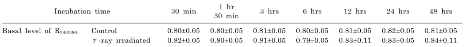
Fig. 1. Effects of γ-irradiation on NCX in RKO and A549 cells. The activity of NCX in cells was mea- sured in the reverse mode of NCX induced by superfusing 0 mM Na+/ 2.5 mM Ca2+ solution containing 1 μM thapsigargin (Thapsi), 5 mM caffeine (CAF), and 250 μM La3+
and in the forward mode of NCX by 140 mM Na+/0 mM Ca2+ solution.
(A) In RKO control cells, R340/380
increased with the reverse mode of NCX and decreased with the for- ward mode. (B) In γ-ray irradiated RKO cells, the second peak was observed. The slope of R340/380
changes by the forward mode of NCX was increased in γ-rays irradiated cells. (C) In A549 control cells, R340/380 changed as in (A). (D) In γ- ray irradiated A549 cells, the slope of R340/380 changes by the forward mode of NCX was increased, confirming the activity of NCX was increased by γ-irradiation (p<
0.0001). Tracings in (A) to (D) represent the average values of R340/380.
Table 2. Comparison of NCX-mediated R340/380 changes in RKO and A549 cells
Cell type Non-irradiated control γ-irradiated
Basal level RKO 0.85±0.05 (n=24) 0.92±0.10 (n=25)
A549 0.73±0.06 (n=35) 0.73±0.06 (n=34)
Plateau value RKO 1.65±0.15 1.66±0.20
A549 1.21±0.12 1.11±0.12
Rate of R340/380 changes/min RKO −0.17±0.05 −0.25±0.10**
A549 −0.13±0.06 −0.24±0.05***
**p<0.001, ***p<0.0001.
Physiological activity of NCX
When the cells were superfused with 0 mM Na+/2.5 mM Ca2+ solution, as described in “METHODS”, R340/380 in RKO cells increased to a plateau value (from 0.85±0.01 to 1.65±0.03) as shown in Fig. 1. Subsequent superfusion of 140 mM Na+/0 Ca2+ solution lowered R340/380 to the resting level with rate of R340/380 changes of −0.17±0.05 /min (Fig.
1A and Table 2). In γ-ray irradiated RKO cells, R340/380 in- creased to a plateau value (from 0.92±0.10 to 1.66±0.20) and an additional increase to 1.87±0.40 was followed.
Subsequent superfusion of 140 mM Na+/0 mM Ca2+ sol- ution lowered R340/380 to the resting level with rate of R340/380
changes of −0.25±0.10 /min (Fig. 1B and Table 2). The de- cay to the basal level of R340/380 in irradiated cells was com- pleted faster than that of control cells (230 sec vs. 290 sec) (p<0.001).
In A549 cells, R340/380 increased to a plateau value (from 0.73±0.06 to 1.21±0.12). Subsequent superfusion of 140 mM Na+/0 mM Ca2+ solution lowered R340/380 to the resting level with rate of R340/380 changes of −0.13±0.06 /min (Fig. 1C and Table 2). In γ-ray irradiated A549 cells, R340/380 in- creased to plateau values (from 0.73±0.06 to 1.11±0.12).
Subsequent superfusion of 140 mM Na+/0 mM Ca2+ sol- ution lowered R340/380 to the resting level with rate of R340/380
changes of −0.24±0.05/min (Fig. 1D and Table 2). The de- cay to the basal level of R340/380 was also completed faster than that of control cells (p<0.0001).
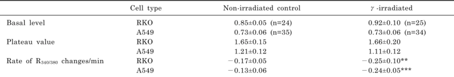
Ca2+ lnflux via SOCI
Ca2+ influx via SOCI were measured by superfusing cells with the normal Tyrode solution after empting the internal Ca2+ stores. When cells were superfused with 0 Ca2+ sol- ution containing 0.1 mM EGTA with 1 μM thapsigargin,
Fig. 2. Effects of γ-irradiation on SOCI in RKO and A549 cells. (A) When RKO control cells were superfused with Tyrode’s solution including 2 mM Ca2+ after depleting intracellular Ca2+ store, R340/380 was increased by Ca2+ influx via SOCI.
(B) Ca2+ influx via SOCI was enhanced in γ-ray irradiated RKO cells, compared to control cells (***p
<0.0001). (C) In A549 control cells, R340/380 was increased by Ca2+ influx via SOCI, as in (A). (D) Ca2+ influx via SOCI was decreased in γ-ray irradiated A549 cells, compared to (C) (*p<0.05). (E) In RKO cells, areas under SOCI response, which approximate the amount of Ca2+
influxed via SOCI, were increased by γ-ray irradiation (***p<0.0001). In A549 cells, areas under SOCI response were decreased by γ-ir- radiation (*p<0.05). The increment of areas under SOCI response in RKO cells after γ-irradiation was significantly different from that in A549 cells, which was decreased by γ-irradiation (###p<0.0001). Tracings in (A) to (D) represent the average values of R340/380.
which empties Ca2+ out of the ER, R340/380 increased and consequently declined below the control level as shown in Fig. 2 and Table 3. In RKO cells, R340/380 increased much greater than that of A549 cells (Fig. 2A vs. 2C). In γ-ray irradiated cells, significant changes in these peaks were not observed in either cell. After the internal Ca2+ stores were emptied, superfusions of 2.5 mM Ca2+ Tyrode solution raised R340/380 from 0.76±0.09 to 1.14±0.14 in RKO cells. In γ-ray irradiated RKO cells, with superfusion of 2.5 mM Ca2+ Tyrode solution, R340/380 increased from 0.74±0.09 to 1.60±0.44 and the increments in R340/380 were much larger than those of control cells (0.86±0.44 vs. 0.38±0.14) (p
<0.0001), as shown in Fig. 2B. Areas under SOCI response which approximate the amount of Ca2+ influxed by SOCI were increased by γ-ray irradiation from 213±112 to 576±304, with statistical significance (p<0.0001), as shown in Fig. 2E.
When A549 cells were superfused with 2.5 mM Ca2+
Tyrode solution, R340/380 increased from 0.77±0.06 to 1.26±0.30 (Fig. 2C). In γ-ray irradiated A549 cells, R340/380
increased from 0.77±0.12 to 1.10±0.24 as shown in Fig. 2D.
The increments in R340/380 in γ-ray irradiated A549 cells were smaller than those of control cells (0.33±0.24 vs.
0.49±0.30) (p<0.05). Areas under SOCI response were de- creased by γ-ray irradiation from 221±116 to 164±95, with statistical significance (p<0.05). The increment of areas under SOCI response in RKO cells by γ-ray irradiation was significantly different from that in A549 cells, which was decreased by γ-ray irradiation, as shown in Fig. 2E (p<0.0001).
ATP-induced Ca2+ release from the ER
Ca2+ release from the ER was measured by applying ATP extracellularly which activates IP3 receptor channels in the ER (Fig. 3 and Table 4). When the Tyrode solution contain-
Table 3. Comparison of SOCI-induced R340/380 changes in RKO and A549 cells
Cell type Non-irradiated control Gamma-irradiated
Basal level RKO 0.76±0.09 (n=22) 0.74±0.09 (n=20)
A549 0.77±0.06 (n=35) 0.77±0.12 (n=35)
Peak value RKO 1.14±0.14 1.60±0.44
A549 1.26±0.30 1.10±0.24
Changes in R340/380 RKO 0.38±0.14 0.86±0.44***
A549 0.49±0.05 0.33±0.24*
*p<0.05, ***p<0.0001.
Fig. 3. Effects of γ-irradiation on ATP-induced [Ca2+]i changes in RKO and A549 cells. (A) Application of 100 μM ATP induced transient changes in R340/380 (ATP-induced Ca2+
responses) in RKO control cells. (B) In γ-ray irradiated RKO cells, transient changes in R340/380 were enhanced, compared to control cells (1.18±0.07 vs. 0.89±0.06; p<0.0001).
(C) In A549 control cells, transient changes in R340/380 were elicited by application of 100 μM ATP. (D) In γ-ray irradiated A549 cells, tran- sient changes in R340/380 were not significantly different from those in control cells. (E) In more than 60%
of A549 cells measured, multiple transient changes in R340/380 were observed with a single application of ATP. (F) The frequency of multiple transient changes was increased in γ-ray irradiated A549 cells (See Table 5). Tracings in (A) to (F) represent the average values of R340/380.
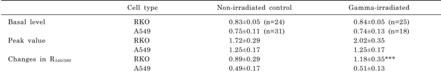
ing 100 μM ATP was applied for 10 sec, R340/380 in RKO cells increased transiently, 0.83±0.05 to 1.72±0.29, and re- turned slowly to the basal level (Fig. 3A). In γ-ray irradi- ated RKO cells, R340/380 increased from 0.84±0.05 to 2.02±0.35. The difference between the peak and basal val- ues for R340/380 in the control cells (n=24) was 0.89±0.29 and that in the γ-ray irradiated cells (n=25) was 1.18±0.35 (p
<0.0001; Table 4).
When A549 cells were superfused with the Tyrode solution containing 100 μM ATP for 10 sec, R340/380 increased tran- siently and returned to the basal level eliciting a single Ca2+ transient, as shown in Fig. 3C, in 50 cells out of 154 cells (32%) measured (Table 4). In other A549 cells, multi- ple Ca2+ transients were observed (Fig. 3E and Table 4).
Table 4. Comparison of ATP-induced R340/380 changes in RKO and A549 cells
Cell type Non-irradiated control Gamma-irradiated
Basal level RKO 0.83±0.05 (n=24) 0.84±0.05 (n=25)
A549 0.75±0.11 (n=31) 0.74±0.13 (n=18)
Peak value RKO 1.72±0.29 2.02±0.35
A549 1.25±0.17 1.25±0.17
Changes in R340/380 RKO 0.89±0.29 1.18±0.35***
A549 0.49±0.17 0.51±0.13
***p<0.0001.
Table 5. Changes in R340/380 induced by ATP in A549 cells
Control (n=154) γ-ray (48 hr) (n=167) Events with 1 peak Number of event
Basal level Peak value Changes in R340/380
50 0.75±0.14 1.25±0.21 0.49±0.21
19 0.74±0.10 1.25±0.13 0.51±0.10
2 peaks Number of event
Basal level Peak value Changes in R340/380
44 0.73±0.07 1.41±0.20 0.68±0.20
28 0.66±0.05 1.28±0.16 0.63±0.16
3 peaks Number of event
Basal level Peak value Changes in R340/380
32 0.73±0.06 1.48±0.17 0.75±0.17
30 0.67±0.05 1.31±0.22 0.64±0.22
Multi peaks Number of event
Basal level Peak value Changes in R340/380
28 0.71±0.05 1.41±0.16 0.69±0.16
90 0.69±0.09 1.33±0.47 0.65±0.38
The amplitudes of Ca2+ transients, the differences between the peak and basal values of R340/380, were not significantly different between control cells and γ-ray irradiated cells (Table 4). However, the frequency of multiple transients was increased in γ-ray irradiated cells (Table 5).
DISCUSSION
Surge of intracellular Ca2+ causing cell death can arise from a variety of sources. [Ca2+]i can be increased by the entry of extracellular Ca2+ via SOCI or the release of stored Ca2+ form the ER via IP3 receptors and ryanodine receptors in ER membranes [14-17]. Ca2+ influx via reverse mode of NCX also contribute to [Ca2+]i increase [17,27,28].
By γ-irradiation, RKO cells begin to exit from G2/M ar- rest to apoptosis by 24 hrs after irradiation. Only small fractions of cells remain in G2/M phase by 48 hrs, implying that the post-mitotic apoptosis occurs by 48 hrs after irradi- ation [29]. During this time span, basal level of [Ca2+]i in RKO cells remained relatively unchanged (Table 1) al- though irradiation elicited enlargement of viable cells (data no shown). Thus experiments were done with cells in- cubated for 48 hrs after γ-ray irradiation.
The change in [Ca2+]i via reverse mode of NCX can be measured by blocking other cellular Ca2+ pathways as pre- viously reported [26,27]. Irradiation does not seem to influ- ence NCX ability to import Ca2+ into the cytosol of both RKO and A549 cells (Fig. 1). The forward mode of NCX plays a major role in clearing Ca2+ out of cytosol and can
be measured by the decline of [Ca2+]i as shown in Fig. 1.
Interestingly, irradiation tends to speed up the pumping activity of NCX forward mode in both cells (Table 2). It is not clear that irradiation-induced pumping activity has any physiological role in cellular metabolism. Meanwhile, to understand the cause for additional increase in [Ca2+]i
via NCX over the plateau region, more information on the irradiation-induced changes in membrane fluidity is need- ed, since an enlargement of cells by irradiation was ob- served (data not shown).
Depletion or depression of Ca2+ content from ER can sig- nal long-term cellular responses such as gene expression and programmed cell death or apoptosis [30,31] and pro- vides a signal that activates Ca2+ entry through the SOCI channels [32,33]. Enhancement of Ca2+ entry via SOCI in RKO cells by irradiation, as shown in Fig. 2, may contribute to promotion of cell death. The enhancement of the SOCI activity may be a consequence of other cellular changes in- duced by irradiation, such as emptying of the ER following the increased Ca2+ release by irradiation (Fig. 3B).
Irradiation may induce direct effects on SOCI-modulating proteins such as STIM and synergistic interaction of SOCI with other cellular components as reported in studies of ir- radiation-induced BAX interaction with SOCI [34,35].
The data of Fig. 2 provide indirect information on the ER content of Ca2+. Pre-emptying the ER to induce Ca2+
influx via SOCI can estimate the size of releasable Ca2+
pool. The results of Fig. 2 and Table 3 imply that the Ca2+
content in the ER of RKO cells is much greater than that of A549 cells. The amounts of Ca2+ released from the ER
by ATP also feature the same character: RKO cells release greater amount of Ca2+ than A549 cells do (Table 4).
Interestingly, γ-irradiation on A549 cells elicited decre- ments of ER Ca2+ content and Ca2+ influx via SOCI, while γ-irradiation on RKO cells resulted in enhancements of Ca2+ influx via SOCI (Fig. 2). These results, along with en- hanced Ca2+ release from the ER by ATP in RKO cells as shown in Fig. 3, can provide possible explanation for dis- tinct difference in cell death between RKO and A549 cells.
Assuming that Ca2+ flux from the ER promotes cell death [20], enhanced Ca2+ release from the ER in RKO cells by γ-irradiation may explain radio-sensitivity of RKO cells.
Unchanged Ca2+ release from the ER may be one of possi- ble mechanisms for radiation resistivity of A549 cells.
These observations are well supported by other reports stating that Ca2+ released from the reduction in ER is not sufficient to produce apoptosis [10,24].
Not surprisingly, it was found that the activity of Ca2+
transporters of A549 cells investigated in this study was not as much affected by γ-irradiation as that of radio- sensitive RKO cells. However, γ-irradiation increased the incidence of multiple Ca2+ peaks in A549 cells which sug- gests that Ca2+-induced Ca2+ release mechanism was acti- vated by γ-irradiation (Table 5). To clarify the involvement of this Ca2+-induced Ca2+ release mechanism in radia- tion-induced Ca2+ deregulation, further study with im- munochemical and molecular biological methods will be needed [36]. However, the resting values of [Ca2+]i were not increased by multiple Ca2+ transients (Table 4). The results of Table 2 and 3 also support the theme that γ- irradiation does not affect intracellular Ca2+ metabolism of A549 cells and these cells may not employ the Ca2+-acti- vated cellular process of cell death. In conclusion, these re- sults suggest that γ-irradiation enhances the cellular Ca2+
metabolism in radiation-sensitive RKO cells and elicits pro- grammed cell death. The results of this study may provide further understanding of the role of Ca2+ in promoting cell death and the opportunities for therapeutic intervention of cancer.
ACKNOWLEDGEMENTS
This study was supported by Inha University Grant.
REFERENCES
1. Miyashita T, Reed JC. Tumor suppressor p53 is a direct transcriptional activator of the human bax gene. Cell. 1995;
80:293-299.
2. Oda E, Ohki R, Murasawa H, Nemoto J, Shibue T, Yamashita T, Tokino T, Taniguchi T, Tanaka N. Noxa, a BH3-only member of the Bcl-2 family and candidate mediator of p53-induced apoptosis. Science. 2000;288:1053-1058.
3. Nakano K, Vousden KH. PUMA, a novel proapoptotic gene, is induced by p53. Mol Cell. 2001;7:683-694.
4. Li P, Nijhawan D, Budihardjo I, Srinivasula SM, Ahmad M, Alnemri ES, Wang X. Cytochrome c and dATP-dependent formation of Apaf-1/caspase-9 complex initiates an apoptotic protease cascade. Cell. 1997;91:479-489.
5. Adams JM, Cory S. The Bcl-2 protein family: arbiters of cell survival. Science. 1998;281:1322-1326.
6. Hengartner MO. The biochemistry of apoptosis. Nature. 2000;
407:770-776.
7. Hajnóczky G, Csordás G, Madesh M, Pacher P. Control of apoptosis by IP(3) and ryanodine receptor driven calcium
signals. Cell Calcium. 2000;28:349-363.
8. Pinton P, Ferrari D, Rapizzi E, Di Virgilio F, Pozzan T, Rizzuto R. A role for calcium in Bcl-2 action? Biochimie. 2002;84:
195-201.
9. Green DR, Kroemer G. The pathophysiology of mitochondrial cell death. Science. 2004;305:626-629.
10. Pinton P, Rizzuto R. Bcl-2 and Ca2+ homeostasis in the endo- plasmic reticulum. Cell Death Differ. 2006;13:1409-1418.
11. Baffy G, Miyashita T, Williamson JR, Reed JC. Apoptosis induced by withdrawal of interleukin-3 (IL-3) from an IL-3- dependent hematopoietic cell line is associated with reparti- tioning of intracellular calcium and is blocked by enforced Bcl-2 oncoprotein production. J Biol Chem. 1993;268:6511-6519.
12. Pinton P, Ferrari D, Magalhães P, Schulze-Osthoff K, Di Virgilio F, Pozzan T, Rizzuto R. Reduced loading of intra- cellular Ca2+ stores and downregulation of capacitative Ca2+
influx in Bcl-2-overexpressing cells. J Cell Biol. 2000;148:
857-862.
13. Foyouzi-Youssefi R, Arnaudeau S, Borner C, Kelley WL, Tschopp J, Lew DP, Demaurex N, Krause KH. Bcl-2 decreases the free Ca2+ concentration within the endoplasmic reticulum.
Proc Natl Acad Sci U S A. 2000;97:5723-5728.
14. Berridge MJ, Lipp P, Bootman MD. The versatility and universality of calcium signalling. Nat Rev Mol Cell Biol.
2000;1:11-21.
15. Carafoli E, Santella L, Branca D, Brini M. Generation, control, and processing of cellular calcium signals. Crit Rev Biochem Mol Biol. 2001;36:107-260.
16. Trebak M, Bird GS, McKay RR, Putney JW Jr. Comparison of human TRPC3 channels in receptor-activated and store- operated modes. Differential sensitivity to channel blockers suggests fundamental differences in channel composition. J Biol Chem. 2002;277:21617-21623.
17. Berridge MJ, Bootman MD, Roderick HL. Calcium signalling:
dynamics, homeostasis and remodelling. Nat Rev Mol Cell Biol.
2003;4:517-529.
18. Kirichok Y, Krapivinsky G, Clapham DE. The mitochondrial calcium uniporter is a highly selective ion channel. Nature.
2004;427:360-364.
19. Rizzuto R, Pozzan T. Microdomains of intracellular Ca2+: molecular determinants and functional consequences. Physiol Rev. 2006;86:369-408.
20. Roderick HL, Cook SJ. Ca2+ signalling checkpoints in cancer:
remodelling Ca2+ for cancer cell proliferation and survival. Nat Rev Cancer. 2008;8:361-375.
21. Skryma R, Mariot P, Bourhis XL, Coppenolle FV, Shuba Y, Vanden Abeele F, Legrand G, Humez S, Boilly B, Prevarskaya N. Store depletion and store-operated Ca2+ current in human prostate cancer LNCaP cells: involvement in apoptosis. J Physiol. 2000;527 Pt 1:71-83.
22. Chen R, Valencia I, Zhong F, McColl KS, Roderick HL, Bootman MD, Berridge MJ, Conway SJ, Holmes AB, Mignery GA, Velez P, Distelhorst CW. Bcl-2 functionally interacts with inositol 1,4,5-trisphosphate receptors to regulate calcium release from the ER in response to inositol 1,4,5-trisphosphate.
J Cell Biol. 2004;166:193-203.
23. Zhong F, Davis MC, McColl KS, Distelhorst CW. Bcl-2 differentially regulates Ca2+ signals according to the strength of T cell receptor activation. J Cell Biol. 2006;172:127-137.
24. Rizzuto R, Pinton P, Ferrari D, Chami M, Szabadkai G, Magalhães PJ, Di Virgilio F, Pozzan T. Calcium and apoptosis:
facts and hypotheses. Oncogene. 2003;22:8619-8627.
25. Huber SM, Butz L, Stegen B, Klumpp D, Braun N, Ruth P, Eckert F. Ionizing radiation, ion transports, and radioresis- tance of cancer cells. Front Physiol. 2013;4:212.
26. Park SI, Park EJ, Kim NH, Baek WK, Lee YT, Lee CJ, Suh CK. Hypoxia delays the intracellular Ca2+ clearance by Na+- Ca2+ exchanger in human adult cardiac myocytes. Yonsei Med J. 2001;42:333-337.
27. Kim YT, Park YJ, Jung SY, Seo WS, Suh CK. Effects of Na+- Ca2+ exchanger activity on the alpha-amino-3-hydroxy-5-methyl- 4-isoxazolone-propionate-induced Ca2+ influx in cerebellar
Purkinje neurons. Neuroscience. 2005;131:589-599.
28. Song M, Chen D, Yu SP. The TRPC channel blocker SKF 96365 inhibits glioblastoma cell growth by enhancing reverse mode of the Na+/Ca2+ exchanger and increasing intracellular Ca2+. Br J Pharmacol. 2014;171:3432-3447.
29. Park HJ, Lyons JC, Ohtsubo T, Song CW. Cell cycle progression and apoptosis after irradiation in an acidic environment. Cell Death Differ. 2000;7:729-738.
30. Berridge MJ. The endoplasmic reticulum: a multifunctional signaling organelle. Cell Calcium. 2002;32:235-249.
31. Rao RV, Ellerby HM, Bredesen DE. Coupling endoplasmic reticulum stress to the cell death program. Cell Death Differ.
2004;11:372-380.
32. Elliott AC. Recent developments in non-excitable cell calcium entry. Cell Calcium. 2001;30:73-93.
33. Putney JW Jr, Broad LM, Braun FJ, Lievremont JP, Bird GS.
Mechanisms of capacitative calcium entry. J Cell Sci. 2001;
114:2223-2229.
34. Scorrano L, Oakes SA, Opferman JT, Cheng EH, Sorcinelli MD, Pozzan T, Korsmeyer SJ. BAX and BAK regulation of endoplasmic reticulum Ca2+: a control point for apoptosis.
Science. 2003;300:135-139.
35. Urashima T, Wang K, Adelstein SJ, Kassis AI. Activation of diverse pathways to apoptosis by (125)IdUrd and gamma- photon exposure. Int J Radiat Biol. 2004;80:867-874.
36. Kim NH, Park KS, Sohn JH, Yeh BI, Ko CM, Kong ID.
Functional Expression of P2Y Receptors in WERI-Rb1 Retinoblastoma Cells. Korean J Physiol Pharmacol. 2011;15:
61-66.