pISSN 2288-6575 • eISSN 2288-6796 https://doi.org/10.4174/astr.2018.94.2.63 Annals of Surgical Treatment and Research
The usefulness of fluorodeoxyglucose-PET/CT for preoperative evaluation of ductal carcinoma in situ
Sungchul Kim1, Seokjae Lee1, Sangwon Kim1, Seokmo Lee2, Hayong Yum3
1Department of Surgery, Mother’s Hospital, Busan, Korea
2Department of Nuclear Medicine, Inje University College of Medicine, Busan, Korea
3Busan PET & Dr Yum’s Thyroid Clinic, Busan, Korea
INTRODUCTION
About 14% of total breast cancer patients were newly diagnosed with ductal carcinoma in situ (DCIS) in Korea, and the incidence rate of DCIS has consistently increased every year [1]. DCIS is defined by malignant epithelial cells confined to the ductal system of the breast without invasion through the basement membrane into the surrounding stroma [2]. Axillary lymph node dissection (ALND) is not recommended for
patients who are diagnosed with DCIS. After definite surgery, the final diagnosis is often upgraded to invasive breast cancer (IBC), thus, it is strongly recommended that patients undergo ALND or sentinel lymph node biopsy (SLNB).
For this reason, several studies have attempted to determine the preoperative clinical, radiologic, or pathologic risk factors of invasive cancer to avoid additional surgical treatment. However, there is still no consensus as to what the correct predictive factors are based on previous studies of the issue [3,4].
Purpose: PET/CT is useful in preoperative evaluation of invasive breast cancer (IBC) to predict axillary metastasis and staging workup. The usefulness is unclear in cases of ductal carcinoma in situ (DCIS) diagnosed at biopsy before surgery, which sometimes is upgraded to IBC after definitive surgery. The aim of this study is to find out the usefulness of PET/CT on DCIS as a preoperative evaluation tool.
Methods: We investigated 102 patients preoperatively diagnosed with DCIS who subsequently underwent definitive surgery between 2010 and 2015. The uptake of 18F-fluorodeoxyglucose was graded by visual and semiquantitative methods.
We analyzed the maximum standardized uptake value (SUVmax) of each patient with clinicopathologic variables. We determined optimal cutoff values for SUVmax by receiver operating characteristic curve analysis.
Results: Fifteen cases out of 102 cases (14.7%) were upgraded to IBC after surgery. The SUVmax was higher in patients upgraded to IBC (mean: 2.56 vs. 1.36) (P = 0.007). The SUVmax was significantly higher in patients who had symptoms, palpable masses, lesions over 2 cm in size and BI-RAD category 5. Both visual and semiquantitative analysis were significant predictors of IBC underestimation. SUVmax of 2.65 was the theoretical cutoff value in ROC curve analysis in predicting the underestimation of IBC. The underestimation rate was significantly higher in patients with SUVmax >2.65 (P
< 0.001), over the moderate enhanced uptake on visual analysis (P < 0.001).
Conclusion: PET/CT can be used as a complementary evaluation tool to predict the underestimation of DCIS combined with the lesion size, palpable mass, symptomatic lesion, and BI-RAD category.
[Ann Surg Treat Res 2018;94(2):63-68]
Key Words: Breast neoplasms, Positron emission tomography computed tomography, Breast carcinoma in situ, SUV, Underestimation
Reviewed January February March April May June July August September October November December
Received May 17, 2017, Revised May 30, 2017, Accepted June 15, 2017 Corresponding Author: Sungchul Kim
Department of Surgery, Mother’s Hospital, 280 Chungnyeol-daero, Dongnae-gu, Busan 47879, Korea
Tel: +82-51-552-0521, Fax: +82-51-552-0233 E-mail: [email protected]
ORCID code: https://orcid.org/0000-0001-9274-9243
Copyright ⓒ 2018, the Korean Surgical Society
cc Annals of Surgical Treatment and Research is an Open Access Journal. All articles are distributed under the terms of the Creative Commons Attribution Non- Commercial License (http://creativecommons.org/licenses/by-nc/4.0/) which permits unrestricted non-commercial use, distribution, and reproduction in any medium, provided the original work is properly cited.
18F-fluorodeoxyglucose (18F-FDG) PET/CT has been used to detect various types of malignant tumors and to evaluate biological aggressiveness. Many studies have reported that the level of standardized uptake value (SUV) in IBC is strongly correlated with clinicopathological parameters and prognostic factors. Few studies have examined the association between the level of SUV and the underestimation of IBC in preoperatively diagnosed DCIS patients [5,6]. We therefore proposed to evaluate visual and semiquantitative assessments of 18F-FDG PET/CT to find out the usefulness of 18F-FDG PET/CT on DCIS patients as a preoperative evaluation tool.
METHODS
Patients
Among 144 patients preoperatively diagnosed with DCIS who subsequently underwent definitive surgery between May 2010 and April 2015, we analyzed 102 cases who had received preoperative 18F-FDG PET/CT retrospectively. All of the patients were asked about their medical history, and received physical examinations, diagnostic mammographies, and ultrasonographic examinations. For the preoperative diagnostic procedure, ultrasound-guided core needle biopsies, ultrasound- guided vacuum-assisted breast biopsies (VABB), and excisional biopsies were performed according to the patient’s symptoms and physician’s choice. The clinicopathological variables, definitive treatments, and associated outcomes were reviewed in the hospital database. The surgical specimens were cut into serial sections of 5-mm thickness, and the final diagnoses were determined based on the results of immunohistochemistry analysis. This study was approved by Inje University Busan Paik Hospital Institutional Review Board (approval number: 16- 0131) which waived the requirement for informed consent from individual patients because this study was a retrospective review of a patient database.
18
F-FDG PET/CT
18F-FDG PET/CT images were obtained using a Discovery PET/CT scanner (GE Healthcare, Chicago, IL, USA). Before administration of 3–5 MBq/kg of 18F-FDG, patients fasted for at least 6 hours to ensure a serum glucose level of less than 150 mg/dL. Sixty minutes after administration of 18F-FDG, PET images were obtained containing 3.3-mm axial section CT scans and 1.25-mm section images were performed in the craniocaudal direction – the parameters were set to 120 kVp and 50 mAs using dose reduction software. 18F-FDG PET/CT was acquired with a 2-minute emission acquisition per bed position from the mid-thigh to the skull base. CT images were used as a transmission map for attenuation correction, and PET images were reconstructed using a conventional iterative algorithm (ordered subsets expectation-maximization, 2 iterations and 8
subsets with time of flight technique).
Imaging assessment
The 18F-FDG PET/CT scans were analyzed visually and semiquantitatively by the same person who performed the CT examinations. Lesions on CT images were localized at the time of analysis. The intensity of 18F-FDG uptake by breast lesions relative to the background activity in the uninvolved adjacent breast parenchyma and the mediastinum was assessed visually, and the intensities were scored with a 4-point scale (none, faint, moderate, or high) as follows: none, not visible on the image display; faint, less intense than mediastinal activity; moderate, same in intensity to mediastinal activity; and high, more intense than mediastinal activity. For semiquantitative analysis, a 10- mm2 circular volume of interest (VOI) was placed over the area exhibiting the peak tracer activity. This VOI was used to derive maximum standardized uptake values (SUVmax). The SUV was calculated using the following formula: SUV = activity in region of interest (MBq/mL) / injected dose (MBq/kg body weight).
The SUVmax of breast was measured on a PET/CT data set. The SUVmax was defined as the maximum SUV in the VOI.
Statistical analyses
Data was expressed as median, range, and mean ± standard deviation. Comparisons between the baseline variables were made with the independent t-test for continuous varia bles and the Pearson chi-square test for categorical variables. Recei- ver operating characteristic (ROC) curves were conducted to determine the optimal cutoff values for detecting underestima- tion of invasive breast cancer and confirming axillary lymph node metastasis. The sensitivity, specificity, and positive and negative predictive values were determined at the optimal cutoff values by use of the ROC curves. All statistical tests were performed 2-sided, and a P-value < 0.05 was considered statistically significant.
RESULTS
Characteristics of the 102 patients included in this study are presented in Table 1. The mean age at diagnosis was 49.6 years (range, 27–82 years). In 7 patients (6.9%), core needle biopsies were performed as preoperative diagnoses. Thirty-eight patients (37.3%) underwent vacuum assisted breast biopsies and 57 patients (55.8%) underwent excision biopsies. Of the 57 patients who underwent excisions, 22 patients underwent needle localized excisions due to clustered microcalcifications visible on the mammography, and 4 patients underwent microdochectomies due to bloody nipple discharge.
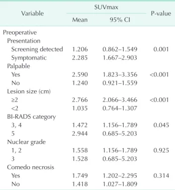
Fifteen patients (14.7%) were diagnosed with IBC at final surgery. We evaluated the association between clinicopatholo- gical variables and the SUVmax of the tumor (Table 2). The
SUVmax was significantly higher in the patients who had symptoms, palpable masses (P = 0.001, P < 0.001), a lesion over 2 cm in size (P < 0.001), and BI-RAD category 5 (P = 0.049).
There were no significant SUVmax differences with regard to nuclear grades and comedo necrosis. The upgrade rate by biopsy method was highest with core needle biopsies (42.9%) as compared with vacuum assisted breast biopsies (10.5%) and excision biopsies (14%) (Table 3). The SUVmax was higher in the patients upgraded to IBC (mean: 2.56 vs. 1.36), (P = 0.007).
A SUVmax of 2.65 was the theoretical cutoff value in ROC curve analysis in predicting the underestimation of IBC (Fig. 1).
The underestimation rate was significantly higher in patients with a SUVmax > 2.65 (P < 0.001), over moderate enhanced uptake on visual analysis (P < 0.001), and positive axillary FDG uptake (P = 0.032) (Table 4). The sensitivity, specificity, and positive and negative predictive values of SUVtumor (>2.65), visual analysis over the moderate grade were 66.7%, 81.6%, 38.4% and 93.4%, and 66.7%, 77.0%, 33.3% and 93.1%, Table 2. Comparison of clinicopathological variables according to SUVmax
Variable SUVmax
Pvalue
Mean 95% CI
Preoperative Presentation
Screening detected 1.206 0.862–1.549 0.001 Symptomatic 2.285 1.667–2.903
Palpable
Yes 2.590 1.823–3.356 <0.001
No 1.240 0.921–1.559
Lesion size (cm)
≥2 2.766 2.066–3.466 <0.001
<2 1.035 0.764–1.307
BIRADS category
3, 4 1.472 1.156–1.789 0.045
5 2.944 0.685–5.203
Nuclear grade
1, 2 1.558 1.156–1.789 0.925
3 1.528 0.685–5.203
Comedo necrosis
Yes 1.749 1.202–2.295 0.314
No 1.418 1.027–1.809
SUVmax, maximum standardized uptake value; CI, confidence interval; BIRADS, Breast Imaging Reporting and Data System.
Table 3. Underestimation rate compared with biopsy methods Biopsy procedure Postoperative pathology Total
(n = 102) DCIS (n = 87) IBC (n = 15)
Core needle 4 (57.1) 3 (42.9) 7 (6.9)
Vacuumassisted 34 (89.5) 4 (10.5) 38 (37.3)
Excision 49 (86.0) 8 (14.0) 57 (55.9)
Values are presented as number (%).
DCIS, ductal carcinoma in situ; IBC, invasive breast carcinoma.
Table 1. Patients characteristics
Characteristic Value
Preoperative
Age (yr) 49.6 (27–82)
Presentation
Screening detected 70 (29.4)
Symptomatic 32 (70.6)
Palpable
Yes 23 (22.5)
No 79 (77.5)
Lesion size (cm) 1.8 (0.3–7.0)
≥2 30 (29.4)
<2 72 (70.6)
BIRADS category
3, 4 97 (95.1)
5 5 (4.9)
Biopsy procedure
Core needle biopsy 7 (6.9)
Vacuumassisted biopsy 38 (37.3)
Excisional biopsy 57 (55.8)
Nuclear grade
1, 2 56 (54.9)
3 46 (45.1)
Comedo necrosis
Yes 39 (38.2)
No 63 (61.8)
SUVmax 1.54 (0–8.2)
Axillary FDG uptake
Positive 20 (19.6)
Negative 82 (80.4)
Visual analysis
None & faint 72 (70.6)
Moderate 17 (16.7)
High 13 (12.7)
Resection procedure
Breast conserving surgery 64 (62.7)
Mastectomy 34 (33.3)
Nipple sparing mastectomy 4 (3.9) Axillary lymphnode staging
Sentinel L/N biopsy 38 (37.3)
Axillary L/N dissection 61 (59.8)
Not done 3 (2.9)
Postoperative Pathology
DCIS 15 (14.7)
IBC 87 (85.3)
Axillary L/N metastasis 6 (5.9)
Values are presented as mean (range) or number (%).
BIRADS, Breast Imaging Reporting and Data System; SUVmax, maximum standardized uptake value; FDG, fluorodeoxyglucose;
L/N, lymph node; DCIS, ductal carcinoma in situ; IBC, invasive breast carcinoma.
respectively. When SUVmax was added to clinical risk factors such as symptom presentation, palpability, and more than 2-cm-sized lesion, area under curve increased from 67% to 78%
in multiple logistic regression analysis (Table 5).
We undertook 64 breast conserving surgeries (62.7%), 34 mastectomies (33.3%), and 4 nipple sparing mastectomies. Of 15 patients who were diagnosed with IBC, 6 patients were detected to have axillary lymph node involvement.
DISCUSSION
This study showed that 18F-FDG PET/CT would be helpful in predicting IDC in patients diagnosed with DCIS. The underestimation rate was significantly higher in patients with a SUVmax > 2.65 and patients with over moderate uptake and positive axillary FDG uptake on visual analysis.
It is widely accepted that axillary lymph node staging operation (ALND or SLNB) is not necessary during definitive surgery in pure DCIS patients. However, a proportion of DCIS patients are upgraded to IBC after definitive surgery because preoperative biopsies cannot represent the whole lesion [7,8].
ALND sometimes leads to arm lymphedema, though less frequently in SLNB. Therefore, it would be more ideal to avoid axillary staging operations when we can reveal that the primary breast lesion is pure DCIS.
The DCIS underestimation rate of preoperatively diagnosed patients showed a widely variable range depending on imaging modality, needle gauges, and biopsy procedures. DCIS underestimation rates of 11%–59% have been reported and a meta-analysis study identified that the random-effects pooled estimate was 25.9% [4,9-11]. The overall DCIS underestimation rate of our study was 14.7% (15 of 102). This rate was relatively lower than other studies. The reason is that our study contained a large proportion of VABB and excisional biopsies.
Underestimation of IBC is known to be lower in VABB and excisional biopsies in which the volume of biopsy is much higher than that of core needle biopsies.
Much effort has been made to determine the key factors in predicting the underestimation of IBC, such as analyzing clinicopathologic factors, sonography, and MRI. Many studies have reported that the underestimation of IBC was associated
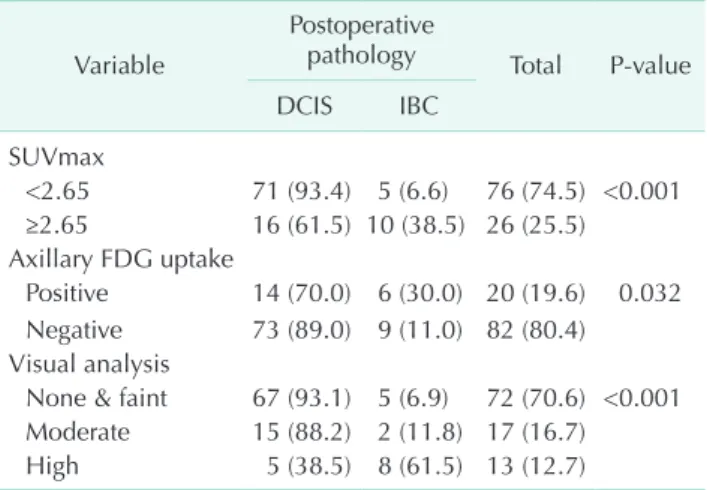
Table 4. Comparison of SUVmax, axillary FDG uptake, and visual analysis between DICS and IBC
Variable
Postoperative
pathology Total Pvalue
DCIS IBC
SUVmax
<2.65 71 (93.4) 5 (6.6) 76 (74.5) <0.001
≥2.65 16 (61.5) 10 (38.5) 26 (25.5) Axillary FDG uptake
Positive 14 (70.0) 6 (30.0) 20 (19.6) 0.032 Negative 73 (89.0) 9 (11.0) 82 (80.4) Visual analysis
None & faint 67 (93.1) 5 (6.9) 72 (70.6) <0.001 Moderate 15 (88.2) 2 (11.8) 17 (16.7) High 5 (38.5) 8 (61.5) 13 (12.7) Values are presented as number (%).
SUVmax, maximum standardized uptake value; CI, confidence interval; BIRADS, Breast Imaging Reporting and Data System.
Table 5. Multiple logistic regression analysis of clinical risk factors with and without SUVmax on the underestimation of IBC
Variable Odds ratio (95% CI) Pvalue Odds ratio (95% CI) Pvalue
Presentation, yes/no 1.058 (0.113–9.926) 0.961 0.917 (0.085–9.944) 0.943
Palpable, yes/no 3.122 (0.311–31.349) 0.334 2.066 (0.172–24.870) 0.567
Lesion size, >2 cm 1.390 (0.367–5.269) 0.628 0.565 (0.122–2.607) 0.464
SUVmax, >2.6 9.270 (2.076–41.391) 0.004
AUC (95% CI) 0.669 (0.569–0.759) 0.779 (0.686–0.855)
SUVmax, maximum standardized uptake value; IBC, invasive breast carcinoma; CI, confidence interval; FDG, fluorodeoxyglucose;
AUC, area under cure.
0 100
Sensitivity
100-Specificity 0
100 80 60 40 20
SUVmax
80 60
40 20
Fig. 1. Receiver operating characteristic curve of maximum standardized uptake valvue (SUVmax) for predicting underestimation of invasive breast cancer. Optimal cutoff of SUVmax was 2.65 (n = 102).
with preoperative clinical, radiologic or pathologic variables [12]. A higher underestimation rate was significantly related to symptoms, lesion palpability, thin biopsy devices, high BI-RADS scores, high nuclear grades, and comedo necrosis. Our study showed that upgraded rates of IBC were significantly higher in patients with symptoms, palpable masses, a lesion size of over 2 cm, and high BI-RAD scores. However, there were no significant difference with nuclear grade and comedo necrosis. These variables related to underestimation are insufficient to skip the ALND or SLNB in patients diagnosed with DCIS preoperatively because they lack objectivity. To supplement this problem, recent studies have focused on the clinical significance of the biological aggressiveness of DCIS.
18F-FDG PET/CT is an imaging tool, which can evaluate the glucose metabolism of tumors. It, therefore, is widely used for diagnosing tumors, assessing treatment responses, and patient follow-up in various kinds of tumors including breast cancer. Furthermore, metabolic parameters such as SUVmax, metabolically active tumor volume, and total lesion glycolysis indicating the glucose metabolism of tumors are known as independent prognostic factors [5,13]. Previous studies have revealed that the high level of SUVmax was significantly associated with large tumor size, lymph node involvement, high nuclear grade, lymphovascular invasion, negative hormone receptor status, and positive HER2 status in IBC patients. Thus, the SUVmax of 18F-FDG PET/CT has been a poor prognostic factor [14,15].
Many studies have placed more emphasis on IBC rather than DCIS. Recently, Shigematsu et al. [16] conducted a study that showed 18F-FDG PET/CT played a major role in predicting the underestimation of IBC in cases of DCIS at needle biopsy.
They showed that a high SUVmax was significantly related to symptomatic presentation, palpation, mass formation, high BI-RADS scores, and core needle biopsy. They concluded that SUVmax is a significant predictive factor of the underestimation of IBC. This finding is similar to our results and our study shows there were no significant differences with nuclear grade and comedo necrosis. When a proper cutoff value on 18F-FDG PET/CT was identified using an ROC curve, high SUVmax values had a strong influence on predicting underestimation of IBC including conventional clinical risk factors, such as symptomatic presentation, palpability and larger lesion size.
Our optimal SUVmax cutoff value on ROC curve for predicting underestimation of IBC was 2.65. SUVmax of more than 2.65
was the only significant predictive value of underestimation of IBC, and clinical risk group with SUVmax had more influence on predicting underestimation of IBC than without SUVmax in multiple logistic regression analysis. Our SUVmax cutoff value was higher than in other studies. In our institution, VABB and excisional biopsies were performed more frequently to achieve correct diagnosis. VABB and excisional biopsies increase inflammatory reaction after procedure and cause increases in SUVmax values.
In general, 18F-FDG PET/CT scans had been analyzed visually and semiquantitatively. Visual and semiquantitative (SUVmax) assessments can differentiate malignant from benign lesions equally regarding other organs [17,18]. We therefore added visual analysis as a qualitative method and axillary FDG uptake to increase the negative predictive value. Because breast physiological activities are variable according to the patient, we attempted to assess the visual analysis by comparing it with normal parenchymal activity. There were no underestimated cases below the moderate uptake cases. Qualitative assessments by visual analysis would also be a useful predictive factor in the underestimation of IBC.
Our study has some limitations. First, our current study was a retrospective review. Second, a relatively small sample size at a single institution was examined. Therefore, further investigation will be required to include a larger sample size at multiple institutions prospectively. Finally, there was the limitation of using 18F-FDG PET/CT on DCIS patients. Using
18F-FDG PET/CT on DCIS patients is currently still open to dispute. Therefore, further prospective studies are necessary to establish the proper indications among the clinicopathological risk factors.
In conclusion, 18F-FDG PET/CT can be a complementary diagnostic tool in predicting the underestimation of IBC in preoperative DCIS patients. Especially high SUVmax values with symptom presentation, palpability, and larger lesion size can help clinicians decide whether these patients should undergo ALND. SUVmax can vary depending on preoperative diagnostic methods, so further studies are necessary to establish the appropriate cutoff value of SUVmax on 18F-FDG PET/CT.
CONFLICTS OF INTEREST
No potential conflict of interest relevant to this article was reported.
REFERENCES
1. Min SY, Kim Z, Hur MH, Yoon CS, Park EH, Jung KW, et al. The basic facts of Korean breast cancer in 2013: results of
a Nationwide Survey and Breast Cancer Registry Database. J Breast Cancer 2016;19:1-7.
2. Siziopikou KP. Ductal carcinoma in situ of the breast: current concepts and future directions. Arch Pathol Lab Med 2013;137:462-6.
3. Park AY, Gweon HM, Son EJ, Yoo M, Kim JA, Youk JH. Ductal carcinoma in situ diagnosed at US-guided 14-gauge core-needle biopsy for breast mass: preo- perative predictors of invasive breast cancer. Eur J Radiol 2014;83:654-9.
4. Brennan ME, Turner RM, Ciatto S, Marinovich ML, French JR, Macaskill P, et al. Ductal carcinoma in situ at core-needle biopsy: meta-analysis of underestimation and predictors of invasive breast cancer.
Radiology 2011;260:119-28.
5. Ueda S, Tsuda H, Asakawa H, Shigekawa T, Fukatsu K, Kondo N, et al. Clinicopa- thological and prognostic relevance of uptake level using 18F-fluorodeoxyglucose positron emission tomography/computed tomography fusion imaging (18F-FDG PET/CT) in primary breast cancer. Jpn J Clin Oncol 2008;38:250-8.
6. Kadoya T, Aogi K, Kiyoto S, Masumoto N, Sugawara Y, Okada M. Role of maxi- mum standardized uptake value in fluorodeoxyglucose positron emission tomography/computed tomography pre- dicts malignancy grade and prognosis of operable breast cancer: a multi-institute study. Breast Cancer Res Treat 2013;141:269- 75.
7. Chin-Lenn L, Mack LA, Temple W,
Cherniak W, Quinn RR, Ravani P, et al.
Predictors of treatment with mastectomy, use of sentinel lymph node biopsy and upstaging to invasive cancer in patients diagnosed with breast ductal carcinoma in situ (DCIS) on core biopsy. Ann Surg Oncol 2014;21:66-73.
8. Sato Y, Kinoshita T, Suzuki J, Jimbo K, Asaga S, Hojo T, et al. Preoperatively di- agnosed ductal carcinoma in situ: risk prediction of invasion and effects on axillary management. Breast Cancer 2016;23:761-70.
9. Park AY, Gweon HM, Son EJ, Yoo M, Kim JA, Youk JH. Ductal carcinoma in situ diagnosed at US-guided 14-gauge core-needle biopsy for breast mass:
preoperative predictors of invasive breast cancer. Eur J Radiol 2014;83:654-9.
10. Park SH, Kim MJ, Kim SJ, Kim EK. Ductal carcinoma in situ diagnosed using an ultrasound-guided 14-gauge core needle biopsy of breast masses: can underesti- mation be predicted preopera tively? Ultra- so nography 2014;33:128-35.
11. Shin SH, Kim BC, Song YJ, Yoon HC, Cho JS, Park MH, et al. Risk factor of invasive breast cancer in patients with preoperative diagnosis of ductal carcinoma in situ. J Korean Surg Soc 2011;80:90-5.
12. Nori J, Meattini I, Giannotti E, Abdulcadir D, Mariscotti G, Calabrese M, et al. Role of preoperative breast MRI in ductal carcinoma in situ for prediction of the presence and assessment of the extent of occult invasive component. Breast J 2014;
20:243-8.
13. Berriolo-Riedinger A, Touzery C, Riedinger JM, Toubeau M, Coudert B, Arnould L, et al. [18F]FDG-PET predicts complete pathological response of breast cancer to neoadjuvant chemotherapy. Eur J Nucl Med Mol Imaging 2007;34:1915-24.
14. Ohara M, Shigematsu H, Tsutani Y, Emi A, Masumoto N, Ozaki S, et al. Role of FDG- PET/CT in evaluating surgical outcomes of operable breast cancer: usefulness for malignant grade of triple-negative breast cancer. Breast 2013;22:958-63.
15. Morris PG, Ulaner GA, Eaton A, Fazio M, Jhaveri K, Patil S, et al. Standardized uptake value by positron emission tomo- graphy/computed tomography as a prog- nostic variable in metastatic breast cancer.
Cancer 2012;118:5454-62.
16. Shigematsu H, Kadoya T, Masumoto N, Matsuura K, Emi A, Kajitani K, et al. Role of FDG-PET/CT in prediction of underes- timation of invasive breast cancer in cases of ductal carcinoma in situ diagnosed at needle biopsy. Clin Breast Cancer 2014;14:
358-64.
17. Hashimoto Y, Tsujikawa T, Kondo C, Maki M, Momose M, Nagai A, et al. Accuracy of PET for diagnosis of solid pulmonary lesions with 18F-FDG uptake below the standardized uptake value of 2.5. J Nucl Med 2006;47:426-31.
18. Herrmann K, Czernin J, Cloughesy T, Lai A, Pomykala KL, Benz MR, et al. Comparison of visual and semiquantitative analysis of 18F-FDOPA-PET/CT for recurrence detection in glioblastoma patients. Neuro Oncol 2014;16:603-9.