Effects of Smoking on Tear Film and Ocular Surface
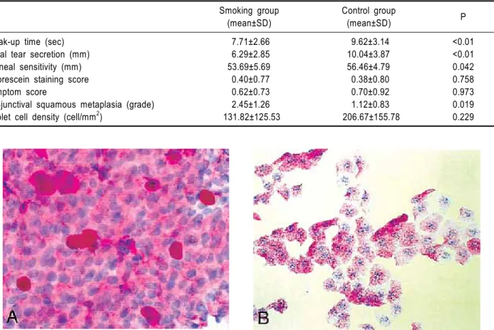
5
0
0
전체 글
(2)
(3)
(4)
(5)
수치
관련 문서
To evaluate the effectiveness of 3% diquafosol, we measured the tear film break-up time (tBUT), performed the Schirmer I test, and used the corneal staining score as an objective
Schirmer’s 1 test, tear film BUT, keratoepitheliopathy score with fluorescein, conjunctival staining score with lissamine green, and Ocular sur- face disease index (OSDI) score
Corneal staining score, tear film break-up time (TF-BUT), Schirmer test and ocular surface dis- ease index (OSDI) were evaluated before surgery and 2, 4, 8, 12 and 16 weeks
Tear film break-up time (BUT), Schirmer’s test I, Oxford scheme, Ocular surface disease index (OSDI), and corneal aberrations were evaluated before surgery and at 1 and 3 months
In this study, we performed tear film osmolarity measurements using the TearLab Osmolarity System and compared it with the TBUT and Schirmer tests in patients with DM.. Although
Results: Significant improvement in the tear break-up time, corneal ocular staining score, and ocular surface disease index score in the study group compared with the control group
In the present study, we evaluated the effect of mixed 0.1% HA and MO eye drops on tear production, ocular surface irregularity, TBUT, corneal fluorescein staining score,
In our results, all tear function para- meters, including BUT, and total and basal tear secretions, were lower in the diabetic group, and these abnormalities were
