© 2017 The Korean Ophthalmological Society
This is an Open Access article distributed under the terms of the Creative Commons Attribution Non-Commercial License (http://creativecommons.org/licenses /by-nc/3.0/) which permits unrestricted non-commercial use, distribution, and reproduction in any medium, provided the original work is properly cited.
Original Article
Sjögren’s syndrome (SS) is a chronic systemic autoim- mune disease affecting mainly middle-aged women [1]. It has a wide clinical spectrum that extends from sicca symp- toms on the mucosal surfaces to extraglandular manifesta- tions [2]. Lymphocytic infiltration and destruction of exo- crine glands (mainly the salivary and lacrimal glands) is the histologic hallmark of SS, resulting in dry eye and dry
Correlation between Tear Osmolarity and Other Ocular Surface Parameters in Primary Sjögren’s Syndrome
Mirinae Kim, Hyun Seung Kim, Kyung-Sun Na
Department of Ophthalmology and Visual Science, Yeouido St. Mary’s Hospital, College of Medicine, The Catholic University of Korea, Seoul, Korea
Purpose: To investigate the relationships between tear osmolarity and other ocular surface parameters and to determine the diagnostic value of tear osmolarity in primary Sjögren’s syndrome (SS) using tear film break-up time, Schirmer I test, and cornea/conjunctiva staining.
Methods: We included 310 eyes of 155 patients diagnosed with dry eye disease (39 primary SS and 116 non- Sjögren dry eye disease) at Seoul St. Mary’s Hospital from August 2010 to January 2015. All subjects complet- ed the Ocular Surface Disease Index (OSDI) questionnaire and underwent ocular examinations including tear osmolarity (TearLab Osmolarity System), Schirmer I test, slit lamp examination for tear film break-up time, and corneal and conjunctival fluorescein staining. We used the mean value of both eyes for all parameters. Fluo- rescein staining was assessed using the Sjögren’s International Collaborative Clinical Alliance ocular staining score (OSS).
Results: In primary SS patients (n = 39), the mean subject age was 52.5 ± 11.9 years, and 94.9% of the subjects were women. Mean tear osmolarity in SS was 311.1 ± 16.4 mOsm/L, with 16 (41.0%) subjects having values
≥316 mOsm/L. In SS, there was a positive correlation between mean tear osmolarity and OSDI score ( ρ = 0.405, p = 0.011) and OSS (ρ = 0.592, p < 0.001). There was a negative correlation between mean tear osmolarity and the Schirmer I test ( ρ = –0.625, p < 0.001). There was no significant correlation between mean tear osmo- larity and tear film break-up time in SS ( ρ = 0.110, p = 0.505).
Conclusions: Tear osmolarity measurements using the TearLab Osmolarity System can reflect both symptom severity (OSDI) and objective signs (Schirmer test and OSS) in SS.
Key Words: Dry eye syndrome, Sjögren’s syndrome, Tear film osmolarity
Received: June 23, 2016 Accepted: November 1, 2016
Corresponding Author: Kyung-Sun Na, MD, PhD. Department of Oph- thalmology, Yeouido St. Mary’s Hospital, College of Medicine, The Catholic University of Korea, #10 63-ro, Yeongdeungpo-gu, Seoul 07345, Korea. Tel: 82-2-3779-1052, Fax: 82-2-761-6869, E-mail: drna@catholic.
ac.kr
This study was presented as a narration at the 115th annual meeting of
the Korean Ophthalmological Society in Busan, Korea, in 2016.
mouth [3]. In addition, systemic features of a cutaneous, respiratory, renal, hepatic, neurologic, and vascular nature often occur [2,4,5]. Primary SS occurs alone, while sec- ondary SS occurs in conjunction with another autoimmune disease [6,7]. Occasionally, ocular symptoms precede other features, but the diagnosis of SS is often delayed owing to its ambiguous symptoms. In one study [8], time from the first symptom to a diagnosis of SS was a mean of 7 years, with considerable psychological distress caused by unex- plained symptoms during that time. Therefore, early non-invasive diagnosis is necessary for SS.
Several tools are used to evaluate the severity of dry eye disease, including tear film break-up time, fluorescein or lissamine green staining of the cornea and conjunctiva, and the Schirmer test, but these tools are limited by their low specificity and interobserver variation [9]. Moreover, sometimes these tools have a low correlation with subjec- tive symptoms. Much effort has been made to find an ideal diagnostic tool that is simple, specific, sensitive, objective, non-invasive, reproducible, and has a clear cut-off value. In recent years, tear osmolarity has been considered a reliable diagnostic marker of dry eye disease [10-13]. Further, tech- nology has become available to measure tear osmolarity in the office setting with a microfluidic lab-on-a-chip device based on electrical impedance [14]. The TearLab Osmolari- ty System (TearLab, San Diego, CA, USA) can collect a tear sample atraumatically and provides an absolute nu- meric score. It provides results in a minute, and there is no interobserver variation.
Data on the correlation between tear osmolarity and oth- er dry eye severity markers is conflicting in the literature.
Recent studies show that tear osmolarity has a correlation with the severity of dry eye in rheumatoid arthritis [15], ocular mucous membrane pemphigoid [16], and ocular graft-versus-host disease [17]. However, Amparo et al. [18]
reported that changes in tear osmolarity did not contribute to prediction of dry eye symptoms or corneal staining after treatment. Further, other authors have reported that the ap- plicability of TearLab is restricted by low reproducibility and wide variation in repeat measurements [12,19,20].
In this study, we focused on the correlations between tear osmolarity and other ocular surface parameters to fur- ther analyze the diagnostic value of tear osmolarity in SS.
Bunya et al. [21] have already investigated the relationship between tear osmolarity, the Schirmer I test, and dry eye symptoms in SS. However, their study did not include cur-
rently available tools, such as tear film break-up time and cornea/conjunctiva staining. Therefore, we included these parameters in our study.
Materials and Methods
This cross-sectional study comprised 310 eyes in 155 pa- tients. We enrolled 39 patients diagnosed as having prima- ry SS and 116 with non-Sjögren dry eye disease who were matched for age and sex. Inclusion criteria were age over 20 years and a proven diagnosis of dry eye disease at Seoul St. Mary’s Hospital from August 2010 to January 2015. SS was diagnosed according to the proposed international cri- teria by American-European Consensus Group (2002) [22]
and confirmed by both an ophthalmologist and a rheuma- tologist.
Exclusion criteria included active ocular inflammation or infection not associated with dry eye, drug toxicity, a history of ocular surgery or trauma, and wearing of con- tact lenses. Subjects using systemic medications that could interfere with tear production such as diuretics, beta-block- ers, benzodiazepines, and antihistamines were excluded, but patients taking systemic immunosuppressive agents for SS were included. Subjects who had used any eye drops in the 6 hours before the study were also excluded.
The study design followed the standards for biomedical research laid down in the Declaration of Helsinki, and the protocols used were approved by the institutional review board of the Catholic University of Korea.
Demographic information, current systemic or topical treatments, and other medical history were recorded. Ocu- lar examinations including visual acuity, non-contact pneumatic tonometry, and anterior segment slit lamp ex- amination were performed for all subjects. The tests for dry eye were performed in the following order: Ocular Surface Disease Index (OSDI) questionnaire, tear osmolar- ity, Schirmer I test, slit lamp examination for tear film break-up time, and corneal and conjunctival fluorescein staining. We allowed a 5-minute interval between the tests.
We assessed subjective symptoms using the OSDI ques- tionnaire, which included 12 questions regarding dry eye symptoms during the past week; each symptom was grad- ed from 0 to 4, for a final score of 0 (mild) to 100 (severe).
A higher OSDI score represents greater disability.
Tear osmolarity was evaluated using the TearLab Osmo-
larity System. A 50-nL tear sample was collected from the inferior lateral meniscus of each eye, and the mean value of both eyes was used for the statistical analysis. Tear os- molarity ≥316 mOsm/L has been reported to identify dry eye disease with high sensitivity, specificity, and predictive accuracy [11].
A 5-minute Schirmer I test (without anesthesia) was per- formed before instillation of any eye drops. The standard- ized Schirmer test strip (Eagle Vision, Memphis, TN, USA) was bent and placed at the inferior outer fornix.
Each patient was instructed to keep his/her eyes closed during the test. The length of maximal wetting was mea- sured after 5 minutes. A Schirmer test value ≤5 mm was considered abnormal.
To measure tear film break-up time, we placed a fluores- cein-impregnated strip in the lateral part of inferior fornix.
Each patient was asked to blink, and the time before the corneal dry spot appeared in the stained tear film was re- corded as the tear film break-up time. Tear film break-up time ≤5 seconds was considered abnormal.
Corneal and conjunctival staining was evaluated after fluorescein staining under a yellow-barrier filter and cobalt blue illumination. Corneal and conjunctival staining was graded according to the Sjögren’s International Collabora- tive Clinical Alliance ocular staining score (OSS) [23].
All statistical analyses were performed using SPSS for Windows ver. 19.0 (IBM Co., Armonk, NY, USA). We
used the mean value of both eyes for all parameters. The Mann-Whitney test was used to compare parameters be- tween the two groups. The Spearman rank-order correla- tion test was used to evaluate the relationships between variables in the patients with Sjögren dry eye disease. A p-value <0.05 was considered to be statistically significant in all analyses.
Results
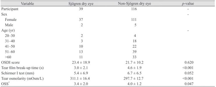
Table 1 shows the clinical characteristics of the 155 study participants. Their mean age was 52.7 ± 12.0 years, and 148 (95.5%) were female. Table 1 summarizes the mean (±stan- dard deviation) values for the ocular surface parameters, i.e., OSDI score, tear film break-up time, Schirmer I test, tear osmolarity, and OSS. In the Sjögren dry eye group (n
= 39), mean tear osmolarity in SS was 311.1 ± 16.4 mOsm/
L, and 16 measurements (41.0%) were over 316 mOsm/L.
Mean tear film break-up time was 3.0 ± 2.1 seconds, and 35 measures (89.7%) were ≤5 mm. The mean Schirmer I test value was 5.4 ± 6.9, and 24 measures (61.5%) were ≤5 mm, indicating significant dry eye. In the non-Sjögren dry eye group (n = 116), mean tear osmolarity was 297.7 ± 12.7 mOsm/L, and 11 measures (9.5%) were over 316 mOsm/L.
Mean tear film break-up time was 4.6 ± 1.9 seconds, and 76 measures (65.5%) were ≤5 mm. The mean Schirmer I
Table 1. Clinical characteristics of study participants
Variable Sjögren dry eye Non-Sjögren dry eye p-value
Participant 39 116 -
Sex -
Female 37 111
Male 2 5
Age (yr) -
20–30 2 4
31–40 3 18
41–50 10 22
51–60 13 39
>60 11 33
OSDI score 23.4 ± 18.9 21.7 ± 10.2 0.620
Tear film break-up time (s) 3.0 ± 2.1 4.6 ± 1.9 <0.001
Schirmer I test (mm) 5.4 ± 6.9 6.7 ± 6.5 0.052
Tear osmolarity (mOsm/L) 311.1 ± 16.4 297.7 ± 12.7 <0.001
OSS
*3.4 ± 2.0 4.0 ± 1.2 0.047
The Mann-Whitney test was used to compare parameters between the two groups. Values are presented as the mean ± standard deviation.
OSDI = Ocular Surface Disease Index; OSS = ocular staining score.
*
By Sjögren’s International Collaborative Clinical Alliance [23].
test value was 6.7 ± 6.5, and 61 measures (52.6%) were ≤5 mm. Tear osmolarity value and OSS were higher in the Sjögren dry eye group. In contrast, the tear film break-up time value was higher in the non-Sjögren dry eye group.
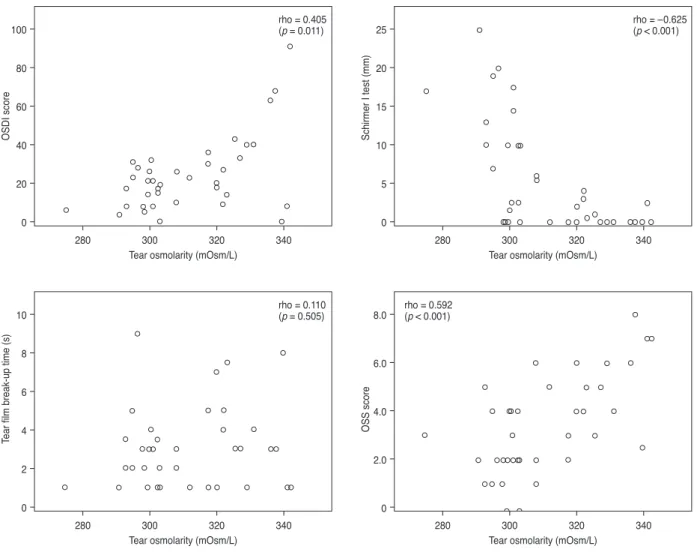
The scatter plots in Fig. 1 show correlations between tear osmolarity and other ocular surface parameters in the Sjögren dry eye group. There was a statistically significant positive correlation between mean tear osmolarity and OSDI score (ρ = 0.405, p = 0.011) and OSS (ρ = 0.592, p <
0.001). There was also a statistically significant negative correlation between mean tear osmolarity and the Schirm- er I test (ρ = –0.625, p < 0.001). However, there was no sig- nificant correlation between mean tear osmolarity and tear film break-up time (ρ = 0.110, p = 0.505).
In the non-Sjögren dry eye group, there were no signifi-
cant correlation between mean tear osmolarity and OSDI score (ρ = 0.101, p = 0.279), Schirmer I test (ρ = 0.119, p = 0.205), tear film break-up time (ρ = 0.133, p = 0.165), or OSS (ρ = 0.145, p = 0.119).
Discussion
The dry eye disease associated with SS is multifactorial in nature but is mainly aqueous deficient-type. In 2002, a revised international classification of SS was established by the American-European Consensus Group [22]. The criteria used include two objective measures of ocular in- volvement, i.e., a Schirmer I test result ≤5 mm in 5 minutes and a Rose Bengal or other ocular dye score ≥4 according
Fig. 1. Scatter plot showing significant correlations between Ocular Surface Disease Index (OSDI) score and average tear film osmolarity (ρ
= 0.405, p = 0.011), average Schirmer I test and average tear film osmolarity (ρ = –0.625, p < 0.001), and average ocular staining score (OSS) and average tear film osmolarity (ρ = 0.592, p < 0.001). The Spearman rank-order correlation test was used to evaluate the relationship between variables. OSS was by Sjögren’s International Collaborative Clinical Alliance [23].
Tear osmolarity (mOsm/L) 280
25 20 15 10 5 0
Schirmer I test (mm)
300 320 340
rho = –0.625 (p < 0.001)
280 100
80 60 40 20
0
Tear osmolarity (mOsm/L)
OSDI score
300 320 340
rho = 0.405 (p = 0.011)
280 10
8 6 4 2
0
Tear osmolarity (mOsm/L)
Tear film break-up time (s)
300 320 340
rho = 0.110 (p = 0.505)
280 8.0
6.0 4.0 2.0
0
Tear osmolarity (mOsm/L)
OSS score
300 320 340
rho = 0.592 (p < 0.001)