© 2019 The Korean Ophthalmological Society
This is an Open Access article distributed under the terms of the Creative Commons Attribution Non-Commercial License (http://creativecommons.org/licenses /by-nc/3.0/) which permits unrestricted non-commercial use, distribution, and reproduction in any medium, provided the original work is properly cited.
Original Article
Purpose: To evaluate tear film function in patients with diabetes mellitus (DM) using tear film osmolarity (TFO) measurements compared to other tear film function tests.
Methods: DM patients without any history of ocular surface disorder but with potential effects on the tear film were enrolled in this cross-sectional study. Data including dry eye symptoms, duration of DM, stage of diabetic retinopathy and blood hemoglobin A1c levels were recorded. Tear film break-up time (TBUT) and basic tear secretion (Schirmer test) were assessed. TFO was determined using the Tearlab Osmolarity System. The out- come measures were the difference between the mean values of TBUT, basic tear secretion and TFO in both the study and control groups.
Results: We recruited 51 DM patients and 20 control subjects with a mean age of 51.2 (range, 21 to 70) and 48.5 (range, 24 to 70) years, respectively. A total of 27 patients (53%) and 11 controls (55%) reported dry eye symptoms (p = 0.668). The mean TBUT was 10.2 + 4.8 seconds in the study group versus 10.5 + 2.8 seconds in controls, which was not significantly different (p = 0.747). The mean Schirmer test score was 8.1 + 4.3 mm in the patients versus 10.1 + 3.0 mm in the controls (p = 0.069). The mean TFO was 294.1 + 12.9 mosmol/L in the patients versus 291.4 + 14.5 mosmol/L in the controls (p = 0.456). It was significantly higher in patients with poor glycemic control determined by hemoglobin A1c > 8% (p = 0.003). TFO had a positive correlation with the duration of DM (p = 0.030) but not with the stage of diabetic retinopathy (p = 0.944). However, TFO showed a significant relationship with dry eye symptoms (p = 0.001).
Conclusions: TFO is impaired in patients with uncontrolled DM and is better correlated with glycemic control and dry eye symptoms than the TBUT and Schirmer tests.
Key Words: Diabetes mellitus, Osmolar cocentration
Received: December 2, 2013 Final revision: May 19, Accepted: May 21, 2014
Corresponding Author: Arash Omidtabrizi, MD. Retina Research Center, Khatam-Al-Anbia Eye Hospital, Mashhad University of Medical Sciences, Abu- talib Crossroad, Kolahdouz Blvd, Mashhad, Iran. Tel: 98-5117281401, Fax: 98-5117245363, E-mail: [email protected]
This article was presented at the 23rd Annual Congress of the Iranian Society of Ophthalmology, 28-31 October 2013, Razi Convention Center, Tehran, Iran
Comparison between Tear Film Osmolar Cocentration and Other Tear Film Function Parameters in Patients with Diabetes Mellitus
Akbar Derakhshan1, Majid Abrishami1, Mohamad Khajedaluee2, Arash Omidtabrizi1, Somayeh Ghassemi Moghaddam3
1Eye Research Center, Mashhad University of Medical Sciences, Mashhad, Iran
2School of Medicine, Mashhad University of Medical Sciences, Mashhad, Iran
3School of Paramedicine, Mashhad University of Medical Sciences, Mashhad, Iran
2014
A number of ocular complications are known to be asso- ciated with diabetes mellitus (DM) [1]. Although some dia- betic ocular complications such as chronic inflammation of the eyelids, acute orbital infections, cataract and retinopa- thy have been discussed previously, corneal complications have only been recently studied [1]. It has been reported that 47% to 64% of patients with DM will experience a primary corneal disorder during their life time [2]. Corneal disorders related to diabetes may include epithelial fragili- ty, microcystic edema and bleb formation, superficial punctate keratopathy, persistent epithelial defects, recur- rent corneal erosions, delayed epithelial healing [3,4], de- creased corneal sensitivity, decreased attachment of the epithelium to underlying layers, neurotrophic corneal ul- ceration, dry eye (tear film function disorders) and fila- mentary keratitis [5-13].
Tear film dysfunction and dry eye have been investigat- ed in several studies using different methods such as tear film break-up time (TBUT), Schirmer test and pathologic evaluations [11-13]. Many of these studies have concluded that dry eye syndrome is more common in patients with DM than in the normal population [11-13]. Nevertheless, few attempts have been made to assess tear film osmolari- ty changes in DM patients, which is considered very sensi- tive for dry eye evaluation [14]. In this study, we describe tear film osmolarity changes in DM patients and compare it with the TBUT and Schirmer tests, which evaluate basic tear secretion.
Materials and Methods
This was a cross-sectional study that compared DM pa- tients and normal individuals for pre-corneal tear film changes. The protocol was approved by the Institutional Review Board and Ethics Committee of Mashhad Univer- sity of Medical Sciences (approval no. 6176) and was per- formed from September 2011 to 2012 in Khatam-al-Anbia Eye Hospital, Mashhad, Iran. All the patients provided an informed consent. Patients with diabetes who were re- ferred to our center for evaluation of diabetic retinopathy were recruited. Twenty age and sex matched individuals without DM were randomly selected from refraction and retina clinics as the control group. Fasting blood sugar was checked in the control group to rule out DM. A detailed history of previous systemic and ocular diseases and medi-
cations that may have had adverse effects on tear film function was taken from each patient. None of the patients and control subjects had a history of dry eye treatment.
The exclusion criteria was as follows: history of any con- junctival and/or corneal diseases, severe meibomian gland dysfunction (MGD), previous ocular surgery and any sys- temic disorders that could influence tear production such as renal failure and certain endocrine diseases. Patients us- ing anti-histamines, tricyclic anti-depressants and topical ophthalmic medications were also excluded. Those patients whose clinical examination revealed significant differences between the right and left eyelid margins were not enrolled in the study.
Demographic data and duration of diabetes were docu- mented. The blood level of glycosylated hemoglobin (Hb) A1C was recorded for every patient. All enrolled patients underwent an ophthalmic examination, which included best-corrected visual acuity, slit-lamp examination and di- lated pupil ophthalmoscopy for grading of diabetic reti- nopathy according to the international classification system developed by Gangaputra et al. [15]. TBUT, Schirmer test and tear film osmolarity measurements were performed before pupil dilation in order to prevent ocular surface ex- posure to preservatives. To measure TBUT, a fluorescein strip was introduced into the conjunctival sac with mini- mal conjunctival stimulation and the patient was asked to keep his or her eyes open after a few blinks. The tear film was observed with a wide slit lamp beam and cobalt blue filter. A dry spot appearance in less than 10 seconds was considered abnormal. Basic tear secretion was tested by Schirmer strips. One drop of tetracaine 0.5% (Sina Daru, Tehran, Iran) was instilled twice within a 1-minute interval and then a Schirmer strip was placed into the inferior for- nix for 5 minutes and the length of the wet tape was re- corded in millimeters. Wetting less than 10 millimeters was considered abnormal. Tear film osmolarity was mea- sured with the TearLab Osmolarity System (TearLab, San Diego, CA, USA). This device only requires 0.5 mm3 of tears for osmolarity measurements, which is obtained by touching the tip of the testing card of the handle with the tear meniscus at the temporal third of the lower eyelid margin. Because the Schirmer and TBUT tests did not show any significant difference between the right and the left eyes, tear film osmolarity test was done on the right eye of the participants only. Hb A1c examination was per- formed at the laboratory of our clinic on the same day as
the ophthalmic examinations. A value greater than 8% was considered the cutoff point of poorly controlled diabetes [16]. All of the above ocular examinations were also per- formed on 20 matched individuals without diabetes who met the exclusion criteria and were categorized as the con- trol group. These subjects were selected from the refrac- tion and retina clinics.
Statistical analysis was performed using the SPSS ver. 13 (SPSS Inc., Chicago, IL, USA). Qualitative variables were expressed as percentages, and quantitative data were ex- pressed as mean values with standard deviation. ANOVA and t-test were used for analysis. Normal distribution of quantitative data was assessed using the Kolmogor- ov-Smirnov test. A p-value less than 0.05 was regarded as statistically significant.
Results
In this study, 51 DM patients and 20 controls were en- rolled. Demographic data of the patients are presented in Table 1. There were 17 eyes (33.3%) with no diabetic reti- nopathy, 27 (53%) had non-proliferative diabetic retinopa- thy, and seven eyes (13.6%) had proliferative diabetic reti- nopathy. In total, 27 patients with DM (53%) and 11 controls (55%) reported symptoms of dry eye syndrome, such as burning, dryness and foreign body sensation.
There was no statistically significant difference between the two eyes of the patients in the TBUT and Schirmer test results (t-test, p > 0.05). No age or sex predilection was ob-
served in the ocular surface parameters (Table 2, 3). Twen- ty-three patients underwent a Hb A1c level examination.
Of these, eight patients had good glycemic control (≤8%) and the other 15 had poor glycemic control (>8%).
The mean TBUT in the patient group was 10.2 ± 4.8 sec- onds (Table 1), with no significant statistical difference compared to 10.5 ± 2.8 seconds in the control group (t-test, p = 0.747). Although the patients with poorly controlled DM had a shorter TBUT compared to patients with good glycemic control, the difference was not statistically sig- nificant (p = 0.132). However, the mean TBUT in both the good and poorly controlled DM subgroups was in the nor- mal range (Table 4). Neither the duration of DM, nor the stage of retinopathy affected the TBUT significantly (p = 0.372 and p = 0.936, respectively) (Table 4).
The mean Schirmer test value was 8.1 ± 4.3 mm in pa- tients with diabetes versus 10.1 ± 3.0 mm in the control subjects (p = 0.069). Thirty-five percent of patients had impaired Schirmer test (values measuring less than 5 mm), whereas none of the controls had impaired test results. The Schirmer test results were better in patients with well con- trolled DM (Hb A1C ≤8%), compared to poor glycemic control patients; however, the difference was not statisti- cally significant (t-test, p = 0.314) (Table 4). The Schirmer test score was neither related to the duration of DM nor to the status of diabetic retinopathy (p = 0.921 and p = 0.807, respectively).
The mean tear osmolarity values were 294.1 ± 12.9 mos- mol/L in the study group versus 291.4 ± 14.6 mosmol/L in the control group (t-test, p = 0.456). Patients with poorly
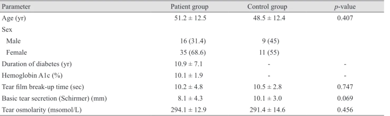
Table 1. Demographic data and ocular surface parameters of the patient and control groups and comparisons using an independent samples t-test
Parameter Patient group Control group p-value
Age (yr) 51.2 ± 12.5 48.5 ± 12.4 0.407
Sex
Male 16 (31.4) 9 (45)
Female 35 (68.6) 11 (55)
Duration of diabetes (yr) 10.9 ± 7.1 - -
Hemoglobin A1c (%) 10.1 ± 1.9 - -
Tear film break-up time (sec) 10.2 ± 4.8 10.5 ± 2.8 0.747
Basic tear secretion (Schirmer) (mm) 8.1 ± 4.3 10.1 ± 3.0 0.069
Tear osmolarity (msomol/L) 294.1 ± 12.9 291.4 ± 14.6 0.456
Values are presented as mean ± standard deviation or number (%).
Table 3. Comparison of ocular surface parameters among age groups in the patient and control groups using the analysis of vari- ance test
Parameter Age <50 yr Age 50–60 yr Age >60 yr p-value
Patient group (n = 20) (n = 16) (n = 15)
Tear film break-up time (sec) 11.1 ± 6.6 10.0 ± 3.1 9.0 ± 3.0 0.453
Schirmer (sec) 8.1 ± 4.5 7.7 ± 4.5 8.6 ± 4.1 0.866
Tear film osmolarity (mosmol/L) 294.4 ± 12.7 295.3 ± 14.0 292.3 ± 12.5 0.811
Control group (n = 12) (n = 2) (n = 6)
Tear film break-up time (sec) 10.7 ± 2.8 9.5 ± 0.7 10.5 ± 3.4 0.860
Schirmer (sec) 10.7 ± 3.1 9.0 ± 4.2 9.1 ± 2.5 0.528
Tear film osmolarity (mosmol/L) 294.0 ± 16.0 291.5 ± 13.4 291.5 ± 14.2 0.960
Values are presented as mean ± standard deviation.
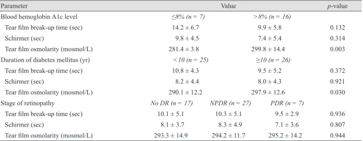
Table 4. Relationship between ocular surface parameters and glycemic control, duration of diabetes mellitus, and DR stage using independent sample t-test and ANOVA
Parameter Value p-value
Blood hemoglobin A1c level ≤8% (n = 7) >8% (n = 16)
Tear film break-up time (sec) 14.2 ± 6.7 9.9 ± 5.8 0.132
Schirmer (sec) 9.8 ± 4.5 7.4 ± 5.4 0.314
Tear film osmolarity (mosmol/L) 281.4 ± 3.8 299.8 ± 14.4 0.003
Duration of diabetes mellitus (yr) <10 (n = 25) ≥10 (n = 26)
Tear film break-up time (sec) 10.8 ± 4.3 9.5 ± 5.2 0.372
Schirmer (sec) 8.2 ± 4.4 8.0 ± 4.3 0.921
Tear film osmolarity (mosmol/L) 290.1 ± 12.2 297.9 ± 12.6 0.030
Stage of retinopathy No DR (n = 17) NPDR (n = 27) PDR (n = 7)
Tear film break-up time (sec) 10.1 ± 5.1 10.3 ± 5.1 9.5 ± 2.9 0.936
Schirmer (sec) 8.1 ± 3.7 8.3 ± 4.9 7.1 ± 3.6 0.807
Tear film osmolarity (mosmol/L) 293.3 ± 14.9 294.2 ± 11.7 295.2 ± 14.2 0.944
Values are presented as mean ± standard deviation.
DR = diabetic retinopathy; NPDR = non-proliferative diabetic retinopathy; PDR = proliferative diabetic retinopathy.
Table 2. Comparison of ocular surface parameters between males and females in the patient and control groups using an indepen- dent sample t-test
Parameter Male Female p-value
Patient group (n = 16) (n = 35)
Tear film break-up time (sec) 10.3 ± 5.3 10.1 ± 4.6 0.894
Schirmer (sec) 7.5 ± 3.2 8.4 ± 4.7 0.529
Tear film osmolarity (mosmol/L) 294.5 ± 13.0 293.8 ± 13.0 0.864
Control group (n = 9) (n = 11)
Tear film break-up time (sec) 11.5 ± 3.1 9.7 ± 2.3 0.989
Schirmer (sec) 10.1 ± 2.8 10.0 ± 3.3 0.157
Tear film osmolarity (mosmol/L) 286.8 ± 13.2 295.1 ± 15.0 0.213
Values are presented as mean ± standard deviation.
controlled disease (Hb A1C >8%) had a significantly high- er mean tear film osmolarity than well controlled patients (299.8 vs. 281.4, respectively; p = 0.003). The mean tear film osmolarity had a significant positive correlation with the duration of DM suggesting that the longer the duration of diabetes the higher the tear film osmolarity (t-test, p = 0.03). There was no statistical correlation between the mean tear film osmolarity and the stage of diabetic reti- nopathy (ANOVA, p = 0.944). In both the study and con- trol groups tear film osmolarity was higher in symptomatic compared to asymptomatic subjects (p = 0.001). Converse- ly, there was no relationship between the Schirmer or TBUT tests scores and dry eye symptoms in any of the study and control groups (Table 5).
Discussion
Several clinical and experimental studies have reported structural, metabolic, and functional abnormalities in the conjunctiva and cornea of patients with DM and have sug- gested that these abnormalities may be responsible for the corneal complications of diabetes [16-22]. In this study, we performed tear film osmolarity measurements using the TearLab Osmolarity System and compared it with the TBUT and Schirmer tests in patients with DM. Although we found no significant difference in the tear film osmo- larity between patients with DM and the normal popula- tion, the duration of DM had a significant influence on tear film osmolarity. Our study revealed a significantly higher tear film osmolarity in patients with high levels of Hb A1c (>8%) and in patients with a longer duration of DM. In a
recent study, Sagdik et al. [14] showed that the patients with DM had a higher Hb A1c level than the normal popu- lation and suggested a positive correlation between tear film osmolarity and the duration of DM. However, they did not find a relationship between Hb A1c levels and tear film osmolarity. It should be mentioned that in our study, de- spite significant difference between tear film osmolarity in poorly controlled and well-controlled diabetes, both values were within the normal range. This result contradicts that of the Sagdik study, in which both poor and well-con- trolled patients had a mean tear film osmolarity higher than normal limits. Thus, we can conclude that there may be other factors that affect tear film osmolarity in patients with diabetes, such as MGD.
Our findings showed that the lower Schirmer test scores and TBUT in DM patients were not significantly different from controls. Also, the results of these tests were not af- fected by the status of glycemic control in the patient group. Previous studies have suggested a significant rela- tionship between the presence/duration of DM and Schirmer test scores and TBUT [11-13]. As we see, our study does not confirm the results of the previous studies, which concluded that the tear film osmolarity test, Schirm- er test and TBUT are impaired in patients with DM com- pared to the normal population. One explanation may be the fact that we excluded patients with severe MGD. Se- vere MGD, which is more common in DM, affects both tear film stability (evaluated by TBUT) and tear secretion (evaluated by Schirmer test) and is a known cause of tear film dysfunction. Hence, it can act as a confounding vari- able. Our study confirmed the Goebbels [23] and also Su- zuki et al. [24] study result that showed a positive correla- Table 5. Relationship between ocular surface parameters and the presence of dry eye symptoms in the patient and control groups using an independent samples t-test
Parameter Dry eye symptoms No dry eye symptom p-value
Patient group (n = 27) (n = 24)
Tear film break-up time (sec) 9.5 ± 5.1 10.8 ± 4.4 0.366
Schirmer (sec) 7.3 ± 4.8 9.0 ± 3.6 0.164
Tear film osmolarity (mosmol/L) 301.1 ± 10.8 286.1 ± 10.2 0.0001
Control group (n = 11) (n = 9)
Tear film break-up time (sec) 10.3 ± 3.5 10.7 ± 1.9 0.755
Schirmer (sec) 9.4 ± 2.9 10.8 ± 3.1 0.307
Tear film osmolarity (mosmol/L) 298.4 ± 15.4 282.8 ± 7.3 0.013
Values are presented as mean ± standard deviation.
tion between tear film osmolarity and ocular surface symptoms in cases of dry eye. Both the study and control groups maintained this relationship (p = 0.001). However, unlike their study, the TBUT and Schirmer tests failed to show a statistically significant correlation with dry eye symptoms.
Moreover, the grade of diabetic retinopathy did not have any influence on tear film osmolarity, a finding that con- firms the results of previous research by Manaviat et al.
[13]. Akinci et al. [12] and Manaviat et al. [13] both sug- gested an association between diabetes duration and dry eye using TBUT and Schirmer tests, which was not con- firmed in our study. Our findings suggest that poor glyce- mic control, not merely having DM, is an important deter- minant of the tear film osmolarity and ocular surface health. Tear film osmolarity measurements have a stronger relationship with dry eye symptoms in patients with diabe- tes and can be used to evaluate tear film function in cases where other test results are normal. One of the limitations of our study was not being able to evaluate corneal neu- ropathy, which is an important part of the tear secretion reflex. The second limitation was a relatively small study population. Further studies including neuropathic assess- ment with a larger study population should be conducted in patients with DM to enhance our understanding of the pathogenesis of dry eye in this disease.
Conflict of Interest
No potential conflict of interest relevant to this article was reported.
Acknowledgements
The authors wish to thank Dr. M. Sedaghat for his assis- tance with invaluable comments and for providing the Tearlab Osmolarity System.
References
1. Dabbs CK, Meredith TA. Diabetic eye disease. In: David- son JK, editor. Clinical diabetes mellitus: a problem orient- ed approach. 2nd ed. New York: Thieme; 1991. p. 427-43.
2. Schultz RO, Van Horn DL, Peters MA, et al. Diabetic kera- topathy. Trans Am Ophthalmol Soc 1981;79:180-99.
3. Saini JS, Khandalavla B. Corneal epithelial fragility in dia- betes mellitus. Can J Ophthalmol 1995;30:142-6.
4. Foulks GN, Thoft RA, Perry HD, Tolentino FI. Factors re- lated to corneal epithelial complications after closed vitrec- tomy in diabetics. Arch Ophthalmol 1979;97:1076-8.
5. Hyndiuk RA, Kazarian EL, Schultz RO, Seideman S. Neu- rotrophic corneal ulcers in diabetes mellitus. Arch Oph- thalmol 1977;95:2193-6.
6. Henkind P, Wise GN. Descemet’s wrinkles in diabetes. Am J Ophthalmol 1961;52:371-4.
7. Sanchez-Thorin JC. The cornea in diabetes mellitus. Int Ophthalmol Clin 1998;38:19-36.
8. Tabatabay CA, Bumbacher M, Baumgartner B, Leuenberg- er PM. Reduced number of hemidesmosomes in the corne- al epithelium of diabetics with proliferative vitreoretinopa- thy. Graefes Arch Clin Exp Ophthalmol 1988;226:389-92.
9. Azar DT, Spurr-Michaud SJ, Tisdale AS, Gipson IK. De- creased penetration of anchoring fibrils into the diabetic stroma. A morphometric analysis. Arch Ophthalmol 1989;107:1520-3.
10. Taylor HR, Kimsey RA. Corneal epithelial basement mem- brane changes in diabetes. Invest Ophthalmol Vis Sci 1981;20:548-53.
11. Modulo CM, Jorge AG, Dias AC, et al. Influence of insulin treatment on the lacrimal gland and ocular surface of dia- betic rats. Endocrine 2009;36:161-8.
12. Akinci A, Cetinkaya E, Aycan Z. Dry eye syndrome in di- abetic children. Eur J Ophthalmol 2007;17:873-8.
13. Manaviat MR, Rashidi M, Afkhami-Ardekani M, Shoja MR. Prevalence of dry eye syndrome and diabetic retinop- athy in type 2 diabetic patients. BMC Ophthalmol 2008;8:10.
14. Sagdik HM, Ugurbas SH, Can M, et al. Tear film osmolari- ty in patients with diabetes mellitus. Ophthalmic Res 2013;50:1-5.
15. Gangaputra S, Lovato JF, Hubbard L, et al. Comparison of standardized clinical classification with fundus photograph grading for the assessment of diabetic retinopathy and dia- betic macular edema severity. Retina 2013;33:1393-9.
16. Action to Control Cardiovascular Risk in Diabetes Study Group, Gerstein HC, Miller ME, et al. Effects of intensive glucose lowering in type 2 diabetes. N Engl J Med 2008;358:2545-59.
17. Tsubota K, Chiba K, Shimazaki J. Corneal epithelium in
diabetic patients. Cornea 1991;10:156-60.
18. Shimazaki J, Tsubota K, Yoshida A, et al. Changes of cor- neal redox state in diabetic animal models. Cornea 1995;14:196-201.
19. Friend J, Ishii Y, Thoft RA. Corneal epithelial changes in diabetic rats. Ophthalmic Res 1982;14:269-78.
20. Chang SW, Hsu HC, Hu FR, Chen MS. Corneal autofluo- rescence and epithelial barrier function in diabetic patients.
Ophthalmic Res 1995;27:74-9.
21. Gobbels M, Spitznas M, Oldendoerp J. Impairment of cor-
neal epithelial barrier function in diabetics. Graefes Arch Clin Exp Ophthalmol 1989;227:142-4.
22. Seifart U, Strempel I. The dry eye and diabetes mellitus.
Ophthalmologe 1994;91:235-9.
23. Goebbels M. Tear secretion and tear film function in insu- lin dependent diabetics. Br J Ophthalmol 2000;84:19-21.
24. Suzuki M, Massingale ML, Ye F, et al. Tear osmolarity as a biomarker for dry eye disease severity. Invest Ophthalmol Vis Sci 2010;51:4557-61.