© 2015 The Korean Ophthalmological Society
This is an Open Access article distributed under the terms of the Creative Commons Attribution Non-Commercial License (http://creativecommons.org/licenses /by-nc/3.0/) which permits unrestricted non-commercial use, distribution, and reproduction in any medium, provided the original work is properly cited.
Original Article
Efficacy of the Mineral Oil and Hyaluronic Acid Mixture Eye Drops in Murine Dry Eye
Jung Han Choi
1, Jung Han Kim
1,2, Zhengri Li
1, Han Jin Oh
1, Kyu Youn Ahn
3, Kyung Chul Yoon
11Department of Ophthalmology and Research Institute of Medical Sciences, Chonnam National University Hospital, Chonnam National University Medical School, Gwangju, Korea
2Kim’s Eye Clinic of the 21st Century, Seoul, Korea
3Department of Anatomy, Chonnam National University Medical School, Gwangju, Korea
Purpose: To investigate the therapeutic effects of mineral oil (MO) and hyaluronic acid (HA) mixture eye drops on the tear film and ocular surface in a mouse model of experimental dry eye (EDE).
Methods: Eye drops consisting of 0.1% HA alone or mixed with 0.1%, 0.5%, or 5.0% MO were applied to desic- cating stress-induced murine dry eyes. Tear volume, corneal irregularity score, tear film break-up time (TBUT), and corneal fluorescein staining scores were measured at 5 and 10 days after treatment. Ten days after treatment, goblet cells in the conjunctiva were counted after Periodic acid-Schiff staining.
Results: There was no significant difference in the tear volume between desiccating stress-induced groups.
The corneal irregularity score was lower in the 0.5% MO group compared with the EDE and HA groups. The 0.5% and 5.0% MO groups showed a significant improvement in TBUT compared with the EDE group. Mice treated with 0.1% and 0.5% MO mixture eye drops showed a significant improvement in fluorescein staining scores compared with the EDE group and the HA group. The conjunctival goblet cell count was higher in the 0.5% MO group compared with the EDE group and HA group.
Conclusions: The MO and HA mixture eye drops had a beneficial effect on the tear films and ocular surface of murine dry eye. The application of 0.5% MO and 0.1% HA mixture eye drops could improve corneal irregulari- ty, the corneal fluorescein staining score, and conjunctival goblet cell count compared with 0.1% HA eye drops in the treatment of EDE.
Key Words: Dry eye, Hyaluronic acid, Mineral oil, Ocular surface
Dry eye disease is a multifactorial disorder of the tears and ocular surface [1]. It leads to various ocular symptoms
and tear film instability induce potential damage of the oc- ular surface and ocular surface inflammation [1]. The resulting diminished vision and quality of life affect mil- lions of people worldwide [2]. Treatments for dry eye dis- ease in increasing order of severity include artificial tears, anti-inflammatory agents, immunosuppressants, punctal plugs, serum eye drops, contact lenses, and surgery [3,4].
Artificial tears are typically used as the first-line manage- ment of dry eye disease. They improve the stability of the tear film and provide symptomatic relief [5].
Received: July 31, 2013 Accepted: January 24, 2014
Corresponding Author: Kyung Chul Yoon, MD, PhD. Department of Ophthalmology, Chonnam National University Hospital, #42 Jebong-ro, Dong-gu, Gwangju 501-757, Korea. Tel: 82-62-220-6741, Fax: 82-62-227- 1642, E-mail: [email protected]
This study was presented as a paper at the 109th Korean Ophthalmologi- cal Society Annual Meeting of 2013 held in Busan, Korea.
Hyaluronic acid (HA) is a linear polymer composed of long chains of repeating disaccharide units of N-acetylglu- cosamine and glucuronic acid [6]. HA is an avid moisturizer and has a higher residency time than other artificial tear components. In addition, HA can protect the ocular surface epithelium by facilitate epithelial healing [7]. 0.1% HA eye drops are commonly used to treat dry eye disease and were found to be effective in improving symptoms as well as signs including corneal epithelial injury [8-10]. However, due to a short duration of action and deficient tear lipid or mucin components, HA eye drops have several limitations includ- ing the need for frequent application.
Mineral oil (MO) is a complex mixture of saturated hy- drocarbons derived from petroleum through various refin- ing steps and subsequent purification [11]. MO-based artifi- cial tears as an ointment formulation prolongs tear retention time in the eye, therefore requiring fewer daily applications [12]. However, no study has been performed on eye drop mixtures of HA and MO as an oil component for dry eye.
The purpose of this study was to evaluate the efficacy of MO and HA combination eye drops for the management of dry eye disease using a desiccating stress-induced mouse model, by evaluating the changes of tear production, tear film break-up time (TBUT), fluorescein staining, ocular sur- face irregularities, and goblet cell count in the conjunctiva.
Materials and Methods
Mouse model of dry eye and experimental procedure The research protocol was approved by the Chonnam National University Medical School Research Institutional Animal Care and Use Committee. All animals were treat- ed according to the standards in the Association for Research in Vision and Ophthalmology Statement for the Use of Animals in Ophthalmic and Vision Research. Six- to eight-week-old female C57BL/6 mice were used in these experiments.
Experimental dry eye (EDE) was induced by subcutane- ous injection of 0.5 mg/0.2 mL scopolamine hydrobromide (Sigma-Aldrich, St. Louis, MO, USA) four times a day (9 a.m., 1 p.m., 5 p.m., and 9 p.m.) with exposure to an air draft and 30% ambient humidity [13-16]. During these ex- periments, the animal’s behavior, food, and water intake were not restricted.
The mice were randomly divided into six groups (n = 5 per group) depending on topical treatment administered:
untreated (UT) mice; EDE mice that received no eye drops;
EDE mice treated with non-preservative 0.1% HA eye drops (Alcon Korea, Seoul, Korea); EDE mice treated with 0.1%
MO and 0.1% HA mixture eye drops; EDE mice treated with 0.5% MO and 0.1% HA mixture eye drops; and EDE mice treated with 5.0% MO and 0.1% HA mixture eye drops. MO and HA mixture eye drops were created by mix- ing MO with 0.1% HA using a surfactant. All treatment groups received 2 μL of eye drops four times a day.
At 5 and 10 days after treatment, tear volume, corneal irregularity score, TBUT, and corneal fluorescein staining score were measured two hours after application of the last eye drops. Ten days after treatment, the mice were eutha- nized, and Periodic acid-Schiff staining was performed.
Each experiment was repeated three times.
Measurement of tear volume
Tear volume was assessed using phenol-red impregnated Zone-Quick cotton threads (Oasis, Glendora, CA, USA) as previously described [17-19]. Cotton threads were placed in the lateral canthus for 20 seconds. The threads length that became wet by tears was measured using a SMZ 1500 stereoscopic zoom microscope (Nikon, Melville, New York, NY, USA). A standard curve was derived to convert distance into volume.
Evaluation of corneal surface irregularity score
The severity of the corneal surface irregularity was grad- ed by measuring the distortion of a white ring from the fi- ber-optic ring illuminator of the stereoscopic zoom micro- scope by two blinded observers [18,19]. The corneal irregularity score was calculated using a 6-point scale (0-5) based on the number of distorted quarters in the reflected ring, as follows: 0, no distortion; 1, distortion in one quarter of the ring; 2, distortion in two quarters; 3, distortion in three quarters; 4, distortion in all four quadrants; and 5, se- vere distortion, in which no ring could be recognized [20].
Measurement of tear film break-up time
One micro-liter of 0.1% liquid sodium fluorescein was
gently applied to the conjunctival sac. After 3 blinks, the
interval between the last complete blink and the appear- ance of the first corneal black spot was recorded in seconds using a slit-lamp microscope equipped with a cobalt blue filter. And the mean of three measurements was calculated [21,22].
Evaluation of the fluorescein staining score
The severity of corneal epithelial damage was graded by two blinded observers who measured the fluorescein stain- ing of the mouse cornea. After 0.1% liquid sodium fluores- cein was dropped into the conjunctival sac, the corneal ep- ithelial damage was graded with a slit-lamp microscope equipped with a cobalt blue filter. The fluorescein staining score was calculated using a 5-point scale (0-4), as follows:
0, no fluorescein staining; 1, slightly punctuate staining
<30 spots; 2, punctuate staining >30 spots, but not diffuse;
3, severe diffuse staining but no positive plaque; and 4, se- vere diffuse staining with positive fluorescein plaque [22].
After scoring all four corneal quadrants, the total score was averaged.
Histology
Eyes and adnexa were surgically excised, fixed in 4%
paraform-aldehyde, and embedded in paraffin. Six-microm- eter sections were stained with Periodic acid-Schiff reagent.
Sections from each group were examined and photographed with a microscope (BX53; Olympus, Tokyo, Japan) equipped with a digital camera (F2; Foculus, Finning, Germany).
Goblet cells in the conjunctiva were counted in three sec- tions from each eye using image analysis software (Media Cybernetics, Silver Spring, MD, USA) and expressed as the number of goblet cells per 100 μm [18,19].
Statistical analysis
Statistical differences in the tear volume, corneal irregu- larity score, TBUT, and fluorescein staining score were evaluated by one-way analysis of variance, with post hoc analysis. Kruskal-Wallis and Mann-Whitney tests were used to compare the levels of cytokines and chemokine be- tween different groups. A p-value <0.05 was considered statistically significant.
Results
Aqueous tear production
The mean tear volumes at 5 days after desiccating stress were 0.035 ± 0.004 μL in the UT group, 0.013 ± 0.003 μL in the EDE group, 0.014 ± 0.002 μL in the HA group, 0.013
± 0.002 μL in the mixed 0.1% MO group, 0.017 ± 0.003 μL in the mixed 0.5% MO group, and 0.013 ± 0.002 μL in the mixed 5.0% MO group. There were no significant differ- ences in the tear volumes between groups. At 10 days, the mean volume in all groups showed similar finding to those at 5 days.
Corneal surface irregularity score
Corneal irregularity scores increased from 0.25 ± 0.45 to 3.92 ± 0.90 (p < 0.01) at 5 days after desiccating stress. The mean corneal irregularity scores at 5 days after treatment were 3.46 ± 0.52 in the HA group (p = 0.51 compared with the EDE group), 3.36 ± 0.51 in the mixed 0.1% MO group (p = 0.30 vs. the EDE group; p = 0.99 vs. the HA group), 1.27 ± 0.47 in the mixed 0.5% MO group (p < 0.01 vs. the EDE and HA groups), and 3.27 ± 0.79 in the mixed 5.0%
MO group (p = 0.16 vs. the EDE group; p = 0.98 vs. the HA group). The results for corneal irregularity scores in all groups at 10 days after desiccating stress were similar to those at 5 days (Fig. 1).
Tear film break-up time
In the UT group, TBUT was 4.13 ± 0.58 and 3.75 ± 1.04 seconds at 5 and 10 days respectively. After desiccating stress, the mean TBUT in the EDE group was 1.00 ± 1.04 and 1.26 ± 0.54 seconds at 5 and 10 days, respectively (p <
0.05 compared with the UT group for both). In the treat- ment group, the TBUT values at 5 days were 1.76 ± 1.04 seconds in the HA group (p = 0.31 vs. the EDE group), 1.67
± 1.16 seconds in the mixed 0.1% MO group (p = 0.40 vs.
the EDE group; p = 0.10 vs. the HA group), 2.13 ± 0.60 sec- onds in the mixed 0.5% MO group (p = 0.02 vs. the EDE group; p = 0.93 vs. the HA group), and 2.10 ± 0.76 seconds in the mixed 5.0% MO group (p = 0.01 vs. the EDE group;
p = 0.93 vs. the HA group). At 10 days, TBUT in all groups
showed similar findings to those at 5 days (Fig. 2).
Corneal fluorescein staining score
After desiccating stress, corneal fluorescein staining scores at 5 and 10 days were 3.00 ± 0.93 and 2.86 ± 0.64 in the EDE group (p < 0.01 compared with the UT group for both). After treatment, the mean staining scores were 2.10
± 0.32 in the HA group (p < 0.01 vs. the EDE group), 1.27
± 0.47 in the mixed 0.1% MO group (p < 0.01 vs. the EDE group; p = 0.03 vs. the HA group), 1.18 ± 0.60 in the mixed 0.5% MO group (p < 0.01 vs. the EDE group; p = 0.01 vs.
the HA group), 3.33 ± 0.49 seconds in the mixed 5.0% MO group (p = 0.67 vs. the EDE group; p < 0.01 vs. the HA group). The results for corneal fluorescein staining scores in all groups at 10 days after desiccating stress were simi- lar to those at 5 days (Fig. 3).
Conjunctival goblet cell count
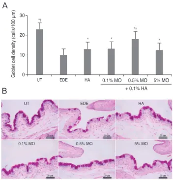
The mean conjunctival goblet cell count significantly de- creased in the EDE group (10.0 ± 0.8 cells/100 μm) com- pared with the UT group (23.0 ± 1.4 cells/100 μm) (p <
0.01). The mean goblet cell counts were 13.0 ± 1.41 cells/100μm in the HA group (p = 0.02 vs. the EDE group), 13.3 ± 0.96 cells/100 μm in the mixed 0.1% MO group (p <
0.01 vs. the EDE group; p = 0.78 vs. the HA group), 18.0 ± 0.82 cells/100 μm in the mixed 0.5% MO group (p < 0.01 vs. the EDE group; p = 0.02 vs. the HA group), and 12.5 ± 1.29 cells/100 μm in the mixed 5.0% MO group (p = 0.02 vs. the EDE group; p = 0.62 vs. the HA group) (Fig. 4).
Discussion
HA can be found naturally in all vertebrates in the ex- tracellular matrix of the skin, synovial fluid, and vitreous body of the eye. It is a biopolymer of disaccharide units composed of N-acetylglucosamine and glucuronic acid in linear chains of varying molecular weights [23]. By form- ing a protective coating that help prevent further irritation and damage of the corneal epithelium, HA can improve corneal fluorescein staining [24]. In addition, dose-depen- dent ability of HA to decrease the size of the wound area has been reported [25]. Topical use of HA provides both objective and subjective symptomatic relief in patients with dry eye disease [8-10]. In addition, there is evidence that hyaluronate may play a role in controlling the local- ized inflammation of ocular surface in patients with kera- toconjuctivitis sicca [26].
HA has several biological effects that are exerted through its direct interaction with a cell surface receptor (CD44). The effects include metastatic potential of tumor cells, secretion of cytokines and chemokines, mediation of inflammatory responses, and cell division [27-29]. In addi- tion, binding of HA to CD44 enhances the growth of the corneal epithelial cells and promotes migration of human corneal epithelial cells [27,28]. The expression of CD44,
Fig. 2. Tear film break-up time in the untreated (UT), experimen-tal dry eye (EDE), hyaluronic acid (HA), mixed 0.1% mineral oil (MO), mixed 0.5% MO, and mixed 5.0% MO groups at 5 and 10 days after desiccating stress. *p < 0.05 compared with the EDE group; †p < 0.05 compared with the HA group.
0 UT EDE HA
+ 0.1% HA 0.1% MO 0.5% MO 5% MO 1
2
†
*
3 4 5
Tear break-up time (sec)
†
*
* *
* * Day 5 Day 10
Fig. 1. Mean corneal irregularity scores (A) and representative figure (B) in the untreated (UT), experimental dry eye (EDE), hyaluronic acid (HA), mixed 0.1% mineral oil (MO), mixed 0.5%
MO, and mixed 5.0% MO groups at 5 and 10 days after desic- cating stress. *p < 0.05 compared with the EDE group; †p < 0.05 compared with the HA group.
Day 5 Day 10
UT EDE HA
+ 0.1% HA 0.1% MO 0.5% MO 5% MO
0 UT EDE HA
+ 0.1% HA 0.1% MO 0.5% MO 5% MO 1
2
†
* 3 4
Corneal smoothness score
*†
†
* *†
Day 5 Day 10
A
B
the receptor of HA, can be increased in corneal and con- junctival cells of patients with dry eye disease, whereas it is decreased following the use of HA eye drops [24,30].
Different types of artificial tear formulation, including gel-based, cellulose-based, carbomer-based and MO-based, have been developed to relieve symptoms of dry eye and have been designed as alternatives to classic artificial tear formulation [12,31,32]. Among these new formulations, MO-based formulations can prolong retention times com- pared with aqueous tear substitutes, but they may cause a sticky sensation and blurred vision [33]. One study demonstrated that a preservative-free oil-in-water emulsion containing 7% soy bean oil, 3% egg yolk phospholipids, and1.8% glycerol could relieve and improve clinical signs such as tear volume and corneal fluorescent staining in a mouse model of dry eye [34]. In addition, MO-based artifi- cial tears were as effective as cellulose-based and carbom- er-based artificial tears in reducing subjective symptoms, and objective signs including TBUT [12].
In the present study, we evaluated the effect of mixed 0.1% HA and MO eye drops on tear production, ocular surface irregularity, TBUT, corneal fluorescein staining score, and conjunctival goblet cell count in an EDE model.
There was no significant difference of tear volume be- tween the stress-induced groups. The corneal irregularity score was lower in the mixed 0.5% MO group compared with the EDE and HA groups. The mixed 0.5% and 5.0%
MO groups showed a significant improvement in TBUT compared with the EDE group. In the mixed 0.1% and 0.5% MO groups, there was a significant improvement of the corneal fluorescein staining score compared with the EDE or HA groups. The conjunctival goblet cell count was higher in the mixed 0.5% MO group compared with the EDE group and the HA group.
Our results showed that HA eye drops containing MO could have a beneficial effect on the tear film and ocular surface of EDE. These results may be attributed to a syn- ergetic effect of HA and MO. The effect of HA as artificial tears which could decrease corneal fluorescein staining by promoting epithelial healing might be enhanced by the addition of MO, and MO itself as a lipid component might improve tear stability and corneal surface irregularity by prolonging the retention time of the tear film. The 0.1%
MO showed an improvement of corneal fluorescein stain- ing scores and goblet cell count, and the 5.0% MO improved the TBUT and goblet cell count, while the 0.5%
Fig. 3. Corneal fluorescein staining scores (A) and representative figure (B) in the untreated (UT), experimental dry eye (EDE), hyaluronic acid (HA), mixed 0.1% mineral oil (MO), mixed 0.5%
MO, and mixed 5.0% MO groups at 5 and 10 days after desic- cating stress. *p < 0.05 compared with the EDE group; †p < 0.05 compared with the HA group.
0 UT EDE HA
+ 0.1% HA 0.1% MO 0.5% MO 5% MO 1
2
†
* 3 4
Corneal staining score
Day 5 Day 10
UT EDE HA
+ 0.1% HA 0.1% MO 0.5% MO 5% MO
†
*
* *
†
* †
† †
† *
* *†
Day 5 Day 10
A
B
Fig. 4. Mean goblet cell count (A) and representative figures (B) of the untreated (UT), experimental dry eye (EDE), hyaluronic acid (HA), mixed 0.1% mineral oil (MO), mixed 0.5% MO, and mixed 5.0% MO groups at 10 days after desiccating stress. *p <
0.05 compared with the EDE group; †p < 0.05 compared with the HA group. Sections were stained with periodic acid-Schiff.
0 UT
UT EDE HA
0.1% MO 0.5% MO 5% MO
EDE HA
+ 0.1% HA 0.1% MO 0.5% MO 5% MO 10
20 30
Goblet cell density (cells/100 μm)
*†
* *
*†
*
A
B
MO ameliorated all measured parameters. These results suggested that the application of a mixture of 0.5% MO and HA eye drops was the best combination for the treatment of EDE compared with the other MO groups.
The application of MO eye drops, however, may cause a sticky, burning sensation and blurred vision [33]. Because this study is based on the dry eye mouse model, further studies based on human will be needed to examine any possible side effects.
In conclusion, the application of eye drops containing a mixture of 0.5% MO and 0.1% HA was more effective in corneal irregularity score, corneal fluorescein staining score, and conjunctival goblet cell count on the ocular sur- face compared with 0.1% HA eye drops in a mouse model of dry eye. These results showed that the MO and HA mixture was more effective than HA agents alone for treating dry eye associated with ocular surface damage. In the near future, clinical studies on the safety and efficacy of MO and HA mixture eye drops for the treatment of dry eye will be needed.
Conflict of Interest
No potential conflict of interest relevant to this article was reported.
Acknowledgements
The study was partially supported by the Chonnam Na- tional University Hospital Biomedical Research Institute (CRI 11076-21 and CRI 13906-22) and Chong Kun Dang Pharmaceutical Co., Seoul, Korea.
References
1. The definition and classification of dry eye disease: report of the Definition and Classification Subcommittee of the International Dry Eye WorkShop (2007). Ocul Surf 2007;5:
75-92.
2. Cuevas M, Gonzalez-Garcia MJ, Castellanos E, et al. Cor- relations among symptoms, signs, and clinical tests in evap- orative-type dry eye disease caused by Meibomian gland dysfunction (MGD). Curr Eye Res 2012;37:855-63.
3. Gupta H, Jain S, Mathur R, et al. Sustained ocular drug de- livery from a temperature and pH triggered novel in situ gel system. Drug Deliv 2007;14:507-15.
4. Behrens A, Doyle JJ, Stern L, et al. Dysfunctional tear syn- drome: a Delphi approach to treatment recommendations.
Cornea 2006;25:900-7.
5. McCann LC, Tomlinson A, Pearce EI, Papa V. Effective- ness of artificial tears in the management of evaporative dry eye. Cornea 2012;31:1-5.
6. McDonald CC, Kaye SB, Figueiredo FC, et al. A ran- domised, crossover, multicentre study to compare the per- formance of 0.1% (w/v) sodium hyaluronate with 1.4% (w/v) polyvinyl alcohol in the alleviation of symptoms associated with dry eye syndrome. Eye (Lond) 2002;16:601-7.
7. Wysenbeek YS, Loya N, Ben Sira I, et al. The effect of so- dium hyaluronate on the corneal epithelium. An ultrastruc- tural study. Invest Ophthalmol Vis Sci 1988;29:194-9.
8. Sand BB, Marner K, Norn MS. Sodium hyaluronate in the treatment of keratoconjunctivitis sicca. A double masked clinical trial. Acta Ophthalmol (Copenh) 1989;67:181-3.
9. DeLuise VP, Peterson WS. The use of topical Healon tears in the management of refractory dry-eye syndrome. Ann Ophthalmol 1984;16:823-4.
10. Stuart JC, Linn JG. Dilute sodium hyaluronate (Healon) in the treatment of ocular surface disorders. Ann Ophthalmol 1985;17:190-2.
11. Rawlings AV, Lombard KJ. A review on the extensive skin benefits of mineral oil. Int J Cosmet Sci 2012;34:511-8.
12. Wang IJ, Lin IC, Hou YC, Hu FR. A comparison of the ef- fect of carbomer-, cellulose- and mineral oil-based artificial tear formulations. Eur J Ophthalmol 2007;17:151-9.
13. Yoon KC, De Paiva CS, Qi H, et al. Desiccating environ- mental stress exacerbates autoimmune lacrimal keratocon- junctivitis in non-obese diabetic mice. J Autoimmun 2008;
30:212-21.
14. Niederkorn JY, Stern ME, Pflugfelder SC, et al. Desiccat- ing stress induces T cell-mediated Sjogren’s Syndrome-like lacrimal keratoconjunctivitis. J Immunol 2006;176:3950-7.
15. De Paiva CS, Corrales RM, Villarreal AL, et al. Cortico- steroid and doxycycline suppress MMP-9 and inflammato- ry cytokine expression, MAPK activation in the corneal epithelium in experimental dry eye. Exp Eye Res 2006;83:
526-35.
16. De Paiva CS, Villarreal AL, Corrales RM, et al. Dry eye-induced conjunctival epithelial squamous metaplasia is modulated by interferon-gamma. Invest Ophthalmol Vis
Sci 2007;48:2553-60.
17. Villareal AL, Farley W, Pflugfelder SC. Effect of topical ophthalmic epinastine and olopatadine on tear volume in mice. Eye Contact Lens 2006;32:272-6.
18. Li Z, Choi W, Oh HJ, Yoon KC. Effectiveness of topical in- fliximab in a mouse model of experimental dry eye. Cor- nea 2012;31 Suppl 1:S25-31.
19. Li Z, Woo JM, Chung SW, et al. Therapeutic effect of topi- cal adiponectin in a mouse model of desiccating stress-in- duced dry eye. Invest Ophthalmol Vis Sci 2013;54:155-62.
20. De Paiva CS, Corrales RM, Villarreal AL, et al. Apical corneal barrier disruption in experimental murine dry eye is abrogated by methylprednisolone and doxycycline. In- vest Ophthalmol Vis Sci 2006;47:2847-56.
21. Dogru M, Erturk H, Shimazaki J, et al. Tear function and ocular surface changes with topical mitomycin (MMC) treatment for primary corneal intraepithelial neoplasia.
Cornea 2003;22:627-39.
22. Xiao X, He H, Lin Z, et al. Therapeutic effects of epider- mal growth factor on benzalkonium chloride-induced dry eye in a mouse model. Invest Ophthalmol Vis Sci 2012;53:
191-7.
23. Vogel R, Crockett RS, Oden N, et al. Demonstration of ef- ficacy in the treatment of dry eye disease with 0.18% sodi- um hyaluronate ophthalmic solution (vismed, rejena). Am J Ophthalmol 2010;149:594-601.
24. Brignole F, Pisella PJ, Dupas B, et al. Efficacy and safety of 0.18% sodium hyaluronate in patients with moderate dry eye syndrome and superficial keratitis. Graefes Arch Clin Exp Ophthalmol 2005;243:531-8.
25. Nakamura M, Hikida M, Nakano T. Concentration and
molecular weight dependency of rabbit corneal epithelial wound healing on hyaluronan. Curr Eye Res 1992;11:981-6.
26. Stern ME, Beuerman RW, Fox RI, et al. The pathology of dry eye: the interaction between the ocular surface and lac- rimal glands. Cornea 1998;17:584-9.
27. Adamia S, Maxwell CA, Pilarski LM. Hyaluronan and hy- aluronan synthases: potential therapeutic targets in cancer.
Curr Drug Targets Cardiovasc Haematol Disord 2005;5:3- 14.
28. Turino GM, Cantor JO. Hyaluronan in respiratory injury and repair. Am J Respir Crit Care Med 2003;167:1169-75.
29. Toole BP. Hyaluronan promotes the malignant phenotype.
Glycobiology 2002;12:37R-42R.
30. Lerner LE, Schwartz DM, Hwang DG, et al. Hyaluronan and CD44 in the human cornea and limbal conjunctiva.
Exp Eye Res 1998;67:481-4.
31. Sullivan LJ, McCurrach F, Lee S, et al. Efficacy and safety of 0.3% carbomer gel compared to placebo in patients with moderate-to-severe dry eye syndrome. Ophthalmology 1997;104:1402-8.
32. Wang TJ, Wang IJ, Ho JD, et al. Comparison of the clinical effects of carbomer-based lipid-containing gel and hy- droxypropyl-guar gel artificial tear formulations in patients with dry eye syndrome: a 4-week, prospective, open-label, randomized, parallel-group, noninferiority study. Clin Ther 2010;32:44-52.
33. Holly FJ. Artificial tear formulations. Int Ophthalmol Clin 1980;20:171-84.
34. Scifo C, Barabino S, De Pasquale G, et al. Effects of a new lipid tear substitute in a mouse model of dry eye. Cornea 2010;29:802-6.