INTRODUCTION
The dry eye syndrome is one of the most prevalent diseases of the eye that affects approximately 10% to 20% of adult population (1). Dry eye develops from multiple etiologies such as a tear deficiency, abnormal evaporation, surface inflamma- tion and abnormal neural regulation (2). The most common medical treatments for dry eye are currently artificial tear su- pplementation, immune suppression with medications such as cyclosporine A (3), and hyaluronic acid (HA) (4) which pr- omotes epithelial healing.
Recently, the role of the P2Y2receptor agonist on dry eye has been investigated in vitro and in vivo (5-7). Topical admin- istration of uracil-cytosine dinucleotide denufosol (Diquafosol) is currently undergoing clinical trials for dry eye disease (8).
Uridine is transformed to UTP (uridine 5-triphosphate) by uridine kinase in vivo after administration. UTP, as a P2Y2 receptor agonist, is known to stimulate mucin secretion in vitro and in vivo in conjunctiva, which increases tear film sta-
bility and protects against desiccation of the ocular surface (9, 10).
In our previous study, we showed that topical uridine could restore the health of the ocular surface in a rabbit corneal wo- und and dry eye model (11). Furthermore, elevated uridine 5′-diphosphate (UDP)-glucose has been shown to contribute to an increase in the synthesis of intrinsic HA (12, 13). Hence, oral uridine may help patients with dry eye by increasing tear secretion or hyaluronic acid. In fact, oral uridine is currently used as a dietary supplement and is commercially available.
Recently, the concept of dry eye has been changed to focus on the importance of dysfunctional tears. The Delphi panel reported on the crucial role of the symptoms as well as the clinical signs for the diagnosis of the dysfunctional tear syn- drome (13). Even without any sign of a damaged ocular sur- face, complaints of dryness or a foreign body sensation can be diagnosed as the dysfunctional tear syndrome based on the guidelines provided by the Delphi panel committee (14-16).
Therefore, the aim of the present study was to investigate
701
Ki Cheol Chang1, Joo Youn Oh1, Youn Seok In1, Mee Kum Kim1, Ki Cheul Shin2, Won Ryang Wee1, Jin Hak Lee1, and Myung Gyu Park3
Department of Ophthalmology1, Seoul National University College of Medicine and Seoul National University Hospital Clinical Research Institute, Seoul;
Department of Ophthalmology2, Kunkuk University Hospital, Seoul; MD Bioalpha3, Suwon, Korea
Address for correspondence Mee Kum Kim, M.D.
Department of Ophthalmology, Seoul National University College of Medicine, 28 Yeongeon-dong, Jongno-gu, Seoul 110-744, Korea
Tel : +82.2-2072-2665, Fax : +82.2-741-3187 E-mail : [email protected]
Presented in part at the ASCRS Annual Meeting, March 2006; San Francisco.
None of the authors except Myung Gyu Park has a financial or proprietary interest in any material or method mentioned.
Supported by Small and Medium Business Administration fund Grant (S1012191) and SNUH Contribution 06-2004-013.
DOI: 10.3346/jkms.2009.24.4.701
Preliminary Effects of Oral Uridine on the Ocular Surface in Dry Eye Patients
We designed a randomized, double blinded, 3-months controlled prospective clini- cal study to investigate effects of oral uridine on the ocular surface in dry eye pati- ents. Twenty-seven patients who diagnosed as dry eye with lower than 5 mm of wetting in the Schirmer strip, with corneal epithelial erosion and who completely followed-up till 3 months were enrolled. Corneal-conjunctival fluorescein staining, non-anesthetic Schirmer test, impression cytology, and Ocular Surface Disease Index (OSDI) were evaluated in the experimental and placebo groups at the base- line, 1 and 3 months after start of medication in a double blinded manner. Fluores- cein stain score of the cornea was markedly decreased in oral uridine group com- pared to the placebo group at 3 months after medication (P=0.032, Mann-Whitney U test). The Schirmer wetting score for the oral uridine group was significantly inc- reased (P=0.001, Wilcoxon signed rank test) at 3 months and its difference between two groups was statistically significant (P=0.030, Mann-Whitney U test). OSDI scores were significantly decreased at 1 and 3 months in treatment group. Oral uridine is effective in treatment of dry eyes.
Key Words : Cornea; Dry Eye Syndromes; Uridine; OSDI
Received : 24 December 2007 Accepted : 25 October 2008
the effect of oral uridine on the ocular surface in dry eye pa- tients, by evaluating the clinical signs and symptoms with the Ocular Surface Disease Index (OSDI).
MATERIALS AND METHODS Patient selection
The study was approved by the institutional review board of Seoul National University Hospital and written consent was obtained from each patient before participation; the study protocol followed the guidelines of the Declaration of Helsin- ki. Patients who were diagnosed with bilateral dry eye syn- drome, showing lower than 5 mm of wetting with the Sc- hirmer strip, superficial punctate keratitis and no improve- ment despite the use of any prescribed medications for at least 2 months in their both eyes were recruited from the Seoul National University Hospital from May 2005 to May 2006.
The patients were initially enrolled in a randomized, dou- ble-blinded, 3-month, controlled clinical study. ‘No improve- ment’ was defined as a case that despite previsously prescribed medications, patient showed no change in their symptoms, Schirmer test and fluorescein score for more than 2 months.
Subjects were excluded if they had any of the following:
dry eye caused by Stevens-Johnson syndrome or cicatricial pemphigoid, a history of intraocular surgery or corneal surgery such as LASIK, or corneal transplant before enrollment. In addition, patients with neurotrophic keratitis, ongoing ocu- lar infection such as a significant blepharitis, herpes infection, or bacterial keratitis were excluded. Furthermore, if the sub- jects could not complete a 3-month follow-up or take their medications accurately, they were excluded.
If they had any of new systemic symptoms after taking me- dication such as indigestion, diarrhea, we recommended them to stop taking medications.
Treatment
Patients were randomly assigned to 3-months of treatment with oral uridine (HALUTECH plus�, KFDA, No.2004- 0060-047, 800 mg/tab, MDbioalpha, Daejeon, Korea) or placebo, 2 tablets 3 times a day. The placebo was composed of L-glutamine, lactose and crystalline cellulose which was the same color and shape as HALUTECH plus�but contained no uridine. HALUTECH plus�which was developed for de- generative arthritis, included 50 mg of uridine in its one ta- blet. The patients were allowed to use their all previously prescribed medications except for the hyaluronic acid eye drops for minimizing the possible confounding effects. The treatment medication or placebo was given to the patients by the medical assistant who was not involved in the evalu- ation of the patients.
Evaluation
All tests were performed in the experimental and placebo groups at the baseline, 1 and 3 months after the start of the study medication in a double-blinded manner.
OSDI was used for the evaluation of the subjective patient symptoms (17). The patients answered the 12 items on the OSDI questionnaire that were graded on a scale of 0 to 4 (0:
none of the time, 1: some of the time, 2: half of the time, 3:
most of the time, 4: all of the time). The OSDI was calcu- lated by ([sum of scores for all questions answered]×100)/
([total number of questions answered]×5). The ophthalmo- logic examination included, in the following order: corneal fluorescein staining, a non-anesthetic Schirmer test and im- pression cytology. The corneal staining was examined with a slit lamp biomicroscope in cobalt blue light 3 min after fluorescein instillation. Punctate staining was recorded with a standardized grading system of 0 to 9. The corneal and con- junctival surface was divided into nine areas, and the num- bers from each of the nine stained divisions were added (20).
For the Schirmer (I) test, Schirmer strip was inserted over the lower lid margin into the cul-de-sac in the temporal one-third of the lid without topical anesthesia. The strip is removed at 5 min and the extent of wetting of the strip was measured.
Conjunctival impression cytology was performed as follows (22). After topical anesthesia with Alcaine�(0.5% propara- caine hydrochloride, Alcon-Couvreur, Puurs, Belgium), MF Millipore membrane filters with a pore size of 0.22 μm were applied onto the temporal bulbar conjunctiva. The membrane filter was cut into 5×5 mm asymmetrical pieces with a poi- nted tip in one corner to guarantee the correct staining sur- face. With an ophthalmodynamometer, a gentle pressure of 60 g was applied onto the micropore filter membrane for 5 sec. Then, the membrane was fixed in 95% ethanol, stained with periodic acid-Schiff, dehydrated in ethanol (70, 80, 90, 100% in sequence), and next with xylene, and, finally cov- erslipped. Goblet cells were counted in 10 consecutive high- power fields (HPFs, ×400). Three observers, who were blind- ed to the patient treatment, performed all pre- and post-treat- ment evaluations. Each observer separately examined each test (fluorescein staining, Schirmer test and impression cytol- ogy). Another observer who was not involved in any other examination counted the goblet cells.
Statistics
Within each group, the changes in tear secretion, corneal staining, OSDI, and goblet cell counts before and after treat- ment were compared using a Wilcoxon signed-rank test. To compare the outcome between the groups, we used the Mann- Whitney U test with ratios of the values at 1 and 3 months compared to the values at baseline for minimizing the statis- tical error caused by individual variation. To know the cor- relation and difference between right and left eye within each
group, Spearman’s rank correlation coefficient and Mann- Whitney U test were used. P<0.05 was considered statistical- ly significant. Statistical analysis was performed using SPSS 14.0 for Windows (SPSS Inc, Chicago, IL, U.S.A.).
RESULTS
Forty-one patients were initially enrolled and randomly assigned to the medication or placebo group. Fourteen pati- ents were excluded from the analysis because they did not complete the 3-month follow-up. The main reasons for the drop-off were mild gastrointestinal symptoms and poor com- pliance. Among the 27 patients who completed the trial, 30 eyes of 15 patients were in the oral uridine group and 24 eyes of 12 patients were in the placebo group. There were 3 men and 24 women, with a mean (±SD) age of 55 (±15.22) yr (range, 13-76). All patients in both groups showed a mod- erate to severe dry eye and their previously prescribed medi- cations included preservative-free artificial tear, topical 0.05%
cyclosporine (Restasis�, Allergan Inc., Irvine, CA, U.S.A.),
autologous serum, and teramycin oint. They used no systemic medication for their dry eye.
Corneal and conjunctival fluorescein staining
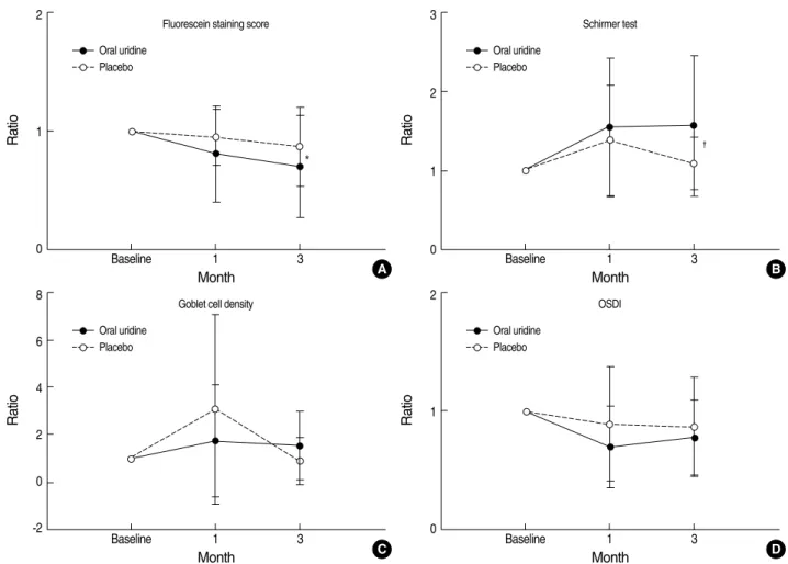
For the oral urdine treatment group, the post-treatment corneal fluorescein staining scores, after 1 and 3 months of medication, were significantly lower than those obtained at the baseline (P=0.004 and P<0.001 for 1 and 3-months of medication, Wilcoxon signed rank test). This reduction was significantly lower in the placebo group (P=0.070 and 0.117 for 1 and 3-months of medication, Wilcoxon signed rank test) (Fig. 1A). The fluorescein staining score was markedly decreased in the oral uridine group compared to the placebo group at 3-months after treatment (P=0.032, Mann-Whit- ney U test) (Fig. 2A). There was a moderate correlation (oral uridine group, ρ=0.449 (P=0.093) and 0.607 (P=0.016) for 1 and 3-months of medication; placebo group, ρ=0.751 (P=
0.008) and 0.653 (P=0.029) for 1 and 3-months of medica- tion) and no significant difference between right and left eye within each both group (oral uridine group, P=0.642 and
Fig. 1. Change within each group of oral uridine and placebo in conjunctival and corneal fluorescein staining (A), aqueous tear production (B), conjunctival goblet cell density (C) and Ocular Surface Disease Index (OSDI) (D). Data are expressed as mean±SD.
*P<0.01; �P=0.044 (Wilcoxon signed rank test).
Fluorescein staining score
9 8 7 6 5 4 3 2 1
0 Baseline 1 3 Baseline 1 3
Oral uridine
*
*
Month
Placebo
A
Schirmer test (mm)
10
8
6
4
2
0 Baseline 1 3 Baseline 1 3
Oral uridine
* *
Month
Placebo
B
Goblet cell density (/×400)
40
30
20
10
0 Baseline 1 3 Baseline 1 3
Oral uridine
Month
Placebo
C
OSDI score
1.0
0.8
0.6
0.4
0.2
0 Baseline 1 3 Baseline 1 3
Oral uridine
*
�
Month
Placebo
D
Fig. 2. Change of ratio of the values at 1 and 3 months of treatment to the values at baseline of treatment. This is for comparison of effica- cy between two groups in conjunctival and corneal fluorescein staining (A), aqueous tear production (B), conjunctival goblet cell density (C) and Ocular Surface Disease Index (OSDI) (D). Data are expressed as meanmean±SD.*P=0.032; �P=0.030 (Mann-Whitney U test).
Ratio
2
1
0 Baseline 1 3
Fluorescein staining score Oral uridine
Month A
*
Placebo
Ratio
3
2
1
0 Baseline 1 3
Schirmer test Oral uridine
Month B
�
Placebo
Ratio
8
6
4
2
0
-2 Baseline 1 3
Goblet cell density Oral uridine
Month C
Placebo
Ratio
2
1
0 Baseline 1 3
OSDI Oral uridine
Month D
Placebo
0.439 for 1 and 3-months of medication; placebo group, P=
0.946 and 0.606 for 1 and 3-months of medication).
Aqueous tear production
The Schirmer wetting score for the oral uridine group was significantly increased (P=0.001, Wilcoxon signed rank test), compared with that for the placebo group (P=0.334, Wilcox- on signed rank test) at 3-months after treatment (Fig. 1B).
The difference between the inter-group ratios between the treatment and control group at 3-months after treatment was also significant (P=0.030, Mann-Whitney U test) (Fig.
2B). There was a moderate correlation between right and left eye in oral uridine group (ρ=0.904 [P<0.001]) and 0.563 (P=
0.023) for 1 and 3-months of medication) but not in place- bo group (ρ=0.362 [P=0.274]and 0.278 [P=0.409]) for 1 and 3-months of medication. There was no significant dif- ference between right and left eye within each both group (oral uridine group, P=0.776 and 0.821 for 1 and 3-months of medication; placebo group, P=0.365 and 0.478 for 1 and 3-months of medication).
Conjunctival goblet cell density
The goblet cell density, for the oral uridine treatment gr- oup, at 1 and 3-months increased; however, the differences were not significant at 1 month (P=0.660, Wilcoxon signed rank test) and showed only marginal significance at 3-months (P=0.053, Wilcoxon signed rank test) (Fig. 1C). The differ- ence between the oral uridine and placebo group, for con- junctival goblet cell density, at 1 and 3-months after treat- ment was not significant (P=0.802 and 0.201 for 1 and 3 months of medication, Mann-Whitney U test) (Fig. 2C).
Ocular surface disease index
The OSDI was significantly decreased in the oral uridine group at 1 and 3-months (P=0.004 and P=0.044 fort 1 and 3-months of medication, Wilcoxon signed rank test) (Fig. 1D).
The change of OSDI in control group after placebo-treatment looked like improved, but it was not significant. However, the difference in the ratios between the two groups, up to 3- months, was not significant (P=0.506 and 0.838 for 1 and
3-months of medication, Mann-Whitney U test) (Fig. 2D).
Side effects of the drug
The most common treatment-related adverse events were gastrointestinal symptoms. Six (four in the oral uridine group, and two in the placebo group) of 14 patients were dropped out from the trial because they complained of mild gastroin- testinal symptoms such as dyspepsia (n=3), indigestion (n=1) and abdominal discomfort (n=2). After treatment discon- tinued, the symptoms resolved. No treatment-related seri- ous adverse events occurred during the study.
DISCUSSION
The objective of the present study was to evaluate the effi- cacy of oral uridine for treatment of the ocular surface in dry eye patients. The results of this study showed that oral uri- dine treatment had a greater effect on improving the tear production and reducing corneal fluorescein staining than did the placebo.
The tear production for the oral uridine group was signif- icantly increased, compared with that for the placebo group at 3-months after treatment. This result of the present study correspond with the previous studies which P2Y receptor agonists such as UTP stimulate active Cl-transport across the excised rabbit conjunctival tissue in vitro (24) and increase tear production in rabbits (25).
The finding of a decrease of corneal staining after the ad- ministration of oral uridine was consistent with the results of our previous study. Previously, we demonstrated that the topical application of uridine enhanced healing of a corneal epithelial defect in a rabbit dry eye model. These findings were thought to be associated with the increased biosynthesis of hyaluronate and glycosaminoglycan, or the reduction of MMP-9 by uridine as described in our previous study (11).
The effect of oral uridine, on the goblet cell counts, appears to be different from our earlier study showing a definite in- crease in the goblet cells with topical uridine. Some possible explanations for these differences are the following. The pre- vious study used rabbit eyes with artificially induced dry eye.
The rabbit is well known for proliferation of epithelial (21) or endothelial cells (22, 23) after damage. The goblet cells of rabbits are more rapidly restored after an insult than they are in humans. Another consideration is the difference of se- verity in dry eye between humans and the rabbits. A dried ocular surface in the rabbits was established by a transient decrease of tear secretion after lacrimal inflammation for 10 days. However, many different factors are involved in the de- velopment of dry eye in humans. In addition, the presence of dry eye in the patients was much longer than in the rab- bits. Our results showed that the goblet cells tended to grad- ually increase until 3-months after the start of treatment with
a marginal significance when compared to the placebo group.
We found that oral uridine treatment was associated with a reduced OSDI. Since the subjective symptoms of dysfunc- tional tear syndrome are used as diagnostic criteria, our results suggest that this treatment may be helpful for patients with dysfunctional tear syndrome even without signs of damage to the ocular surface. Treatment with oral uridine significant- ly improved the subjective symptoms of dry eye. However, placebo group also showed some reduction of OSDI which turned out to be insignificant and was assumed to be caused by “placebo-effect”. It might affect the result which present- ed no significant difference between those groups. To clarify that issue, large number of prospective study is pending.
Diquafosol tetrasodium (INS365) (8), has also demonstrat- ed improvement in tear production in randomized, controlled ongoing clinical trials. Diquafosol, a P2Y2 receptor agonist, which is an analogue of the endogenous compound UTP, is thought to contribute to the restoration of the normal integri- ty of the ocular surface by promoting fluid and mucin secre- tion, regardless of lacrimal gland function (24, 25). Uridine is transformed to UDP or UTP by uridine kinase in vivo. In a similar manner to diquafosol, the results of our study showed that uridine has a similar effect on the ocular surface, as does the P2Y2receptor agonist. Furthermore, uridine administra- tion appears to induce cross-talk for the synthesis of hyalu- ronate, as discussed in our previous study (11). Several other studies showed elevated UDP-glucose with the synthesis of hyaluronic acid (12, 13). Therefore, oral uridine may have a dual role, the increase of hyaluronic acid synthesis and as a P2Y2 receptor agonist, possibly resulting in restoration of healthy ocular surface including increase of goblet cells.
Meanwhile, several studies showed that repeated exposure with purinergic agonist such as UTP causes receptor desen- sitization in vitro (26-28). Therefore, adequate frequency and dosage of UTP derivatives to avoid desensitization seems to be crucial for being effective on ocular surface in dry eye pa- tients. Although we found application of uridine 3 times a day (300 mg/day) for 3 months is likely to be working on ocular surface, we do not know the optimal frequency and dosage of uridine to maximize the effect and whether long term application of daily dosage may induce desensitization or not. Further study would be warranted to evaluate desen- sitization.
The effects of oral uridine seemed to be more prominent at 3-months of treatment than at 1-month. Although uri- dine has been used as a therapeutic agent in the treatment of several other medical disorders (29-31), we do not know the precise bioavailiability of the uridine which we provid- ed to the patients. Regarding the clinical use of oral uridine has been limited by its poor oral bioavailability (7-8%) and short plasma elimination half-life (32, 33), further investi- gation of its bioavailability is needed to improve these limi- tations.
In order to determine the exact mechanism of action of
oral uridine on the ocular surface and proper dosage for dry eye, another study on the relationship between the expression of hyaluronan synthase (HAS) or the P2Y2receptor with ad- ministration of oral uridine, in an in vivo model, is currently under way.
For the safety of oral uridine, prior uridine studies (32-34) using high doses of oral uridine or uridine prodrugs have shown the drug to be well tolerated in humans; osmotic diar- rhea sometimes has been reported as dose-limiting and uri- dine is now commercially available as sugarcane extracts in the world. In our study there were also several patients having mild gastrointestinal symptom after medication. Of them, four patients belong to oral uridine group and two belong to placebo group. In fact, the uridine which we used was not a pure uridine but a combination with other components including L-glutamine, lactose and crystalline cellulose. The- refore, gastrointestinal symptom might be possibly caused by other components as well as uridine. These side effects were mild and resolved after discontinuation of the medication.
This study has several limitations. First, the small num- ber of patients was enrolled. A larger randomized clinical trial is required to confirm the safety and efficacy of oral uri- dine. Second, the tear secretion was measured by the Schirmer test, which has been criticized for its variability and low sen- sitivity (18). However, it is still a useful way of measuring the aqueous tear production; it is a simple test, with low cost and wide availability, and it has been used in many clinical studies on dry eye. Like other studies, Schirmer I test (with- out topical anesthesia) was also used in our study. Schimer I test better reflects the reflex tearing and ocular surface abnor- malities in patients with dry eye as compared to the schirmer test with anesthesia because the reflex tearing is important for the maintenance of the ocular surface (35). Third, the de- sign of the study was, at some point, limited because the pa- tients had been allowed to keep their most previously pre- scribed topical eyedrops except the hyaluronic acid eye drops.
Because the patients suffered from intractable dry eyes even though all those medication, ethically we could not stop giv- ing them those drops. We admit those differences of topical medication between treated and control groups may have a confounding effect on analyzing the influence of oral uridine.
However, we did not directly compare treated group with control group. Considering that the changes of post-treated eye from the pre-treated were indirectly compared using the ratio in each group, we still believe this study shows clinical relevance in some extent.
Despite several limitations, to the best of our knowledge, this is the first study showing that oral uridine may be help- ful for patients with moderate-to-severe dry eye syndrome at least for 3 months.
The results of this study showed that the administration of oral uridine was likely to be safe and effective; it increased tear secretion, decreased fluorescein staining of the damaged cornea and improved patient subjective symptoms.
REFERENCES
1. Schein OD, Munoz B, Tielsch JM, Bandeen-Roche K, West S. Pr- evalence of dry eye among the elderly. Am J Ophthalmol 1997; 124:
723-8.
2. Stern ME, Beuerman RW, Fox RI, Gao J, Mircheff AK, Pflugfelder SC. The pathology of dry eye: the interaction between the ocular sur- face and lacrimal glands. Cornea 1998; 17: 584-9.
3. Sall K, Stevenson OD, Mundorf TK, Reis BL. Two multicenter, ran- domized studies of the efficacy and safety of cyclosporine ophthalmic emulsion in moderate to severe dry eye disease. CsA Phase 3 Study Group. Ophthalmology 2000; 107: 631-9.
4. Aragona P, Papa V, Micali A, Santocono M, Milazzo G. Long term treatment with sodium hyaluronate-containing artificial tears reduces ocular surface damage in patients with dry eye. Br J Ophthalmol 2002; 86: 181-4.
5. Fujihara T, Murakami T, Fujita H, Nakamura M, Nakata K. Improve- ment of corneal barrier function by the P2Y(2) agonist INS365 in a rat dry eye model. Invest Ophthalmol Vis Sci 2001; 42: 96-100.
6. Fujihara T, Murakami T, Nagano T, Nakamura M, Nakata K. INS365 suppresses loss of corneal epithelial integrity by secretion of mucin- like glycoprotein in a rabbit short-term dry eye model. J Ocul Phar- macol Ther 2002; 18: 363-70.
7. Yerxa BR, Mundasad M, Sylvester RN, Garden JC, Cooper M, Kel- lerman DJ. Ocular safety of INS365 ophthalmic solution, a P2Y2ago- nist, in patients with mild to moderate dry eye disease. Adv Exp Med Biol 2002; 506 (Pt B): 1251-7.
8. Tauber J, Davitt WF, Bokosky JE, Nichols KK, Yerxa BR, Schaberg AE, LaVange LM, Mills-Wilson MC, Kellerman DJ. Double-blind- ed, placebo-controlled safety and efficacy trial of diquafosol tetra- sodium (INS365) ophthalmic solution for the treatment of dry eye.
Cornea 2004; 23: 784-92.
9. Ralevic V, Burnstock G. Receptors for purines and pyrimidines. Ph- armacol Rev 1998; 50: 413-92.
10. Jumblatt JE, Jumblatt MM. Regulation of ocular mucin secretion by P2Y2nucleotide receptors in rabbit and human conjunctiva. Exp Eye Res 1998; 67: 341-6.
11. Oh JY, In YS, Kim MK, Ko JH, Lee HJ, Shin KC, Lee SM, Wee WR, Lee JH, Park M. Protective effect of uridine on cornea in a rab- bit dry eye model. Invest Ophthalmol Vis Sci 2007; 48: 1102-9.
12. Pitsillides AA, Wilkinson LS, Mehdizadeh S, Bayliss MT, Edwards JC. Uridine diphosphoglucose dehydrogenase activity in normal and rheumatoid synovium: the description of a specialized synovial lin- ing cell. Int J Exp Pathol 1993; 74: 27-34.
13. Magee C, Nurminskaya M, Linsenmayer TF. UDP-glucose pyrophos- phorylase: up-regulation in hypertrophic cartilage and role in hyal- uronan synthesis. Biochem J 2001; 360: 667-74.
14. Pflugfelder SC, Geerling G, Kinoshita S. Management and therapy of dry eye disease: report of the Management and Therapy Subcom- mittee of the International Dry Eye Workshop (2007). Ocul Surf 2007;
5: 163-78.
15. Wilson SE, Stulting RD. Agreement of physician treatment practices with the international task force guidelines for diagnosis and treat- ment of dry eye disease. Cornea 2007; 26: 284-9.
16. Behrens A, Doyle JJ, Stern L, Chuck RS, McDonnell PJ, Azar DT, Dua HS, Hom M, Karpecki PM, Laibson PR, Lemp MA, Meisler DM, Del Castillo JM, O’Brien TP, Pflugfelder SC, Rolando M, Schein OD, Seitz B, Tseng SC, van Setten G, Wilson SE, Yiu SC; Dysfunc- tional Tear Syndrome Study Group. Dysfunctional tear syndrome:
a Delphi approach to treatment recommendations. Cornea 2006;
25: 900-7.
17. Perry HD, Donnenfeld ED. Dry eye diagnosis and management in 2004. Curr Opin Ophthalmol 2004; 15: 299-304.
18. Norn MS. Lissamine green. Vital staining of cornea and conjuncti- va. Acta Ophthalmol (Copenh) 1973; 51: 483-91.
19. Martinez AJ, Mills MB, Jaceldo KB, Tio FO, Aigbivbalu IB, Hilsen- beck SB, Yee RW. Standardization of conjunctival impression cytol- ogy. Cornea 1995; 14: 515-22.
20. Clinch TE, Benedetto DA, Felberg NT, Laibson PR. Schirmer’s test:
a closer look. Arch Ophthalmol 1983; 101: 1383-6.
21. Moses RA, Parkison G, Schuchardt R. A standard large wound of the corneal epithelium in the rabbit. Invest Ophthalmol Vis Sci 1979;
18: 103-6.
22. Van Horn DL, Sendele DD, Seideman S, Buco PJ. Regenerative ca- pacity of the corneal endothelium in rabbit and cat. Invest Ophthal- mol Vis Sci 1977; 16: 597-613.
23. Olsen EG, Davanger M. The healing of rabbit corneal endothelium.
Acta Ophthalmol (Copenh) 1984; 62: 796-807.
24. Li Y, Kuang K, Yerxa B, Wen Q, Rosskothen H, Fischbarg J. Rab- bit conjunctival epithelium transports fluid, and P2Y2receptor ago- nists stimulate Cl- and fluid secretion. Am J Physiol Cell Physiol 2001; 281: C595-602.
25. Murakami T, Fujihara T, Nakamura M, Nakata K. P2Y(2) receptor stimulation increases tear fluid secretion in rabbits. Curr Eye Res 2000; 21: 782-7.
26. Srinivas SP, Yeh JC, Ong A, Bonanno JA. Ca2+ mobilization in
bovine corneal endothelial cells by P2 purinergic receptors. Cur Eye Res 1998; 17: 994-1004.
27. Pediani JD, McGrath JC, Wilson SM. P2Y receptor-mediated Ca2+
signalling in cultured rat aortic smooth muscle cells. Br J Pharma- col 1999; 126: 1660-6.
28. Clarke LL, Harline MC, Otero MA, Glover GG, Gerrad RC, Krugh B, Walker NM, Gonzalez FA, Turner JT, Weisman GA. Desensiti- zation of P2Y2receptor-activated transepithelial anion secretion. Am J Physiol 1999; 276: C777-87.
29. Walker UA, Venhoff N. Uridine in the prevention and treatment of NRTI-related mitochondrial toxicity. Antivir Ther 2005; 10 (Suppl 2):
M117-23.
30. Kunzelmann K, Mall M. Pharmacotherapy of the ion transport defect in cystic fibrosis: role of purinergic receptor agonists and other poten- tial therapeutics. Am J Respir Med 2003; 2: 299-309.
31. Girot R, Hamet M, Perignon JL, Guesnu M, Fox RM, Cartier P, Du- randy A, Griscelli C. Cellular immune deficiency in two siblings with hereditary orotic aciduria. N Engl J Med 1983; 308: 700-4.
32. Van Groeningen CJ, Peters GJ, Nadal JC, Laurensse E, Pinedo HM.
Clinical and pharmacologic study of orally administered uridine. J Natl Cancer Inst 1991; 83: 437-41.
33. Al Safarjalani ON, Zhou XJ, Naguib FN, Shi J, Schinazi RF, el Kouni MH. Enhancement of the bioavailability of oral uridine by coadmin- istration of 5-(phenylthio)acyclouridine, a uridine phosphorylase in- hibitor: implications for uridine rescue regimens in chemotherapy.
Cancer Chemother Pharmacol 2001; 48: 389-97.
34. Venhoff N, Zilly M, Lebrecht D, Schirmer D, Klinker H, Thoden J, Langmann P, Walker UA. Uridine pharmacokinetics of mitocnol, a sugar cane extract. AIDS 2005; 19: 739-40.
35. Tsubota K, Kaido M, Yagi Y, Fujihara T, Shimmura S. Diseases as- sociated with ocular surface abnormalities: the importance of reflex tearing. Br J Ophthalmol 1999; 83: 89-91.