ISSN 0378-6471 (Print)⋅ISSN 2092-9374 (Online)
http://dx.doi.org/10.3341/jkos.2015.56.3.339
Original Article
눈물막파괴시간이 짧은 건성안에서 3% 디쿠아포솔나트륨 점안액의 임상효과
Clinical Efficacy of Topical 3% Diquafosol Tetrasodium in Short Tear Film Break-Up Time Dry Eye
정현호⋅강연수⋅성미선⋅윤경철
Hyun Ho Jung, MD, Yeon Soo Kang, MD, Mi Sun Sung, MD, Kyung Chul Yoon, MD, PhD
전남대학교 의과대학 안과학교실
Department of Ophthalmology, Chonnam National University Medical School, Gwangju, Korea
Purpose: To evaluate the clinical efficacy of topical diquafosol tetrasodium 3% ophthalmic solution in patients with short tear film break-up time (BUT) dry eye.
Methods: This prospective study involved 30 eyes in 30 patients with dry eye who had tear film BUT values ≤5 seconds and schirmer’s 1 test ≥ 5 mm, and showed no improvement with non-preservative sodium hyaluronate (SH) 0.1% artificial tears. All patients were treated with topical diquafosol tetrasodium 3% 6 times a day, in addition to SH 0.1% artificial tears. Schirmer’s 1 test, tear film BUT, keratoepitheliopathy score with fluorescein, conjunctival staining score with lissamine green, and Ocular sur- face disease index (OSDI) score were evaluated at before treatments, and 1 month and 3 months after treatments.
Results: Significant improvements of tear film BUT and OSDI were observed at 1 month and 3months after diquafosol tetraso- dium 3% administration. At before treatment, and followed up at 1 and 3 months, tear film BUTs were 3.3 ± 1.2, 4.4 ± 1.0 (p <
0.01) and 4.9 ± 1.1 seconds (p < 0.01), respectively, and OSDI scores were 43.5 ± 24.4, 34.6 ± 25.0 (p = 0.01) and 26.7 ± 21.5 (p < 0.01), respectively. There were no significant changes of Schirmer’s score, keratoepitheliopathy, and conjunctival staining score. After diquafosol tetrasodium 3% administration, severe adverse effects were not found in any of the patients.
Conclusions: Topical diquafosol tetrasodium 3% administration was shown to be an effective treatment for improvements of tear film stability and dry eye symptoms.
J Korean Ophthalmol Soc 2015;56(3):339-344
Key Words: Diquafosol tetrasodium 3%, Dry eye, Short tear film break-up time
■Received: 2014. 5. 24. ■ Revised: 2014. 8. 25.
■Accepted: 2015. 3. 1.
■Address reprint requests to Kyung Chul Yoon, MD, PhD Department of Ophthalmology, Chonnam National University Hospital, #42 Jebong-ro, Dong-gu, Gwangju 501-757, Korea Tel: 82-62-220-6741, Fax: 82-62-227-1642
E-mail: [email protected]
ⓒ2015 The Korean Ophthalmological Society
This is an Open Access article distributed under the terms of the Creative Commons Attribution Non-Commercial License (http://creativecommons.org/licenses/by-nc/3.0/) which permits unrestricted non-commercial use, distribution, and reproduction in any medium, provided the original work is properly cited.
건성안은 전 세계적으로 매우 흔한 안과적 질환 중 하나 로,1,2 최근에는 눈물막의 삼투압 증가, 안구표면의 염증 및 눈물막의 불안정성에 의해 눈의 불편감과 시각장애를 유발
하는 다인자성 질환으로 이해되고 있다.3 건성안은 다양한 안구자극 증상을 야기하며, 심한 경우 일상생활에 지장을 줄 정도의 안구불편감이나 시력의 질 저하 등의 다양한 임 상양상을 보인다. 현재 건성안의 치료를 위해 윤활작용을 하는 인공누액과 더불어 스테로이드와 사이클로스포린 제 제와 같은 항염증 점안제의 사용이 보편화되었다.3
건성안은 일반적으로 병인에 따라 수성눈물생성부족 (aqueous deficient) 건성안과 눈물막증발증가(evaporative) 건성안으로 구분할 수 있다.3 눈물막증발증가 건성안 중 눈 물막 파괴시간이 짧은 건성안은 결막배상세포의 감소에 의
한 뮤신 분비 저하와 연관이 있고, 눈물막이 불안정한 반면 염증이나 각결막상피 손상이 심하지 않다는 특징이 있다.4-6 또한, 각결막소견에 비해 심한 건성안 증상을 야기하고, 시 력의 질을 저하시킬 수 있으며, 다수의 환자들에서 인공누 액 등의 보편적인 치료에 효과적으로 반응하지 않는다고 알려졌다.5,6
3% 디쿠아포솔나트륨은 최근 국내에 도입된 P2Y2 수용 체 작용제로서, 결막상피세포에 작용하여 눈물 분비를 촉 진시킬 뿐 아니라 결막배상세포에서 뮤신 당단백질의 분비 를 증가시켜 눈물막의 안정성을 증가시키고, 각결막상피 장애를 개선시킬 수 있는 약제이다.7,8 지금까지 수성눈물생 성부족 건성안을 대상으로 한 여러 연구들에서 디쿠아포솔 나트륨 점안 후 건성안의 증상과 더불어, 눈물막과 안구표 면 인자같은 객관적 징후가 호전됨을 보고하였다.9-13
또한, 디쿠아포솔나트륨 점안액의 사용이 눈물막파괴 시 간이 짧은 건성안이나 폐쇄성 마이봄샘기능장애(meibomian gland dysfunction, MGD)의 치료에도 효과적이라는 연구들 도 보고되었다.14-16 그러나 아직까지 국내에서는 눈물막파 괴시간이 짧은 건성안 환자들을 대상으로 3% 디쿠아포솔 나트륨의 치료효과를 알아본 연구는 발표되지 않았다. 이에 저자들은 기존의 무방부제성 0.1% 히알루론산 인공누액 점 안으로 호전을 보이지 않았던 눈물막파괴시간이 짧은 건성 안 환자들에서 3% 디쿠아포솔나트륨을 점안한 후 눈물막 기능과 안구표면 변화를 분석하고자 하였다.
대상과 방법
2013년 11월부터 2013년 12월까지 본원에 내원한 건성 안 환자들 중 눈물막파괴시간이 5초 이하이고, 기초눈물분 비량이 5 mm 이상이며, 3개월 이상 기존의 무방부제성 0.1% 히알루론산 인공누액 점안으로 호전이 없는 30명을 대상으로 전향적인 연구를 시행하였다. 본 임상시험은 본 원 임상시험심사위원회와 윤리위원회의 심사를 통과하였 으며, 각 환자들은 모두 자발적인 의사 하에 임상 시험에 대한 충분한 설명을 듣고 이해한 후 동의서를 작성하였다.
치료시작 전 3개월 이내부터 경과관찰 기간 동안 안과 수술력 혹은 안외상의 과거력이 있는 환자, 마이봄샘 기능 장애가 있는 환자, 영구적 눈물점 폐쇄술을 시행받은 환자, 활동성 안구 감염이 있는 환자, 최소 7일 이내에 콘택트렌 즈를 착용한 환자 및 만 18세 미만인 환자는 연구 대상에서 제외하였다. 모든 환자들에서 단안 만을 연구에 이용하였 으며, 눈물막파괴시간이 더 짧은 안을 선택하였고, 양안의 눈물막파괴시간이 같은 경우는 우안을 선택하였다.
모든 환자들은 무방부제성 0.1% 히알루론산 인공누액
(Hyalu Mini®, Hanmi Pharm. Co., Seoul, Korea)과 더불어 3%
디쿠아포솔나트륨(Diquas®, Santen Inc., Japan)을 하루 6회 점 안하였다. 치료 전과 치료 후 1개월 및 3개월째 눈물막 기능과 안구표면의 변화를 알기 위해 쉬르머 검사를 통한 기본눈물분 비량 측정, 눈물막파괴시간 측정, 플루레신(fluorescein)을 이 용한 각막표면염색검사 및 리사민그린(lissamine green)을 이용한 결막표면염색검사를 시행하였고, 환자들의 주관적 인 증상을 평가하기 위해 안구표면질환지수(Ocular surface disease index, OSDI) 설문을 시행하였다.
쉬르머 검사는 0.5%로 희석시킨 형광색소(Fluorescite;
Alcon, Fort Worth TX, USA) 10 μl를 하측 구결막에 넣고 5분 후 쉬르머 검사지(Eagle Vision, Memphis TN, USA)를 아래 눈꺼풀의 외측 1/3 부분의 구결막에 5분간 접촉시킨 다음 젖은 부위의 길이를 밀리미터 단위로 측정하였다.17,18 눈물막파괴시간 측정은 형광검사지를 결막낭에 접촉시킨 후 피검자가 수초간 몇 번 눈을 깜박이게 한 후 염색된 눈 물막 층에서 검은 점, 구멍, 또는 줄의 형태로 형광 색소 염 색의 결손이 관찰될 때까지 시간을 세극등현미경의 코발트 블루광원을 이용하여 측정하였다.19-21 각막상피병증의 정도 는 플루레신 형광색소(Haag-Streit AG, Köniz, Switzerland) 염색으로 평가하였다. 염색된 면적과 밀도를 점수화하여 각각 0부터 3까지로 구분하고 이를 곱한 수치로 나타내었 으며, 형광 염색은 각막이 염색되지 않은 경우를 0, 각막의 1/3 미만인 경우를 1, 1/3과 2/3 미만 사이를 2, 2/3 이상을 3으로 하였고, 밀도 점수는 염색이 되지 않는 경우를 0, 경 도를 1, 중증도를 2, 밀도가 높으면서 병변이 서로 겹치는 경우를 3으로 나타내었다.22-25 결막상피손상의 정도 측정은 리사민그린 염색을 이용하였으며, Oxford scheme을 통해 0 부터 5까지 등급을 나누었다.26
OSDI는 건성안 증상(Ocular symptoms) 관련 항목 3가지, 시야(Vision-related function) 관련 항목 6가지 및 환경적인 자극(Environmental triggers) 관련 항목 3가지로 세분하였 고, 각 항목에 대해 0에서 4점으로 하여 각 점수의 총합을 답변한 질문수로 나누어 전체 점수를 계산하였다.27,28
치료 전과 치료 후 경과관찰 동안의 변화를 비교하기 위 해 Wilcoxon signed rank test를 이용하였다. 통계학적 분석 은 SPSS version 17.0 (SPSS Inc., Chicago, IL, USA)을 이 용하였으며, p값이 0.05 미만인 경우를 유의하다고 정의하 였다.
결 과
본 연구에 참여한 대상자들의 평균 나이는 49.6 ± 16.2세 였고, 남자 5명, 여자 25명이었다. 치료 전의 쉬르머검사,
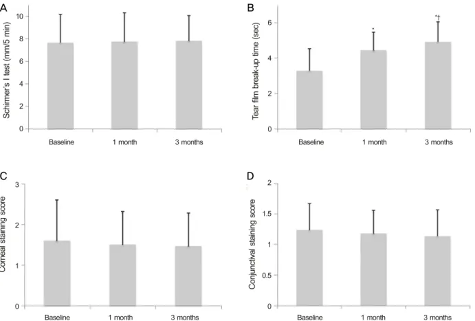
Figure 1. Changes of tear film and ocular surface parameters before and after treatment with topical 3% diquafosol tetrasodium. (A)
Schirmer’s I test. (B) Tear film break-up time. (C) Corneal staining score. (D) Conjunctival staining score. *p < 0.05 compared with baseline; †p < 0.05 compared with 1 month after treatment.Table 1. Demographics of patients with short tear film break-
up time dry eyeAge
Years (range) 49.6 ± 16.2 (28-61)
Sex (M:F) 5:25
Schirmer’s I test (mm/5 min) 7.6 ± 2.5 Tear film break-up time (sec)
Corneal staining score (0-9)
3.3 ± 1.2 1.6 ± 1.0 Conjunctival staining score (0-5)
Overall OSDI score (0-100)
1.2 ± 0.4 43.5 ± 24.4 Values are presented as mean ± SD.
OSDI = ocular surface disease index.
눈물막파괴시간, 각막염색점수, 결막염색점수 및 안구표면 질환지수의 평균값은 각각 7.6 ± 2.5 mm, 3.3 ± 1.2초, 1.6
± 1.0, 1.2 ±0.4 및 43.5 ± 24.4이었다(Table 1).
쉬르머검사는 치료 후 1개월째 7.7 ± 2.6 mm (p=0.26), 3개월째 7.8 ± 2.2 mm (p=0.20)이었다. 눈물막파괴시간은 치료 후 1개월째 4.4 ± 1.0초(p<0.01), 3개월째 4.9 ± 1.1초 (p<0.01)로 치료 전에 비해 유의하게 증가하였고, 치료 1개 월째와 비교했을 때 치료 3개월째에 통계학적으로 유의한 호전을 나타내었다(p=0.03). 각막염색점수와 결막염색점수
는 치료 후 1개월째 각각 1.5 ± 0.8 (p=0.32), 1.2 ± 0.4 (p=0.20)이었고, 치료 후 3개월째 1.5 ± 0.8 (p=0.19), 1.1 ± 0.4 (p=0.11)이었다(Fig. 1).
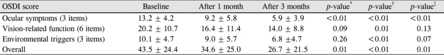
OSDI의 전체 점수는 치료 전에 비해 치료 1개월째와 치 료 3개월째에 통계학적으로 유의한 호전을 보였고(p<0.01 and p<0.01), 치료 1개월째와 비교했을 때 치료 3개월째 또 한 통계학적으로 유의하게 점수가 감소하였다(p<0.01) (Table 2). OSDI를 세부항목으로 나누어 분석하였을 때, 건 성안 증상 관련 항목에서는 치료 전에 비해 치료 1개월째 와 치료 3개월째에 통계학적으로 유의한 호전을 보였고 (p<0.01 and p<0.01), 치료 1개월째와 비교했을 때 치료 3개 월째 또한 통계학적으로 유의하게 점수가 감소하였다 (p<0.01). 반면에, 시야 관련 항목과 환경적인 자극 관련 항 목에서는 치료 전과 비교했을 때 치료 3개월째에서만 유의 한 호전을 보였다(p<0.01 and p<0.01, Table 2).
30명의 환자 중 1명(3.3%)에서 3% 디쿠아포솔나트륨 점 안 후 안구자극감과 충혈이 발생하였으나, 모든 환자에서 안약 점안을 중단할 정도의 심각한 부작용은 발생하지 않 았다.
A B
C D
Table 2. Changes of ocular surface disease index before and after treatment with topical 3% diquafosol tetrasodium
OSDI score Baseline After 1 month After 3 months p-value* p-value† p-value‡
Ocular symptoms (3 items) 13.2 ± 4.2 9.2 ± 5.8 5.9 ± 3.9 <0.01 <0.01 <0.01
Vision-related function (6 items) 20.2 ± 10.7 16.4 ± 11.4 14.0 ± 8.8 0.09 0.01 0.13
Environmental triggers (3 items) 10.1 ± 4.7 9.0 ± 5.7 6.8 ±4.7 0.26 <0.01 0.07
Overall 43.5 ± 24.4 34.6 ± 25.0 26.7 ± 21.5 0.01 <0.01 0.01
Values are presented as mean ± SD.
OSDI = ocular surface disease index.
*Difference between before and 1 month after treatment; †Difference between before and 3 months after treatment; ‡Difference between 1 month and 3 months after treatment.
고 찰
눈물막증발증가 건성안은 수성눈물생성부족 건성안에 비해 눈물분비량이 상대적으로 적지 않으나, 눈물막의 불 안정성으로 인해 눈물막파괴시간이 짧은 특징이 있으며, 대표적인 내인적 요인으로는 마이봄선 기능장애, 눈꺼풀 형태 장애 및 눈깜박임 횟수의 감소 등이 있으나,29,30 아직 까지 정확한 병인은 밝혀지지 않았다. 안구표면은 계속적 으로 생성되는 눈물막으로 덮이게 되며, 눈물막은 크게 지 방층(lipid layer)과 수성-점액층(aqueous-mucin layer)으로 구성되어 있다. 건성안 환자에서 이러한 눈물층 사이의 균 형이 파괴될 때 눈물막의 불안정성이 야기되며, 이 중 뮤신 은 각결막 표면을 보호하고, 눈물막 안정성의 유지에 매우 중요한 역할을 하는 것으로 보고되었고, 크게 분비형 (secretory) 뮤신과 막결합(membrane-bound) 뮤신과 구분할 수 있다.31
3% 디쿠아포솔나트륨은 P2Y2 수용체 작용제로서, 결막 상피세포와 결막배상세포의 소포체(endoplasmic reticulum) 로부터 칼슘 분비를 증가시킴으로써 눈물과 뮤신 분비를 촉진시키는 작용을 한다.5,6,32 특히 결막배상세포에서 분비 형 뮤신인 MUC5AC의 분비를 자극하고,33 각막상피세포에 서 안구표면 당질층(glycocalyx)의 주된 성분인 MUC1, MUC4 및 MUC16 등의 막결합 뮤신의 발현을 증가시킨다 고 보고되었다.34 수성눈물생성부족 건성안을 대상으로 한 다수의 임상연구에서 디쿠아포솔나트륨 점안 후 건성안 증 상, 눈물막과 안구표면 인자 등의 객관적 징후의 호전을 발 표하였으며,9-13 특히 Koh et al13은 디쿠아포솔나트륨을 6개 월 동안 장기간 점안하였을 때 심각한 부작용 없이 건성안 의 증상, 각결막상피손상 및 눈물막파괴시간이 호전되었다 고 보고하였다.
본 연구에서 눈물막파괴시간이 짧은 눈물막 불안정성 건 성안 환자를 대상으로 3% 디쿠아포솔나트륨 하루 6회 점 안하였을 때, 점안 후 1개월째와 3개월째 모두에서 눈물막 파괴시간과 건성안 증상의 유의한 호전이 관찰되었고, 1개 월째와 비교 시 3개월째에서도 또한 유의한 호전을 보임을
알 수 있었다. Shimazaki-Den et al14은 쉬르머검사 결과와 상관없이 건성안 증상이 있고, 눈물막파괴시간이 5초 이하 이며, 각결막상피손상이 적은 환자 39명의 39안을 대상으 로 디쿠아포솔나트륨을 하루 6회 3개월간 사용한 전향적인 연구에서, 본 연구의 결과와 마찬가지로 건성안 증상과 눈 물막파괴시간이 치료 후 1개월째와 3개월째 통계학적으로 유의한 호전을 보였다고 보고하였다. 이들 연구에서는 디 쿠아포솔나트륨 단독 치료군 9안과 인공누액 단독치료군 8 안의 비교분석도 추가적으로 시행하였는데, 디쿠아포솔나 트륨 치료군에서는 치료 후 4주째 눈물막파괴시간의 유의 한 증가가 관찰되었으나, 인공누액 치료군에서는 유의한 호전이 관찰되지 않았다. 또한, 건성안 증상은 치료 후 2주 째 디쿠아포솔나트륨 치료군에서만 유의한 증상 완화가 있 었으며, 치료 후 4주째에는 두 군 모두에서 치료 전에 비해 유의한 호전이 관찰되었다. Kaido et al15은 건성안 증상이 있고 눈물막파괴시간이 5초 이하인 대상자 11명의 11안과 건성안 증상이 없고 눈물막파괴시간이 5초 이하인 대상자 13명의 13안을 대상으로 디쿠아포솔나트륨을 하루 6회 단 독으로 1개월간 점안하였을 때, 두 군 모두에서 눈물막파괴 시간이 유의하게 증가하였고, 건성안 증상이 있는 군의 환 자들 중 75%에서 증상의 완화를 보였다고 보고하였다. 하 지만, 시력과 고위수차를 평가하였을 때는 증상이 있는 군 에서만 유의한 호전을 보였다. 눈물막 불안정성은 안구수 차(ocular aberrations)를 증가시킴으로써 시력의 질을 감소 시킨다고 알려졌다.35,36 본 연구에서는 시력과 고위수차를 평가하지 않았으나, OSDI를 세부항목으로 나누어 분석을 시행하였을 때, 시야관련 항목은 3% 디쿠아포솔나트륨 점 안 후 3개월째에 유의한 호전을 보여, 눈물막 안정성의 호 전으로 인한 시력의 질의 향상이 있었음을 간접적으로 추 론해 볼 수 있다. 하지만, 추후 3% 디쿠아포솔나트륨 점안 전후로 시력이나 고위수차 등을 직접적으로 비교분석하는 연구가 필요할 것으로 생각한다.
본 연구에서 쉬르머검사와 각결막상피손상 정도는 치료 전에 비해 통계적으로 유의한 호전을 보이지는 않았다. 이 는 본 연구에서 눈물막파괴시간이 짧고, 쉬르머검사가 5
mm 이상인 환자들만을 대상으로 포함하였으며, 치료 전 각결막상피손상 정도가 경미하였기 때문이라고 생각한다.
또한, 본 연구의 경과관찰 기간이 3개월로 짧았다는 제한점 도 있어, 향후 연구에서는 디쿠아포솔나트륨을 점안한 후 장기적인 추적관찰이 필요하겠다.
Nakamura et al37은 디쿠아포솔나트륨 점안을 시행했던 연구들의 모든 대상자들을 분석하였을 때, 23.7%에서 점안 후 부작용이 발생하였다고 보고하였다. 주된 부작용으로는 안구 자극감, 안구 분비물, 결막충혈 등이 있었고, 대부분의 부작용은 안약 점안을 중단할 정도로 심하지 않았으며, 안 구 자극감이나 통증은 점안 28일 이내에 대부분에서 호전 되었다. 본 연구에서는 1명(3.3%)에서 안구자극감과 충혈 이 발생하였고, 이전 연구들의 결과와 비슷하게 모든 환자 들에서 안약 점안을 중단할 정도의 심각한 부작용은 발생 하지 않았다. 본 연구에서 3% 디쿠아포솔나트륨 점안과 관 련한 부작용이 적었던 이유는 0.1% 히알루론산 인공누액을 병용하였기 때문이라고 생각한다.
본 연구의 제한점은 대상자의 수가 적었고, 서로 다른 군 사이에서 비교분석을 시행하지 못하였다는 점이다. 또한, 3% 디쿠아포솔나트륨 점안액과 0.1% 히알루론산 인공누액 의 병용 치료를 하였기 때문에 3% 디쿠아포솔나트륨 점안 액의 단독효과에 대해서는 알 수 없었다. 그러나, 기존의 무방부제성 0.1% 히알루론산 인공누액에 호전을 보이지 않 았던 환자들을 대상으로 하였기 때문에, 두 점안액 병용으 로 인한 상승 작용의 가능성을 고려하더라도 3% 디쿠아포 솔나트륨 점안액은 효과적인 치료방법일 것으로 생각한다.
향후 이러한 점을 보완하여 3% 디쿠아포솔나트륨 점안액 단독치료를 시행한 군과 인공누액을 병용한 군 혹은 다른 건성안 치료약제를 사용한 군 간의 비교분석 연구가 필요 할 것으로 생각한다.
결론적으로, 눈물막파괴시간이 짧은 눈물막 불안정성 건 성안 환자에서 3% 디쿠아포솔나트륨 점안은 눈물막 안정 성과 건성안 증상을 개선시키는 효과적인 치료방법이 될 수 있을 것이다.
REFERENCES
1) Schaumberg DA, Sullivan DA, Buring JE, Dana MR. Prevalence of dry eye syndrome among US women. Am J Ophthalmol 2003;
136:318-26.
2) Brewitt H, Sistani F. Dry eye disease: the scale of the problem.
Surv Ophthalmol 2001;45 Suppl 2:S199-202.
3) The definition and classification of dry eye disease: report of the Definition and Classification Subcommittee of the International Dry Eye WorkShop (2007). Ocul Surf 2007;5:75-92.
4) Gipson IK, Hori Y, Argüeso P. Character of ocular surface mucins and their alteration in dry eye disease. Ocul Surf 2004;2:131-48.
5) Toda I, Fujishima H, Tsubota K. Ocular fatigue is the major symp- tom of dry eye. Acta Ophthalmol (Copenh) 1993;71:347-52.
6) Toda I, Shimazaki J, Tsubota K. Dry eye with only decreased tear break-up time is sometimes associated with allergic conjunctivitis.
Ophthalmology 1995;102:302-9.
7) Fujihara T, Murakami T, Nagano T, et al. INS365 suppresses loss of corneal epithelial integrity by secretion of mucin-like glyco- protein in a rabbit short-term dry eye model. J Ocul Pharmacol Ther 2002;18:363-70.
8) Fujihara T, Murakami T, Fujita H, et al. Improvement of corneal barrier function by the P2Y(2) agonist INS365 in a rat dry eye model. Invest Ophthalmol Vis Sci 2001;42:96-100.
9) Matsumoto Y, Ohashi Y, Watanabe H, Tsubota K; Diquafosol Ophthalmic Solution Phase 2 Study Group. Efficacy and safety of diquafosol ophthalmic solution in patients with dry eye syndrome:
a Japanese phase 2 clinical trial. Ophthalmology 2012;119:1954-60.
10) Tauber J, Davitt WF, Bokosky JE, et al. Double-masked, place- bo-controlled safety and efficacy trial of diquafosol tetrasodium (INS365) ophthalmic solution for the treatment of dry eye. Cornea 2004;23:784-92.
11) Kamiya K, Nakanishi M, Ishii R, et al. Clinical evaluation of the additive effect of diquafosol tetrasodium on sodium hyaluronate monotherapy in patients with dry eye syndrome: a prospective, randomized, multicenter study. Eye (Lond) 2012;26:1363-8.
12) Takamura E, Tsubota K, Watanabe H, Ohashi Y; Diquafosol Ophthalmic Solution Phase 3 Study Group. A randomised, dou- ble-masked comparison study of diquafosol versus sodium hyalur- onate ophthalmic solutions in dry eye patients. Br J Ophthalmol 2012;96:1310-5.
13) Koh S, Ikeda C, Takai Y, et al. Long-term results of treatment with diquafosol ophthalmic solution for aqueous-deficient dry eye. Jpn J Ophthalmol 2013;57:440-6.
14) Shimazaki-Den S, Iseda H, Dogru M, Shimazaki J. Effects of di- quafosol sodium eye drops on tear film stability in short BUT type of dry eye. Cornea 2013;32:1120-5.
15) Kaido M, Uchino M, Kojima T, et al. Effects of diquafosol tetraso- dium administration on visual function in short break-up time dry eye. J Ocul Pharmacol Ther 2013;29:595-603.
16) Arita R, Suehiro J, Haraguchi T, et al. Topical diquafosol for pa- tients with obstructive meibomian gland dysfunction. Br J Ophthalmol 2013;97:725-9.
17) Yoon KC, Heo H, Im SK, et al. Comparison of autologous serum and umbilical cord serum eye drops for dry eye syndrome. Am J Ophthalmol 2007;144:86-92.
18) Tsubota K. Tear dynamics and dry eye. Prog Retin Eye Res 1998;
17:565-96.
19) Jeong IY, Park YW, Lee SS, et al. Long term follow-up results of topical 0.05% cyclosporine a in patient with dry eye. Chonnam Med J 2008;44:151-6.
20) Dogru M, Katakami C, Inoue M. Tear function and ocular surface changes in noninsulin-dependent diabetes mellitus. Ophthalmology 2001;108:586-92.
21) Yoon KC, Im SK, Kim HG, You IC. Usefulness of double vital staining with 1% fluorescein and 1% lissamine green in patients with dry eye syndrome. Cornea 2011;30:972-6.
22) Kaido M, Goto E, Dogru M, Tsubota K. Punctal occlusion in the management of chronic Stevens-Johnson syndrome. Ophthalmology 2004;111:895-900.
= 국문초록 =
눈물막파괴시간이 짧은 건성안에서 3% 디쿠아포솔나트륨 점안액의 임상효과
목적: 눈물막파괴시간이 짧은 건성안에서 3% 디쿠아포솔나트륨 점안의 임상적 효과를 알아보고자 하였다.
대상과 방법: 눈물막파괴시간이 5초 이하이고 기초눈물분비량이 5 mm 이상인 건성안 환자들 중 무방부제성 0.1% 히알루론산 인공누 액 점안으로 호전이 없었던 30명을 대상으로 전향적인 연구를 시행하였다. 모든 대상자는 3% 디쿠아포솔나트륨을 하루 6회 추가하여 점안하였다. 쉬르머검사를 통한 기초눈물분비량, 눈물막파괴시간, 플루레신을 이용한 각막염색점수, 리사민그린을 이용한 결막염색점 수 및 안구표면질환지수(ocular surface disease index, OSDI)를 치료 전과 치료 후 1개월, 3개월째에 각각 시행하여 비교 분석하였다.
결과: 눈물막파괴시간은 치료 전과 치료 1개월, 3개월째 각각 3.3 ± 1.2초, 4.4 ± 1.0초(p<0.01), 4.9 ± 1.1초(p<0.01)로 치료 후 유의한 호전을 보였다. OSDI 또한 치료 전과 치료 1개월, 3개월째 각각 43.5 ± 24.4점, 34.6 ± 25.0점(p=0.01), 26.7 ± 21.5점(p
<0.01)으로 치료 전에 비해 유의한 호전을 보였다. 기초눈물분비량, 플루레신 각막염색점수 및 리사민그린을 통한 결막염색점수는 유의한 변화를 보이지 않았다. 모든 환자에서 점안을 중단할 만한 심각한 부작용은 발생하지 않았다.
결론: 3% 디쿠아포솔나트륨 점안은 눈물막파괴시간이 짧은 건성안에서 눈물막 안정성과 건성안 증상을 개선시키는 효과적인 치료방 법으로 생각한다.
<대한안과학회지 2015;56(3):339-344>
23) Yoon KC, Jeong IY, Im SK, et al. Therapeutic effect of umbilical cord serum eyedrops for the treatment of dry eye associated with graft-versus-host disease. Bone Marrow Transplant 2007;39:231-5.
24) Yoon KC, Im SK, Park YG, et al. Application of umbilical cord se- rum eyedrops for the treatment of dry eye syndrome. Cornea 2006;
25:268-72.
25) Yoon KC, Park CS, You IC, et al. Expression of CXCL9, -10, -11, and CXCR3 in the tear film and ocular surface of patients with dry eye syndrome. Invest Ophthalmol Vis Sci 2010;51:643-50.
26) Bron AJ, Evans VE, Smith JA. Grading of corneal and conjunctival staining in the context of other dry eye tests. Cornea 2003;22:
640-50.
27) Schiffman RM, Christianson MD, Jacobsen G, et al. Reliability and validity of the Ocular Surface Disease Index. Arch Ophthalmol 2000;118:615-21.
28) Miller KL, Walt JG, Mink DR, et al. Minimal clinically important difference for the ocular surface disease index. Arch Ophthalmol 2010;128:94-101.
29) Foulks GN, Bron AJ. Meibomian gland dysfunction: a clinical scheme for description, diagnosis, classification, and grading. Ocul Surf 2003;1:107-26.
30) Tsubota K, Nakamori K. Effects of ocular surface area and blink rate on tear dynamics. Arch Ophthalmol 1995;113:155-8.
31) Fahmy AM, Hardten DR. Treating ocular surface disease: new agents in development. Clin Ophthalmol 2011;5:465-72.
32) Nakamura M, Imanaka T, Sakamoto A. Diquafosol ophthalmic sol- ution for dry eye treatment. Adv Ther 2012;29:579-89.
33) Takaoka-Shichijo Y, Sakamoto A, Nakamura M. Effect of diquafo- sol tetrasodium on MUC5AC secretion by rabbit conjunctival tissues. J Eye 2011;28:261-5.
34) Takaoka-Shichijo Y, Nakamura M. Stimulatory effect of diquafo- sol tetrasodium on the expression of membrane-binding mucin genes in cultured human corneal epithelial cells. J Eye 2011;28:425 -9.
35) Lombardo M, Lombardo G. Wave aberration of human eyes and new descriptors of image optical quality and visual performance. J Cataract Refract Surg 2010;36:313-31.
36) Li KY, Yoon G. Changes in aberrations and retinal image quality due to tear film dynamics. Opt Express 2006;14:12552-9.
37) Nakamura M, Imanaka T, Sakamoto A. Diquafosol ophthalmic sol- ution for dry eye treatment. Adv Ther 2012;29:579-89.