Korean Journal of Ophthalmology 2008;22:174-177 ISSN : 1011-8942
DOI : 10.3341/kjo.2008.22.3.174
174
The Effect of Unilateral Medial Rectus Muscle Resection in Patients with Recurrent Exotropia
Sun Hwa Chae, MD, Bo Young Chun, MD, Jung Yoon Kwon, MD, PhD
Department of Ophthalmology, Kyungpook National University, School of Medicine, Daegu, Korea
Purpose: To investigate the effect of unilateral medial rectus muscle resection for recurrent exotropia after bilateral lateral rectus muscle recession for intermittent exotropia
Methods: A retrospective analysis was made of thirtypatients who underwent unilateral medial rectus resection for recurrent exotropia. All had prior bilateral lateral rectus recession for intermittent exotropia. Data were collected for age, the preoperative deviation, the postoperative deviation at 2 weeks, 3 months, 6 months and the last visit, and the amount of medial rectus resection performed.
Results: The average preoperative deviation was 27.0±3.6 PD. After unilateral medial rectus resection, average deviation at distance was 2.8 PD at postoperative 2 weeks, 4.5 PD at 3 months, 5.1 PD at 6 months and 5.8 PD at last visit. The average deviation corrected per millimeter of medial rectus resection was 3.53±0.17 PD/mm.
Conclusions: Considering that deviation angles of recurrent exotropia is smaller than those of primary surgery and the possibility of saving the other medial rectus muscle, unilateral rectus muscle resection could be effective surgical method for recurrent exotropia.
Korean J Ophthalmol 2008;22:174-177 ⓒ 2008 by the Korean Ophthalmological Society.
Key Words: Intermittent exotropia, Recurrence, Unilateral medial rectus muscle resection.
Received: November 5, 2007 Accepted: August 5, 2008
Reprint requests to Jung Yoon Kwon, MD, PhD. Department of Ophthal- mology, Kyungpook National University, School of Medicine, 50 Samduk-2ga, Jung-gu, Daegu 700-721, Korea. Tel: 82-53-420-5812, Fax: 82-53-426-6552, E-mail: [email protected]
* This paper was presented in part at the 97
thAnnual Meeting of the Korean Ophthalmological Society, April 6
th, 2007, Pusan, Korea.
The intermittent exotropia [X(T)] can be cured mainly through surgery but significant portion of patients have undercorrection or recurrence of exotropia at postoperative follow-up.
1,2The rate of recurrence or undercorrection of X(T) is increasing with time and these patients are usually treated by bilateral medial rectus (MR) resection, re-recession of previously recessed lateral rectus (LR) or unilateral LR recession and MR resection (R&R), according to the methods of the previous surgery.
3-6The one muscle surgery has advantages of requiring less time, less anesthesia and placing only one eye at risk for any possible surgical complication.
3Furthermore, there are more risks of overcorrection in the second surgery than the first surgery because of the smallerdeviation of the recurred X(T).
7-9It has been reported that the unilateral MR or LR recession is effective and predictable surgical method for small to moderate angle deviations.
10,11Therefore, we performed this study to evaluate the efficacy
of the unilateral MR resection for the patients of recurred X(T) who had undergone bilateral LR recession as their previous surgery.
Materials and Methods
A retrospective analysis was made of all the patients who had undergone unilateral MR muscle resection between March 1995 and February 2007 for recurred X(T), which developed after bilateral LR recession.
There are 5 exclusion criteria for this retrospective study.
These parameters were: (1) presence of other ocular disease (central nervous system abnormalities or anatomic abnormalities of the eye), (2) presence of amblyopia which include more than two lines of visual acuity difference, (3) presence of paralytic strabismus, (4) presence of dissociated vertical deviation or dysfunction of oblique muscles, and (5) infantile exotropia were excluded from this study.
The following parameters were reviewed and analyzed:
patient age, gender, deviation before 1
stsurgery, deviation before 2
ndsurgery, postoperative deviations according to follow-up duration, ocular motility and the amount of surgery performed.
All patients in this study had a complete ophthalmologic examination prior to surgery. A cycloplegic refraction was performed using 1% cyclopentolate and 0.5% tropicamide.
Visual acuities were measured by Snellen’s chart or ‘E’ chart.
SH Chea, et al. UNILATERAL MEDIAL RECTUS RESECTION FOR RECURRENT X(T)
175
2 7.0
2 .8 4 .5 5.1 5 .8
-5 0 5 10 15 20 25 30 35
Pre-op 2 weeks 3 months 6 months Final Visit Time
De vi a ti o n a n gl e ( P D )
* PD=prism diopters of distant manifest deviation angle.
Fig. 1. Distribution of the mean deviation angle according to follow-up duration after unilateral MR resection for recurrent exotropia.
Number of patients 30
Sex (F/M) 17:13*
Mean age at 1st operation (years) 7.2±5.5 Exodeviation at 1st operation (PD*) 41.2±5.8 Time at the recurrence after 1st operation (months) 15.1±18.6 Mean age at 2nd operation (years) 9.1±4.8 Exodeviation at 2nd operation (PD*) 27.0±3.6
* PD=prism diopters, distant manifest deviation.
Table 1. Summary of clinical data (Mean±SD)
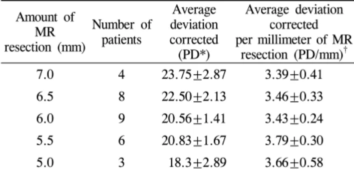
Amount of resection (mm) MR
Number of patients
Average deviation corrected (PD*)
Average deviation corrected per millimeter of MR
resection (PD/mm)
†7.0 4 23.75±2.87 3.39±0.41
6.5 8 22.50±2.13 3.46±0.33
6.0 9 20.56±1.41 3.43±0.24
5.5 6 20.83±1.67 3.79±0.30
5.0 3 18.3±2.89 3.66±0.58
* PD=prism diopters;
†Pearson’s correlation; r=-0.225, p=0.232 Table 2. The surgical effects of unilateral medial rectus resection
The distant and near deviation angles were measured at 6 and 0.33 m by alternate prism cover test, if not possible, by Krimsky methods. The angle measurements of the preoperative deviation and the postoperative deviation at 1 day, 2 weeks, 3 months, 6 months and the last visit were done. Titmus ring test was performed preoperatively and at 3 months postoperatively.
Unilateral MR resections were performed on the non-fixating eye or on the left eye if the patient had an alternative fixation behavior by one of the authors (JYK).
Under the general anesthesia, surgeon made a limbal incision, exposed and resected the MR muscle 5 to 7 mm according to their deviation angle.
We defined the success of the surgery as being orthophoric or having deviation angle less than 10 PD of distant manifest deviation at postoperative 6 months of follow-up. We defined the recurrence as 10 PD or more exodeviation at distance and the overcorrection as 10 PD or more esodeviation at distance at postoperative 6 months of follow-up. The survival was defined as the duration until developing exodeviation more than 10 PD after unilateral MR resection and made Kaplan- Meier survival analysis.
Results
Thirty patients who had undergone unilateral MR resection for recurred XT after bilateral LR recession were included in this study. The descriptive data of these subjects are demonstrated in Table 1. The mean age at bilateral LR recession was 7.2±5.1 year and 9.1±5.4 year at unilateral MR resection. The median time interval between the previous surgery and the recurrence of exodeviation was 9 months (range 3 to 72 months). The mean deviation angle before bilateral LR recession was 41.2±5.8 PD at distance and 39.5±4.8 PD at near. And the mean deviation angle before unilateral MR resection was 27.0±3.6 PD at distance and 24.5±4.2 PD at near. No close relationship was found between deviation angles before bilateral LR recession and those before unilateral MR resection (p>0.05). All of the patients were diagnosed as intermittent exotropia of basic type. No patient demonstrated a noticeable limitation of abduction.
The amount of the unilateral MR resection was decided as 5.0 mm for 20~24 PD, 6.0 mm for 25~29 PD, 6.5 mm for 30~34 PD and 7.0 mm for 35~39 PD. However, there was intraoperative adjustment of the amount of MR resection. For example, if MR muscle seemed to be thinner or looser than usual, the surgeon added 0.5 mm to the planned amount of resection. If MR was found slightly tighter than usual, the surgeon subtracted 0.5 mm from planned resection. In addition, if recurrence of exotropia had occurred early (≤3 months) after previous surgery, the surgeon added 0.5 mm to the planned MR resection and subtracted 0.5 mm when overcorrection was specially concerned due to patients’
young age (≤4 years). Among 30 patients, 19 patients had no adjustment to planned amount of MR resection. The surgeon added 0.5 mm to the amount of planned MR resection to 2 patients having 30~34 PD and 3 patients having 25~29 PD, and subtracted 0.5 mm from 6 patients having 25~29 PD.
Table 2 shows the average amount of corrected
exodeviation per millimeter of resection performed on the
unilateral MR. The average amount of MR resection was 6.1
mm (range 5.0 to 7.0 mm). The average deviation corrected
per millimeter of MR resection was 3.53±0.17 PD/mm.
Korean J Ophthalmol Vol.22, No.3, 2008
176
0 20 4 0 6 0 80 100
Fol lo w- up d ura tio n(months)
0.0 0.2 0.4 0.6 0.8 1.0
Success rates(%)