© 2015 The Korean Ophthalmological Society
This is an Open Access article distributed under the terms of the Creative Commons Attribution Non-Commercial License (http://creativecommons.org/licenses /by-nc/3.0/) which permits unrestricted non-commercial use, distribution, and reproduction in any medium, provided the original work is properly cited.
Original Article
Comparison of Surgical Outcomes with Unilateral Recession and Resection According to Angle of Deviation in Basic Intermittent
Exotropia
Soon Young Cho1, Se Youp Lee2, Jong Hyun Jung3
1Department of Ophthalmology, Dongguk University College of Medicine, Gyeongju, Korea
2Department of Ophthalmology, Dongsan Medical Center, Keimyung University School of Medicine, Daegu, Korea
3Department of Ophthalmology, Myongji Hospital, Goyang, Korea
Purpose: The purpose of this study is to compare the surgical outcomes and near stereoacuities after unilateral medial rectus (MR) muscle resection and lateral rectus (LR) recession according to deviation angle in basic intermittent exotropia, X(T).
Methods: Ninety patients with basic type X(T) were included in this study. They underwent unilateral recession of the LR and resection of the MR and were followed postoperatively for at least 12 months. Patients were divided into three groups according to their preoperative deviation angle: group 1 ≤20 prism diopter (PD), 20 PD< group 2 <40 PD, and group 3 ≥40 PD. Surgical outcomes and near stereoacuities one year after surgery were evaluated. Surgical success was defined as having a deviation angle range within ±10 PD for both near and distance fixation.
Results: Among 90 patients, groups 1, 2, and 3 included 30 patients each. The mean age in groups 1, 2, and 3 was 9.4 years, 9.4 years, and 11.0 years, respectively. The surgical success rates one year after surgery for groups 1, 2, and 3 were 80.0%, 73.3%, and 73.3% (chi-square test, p = 0.769), respectively. The undercorrec- tion rates for groups 1, 2, and 3 were 16.7%, 23.3%, and 26.7%, and the overcorrection rates were 3.3%, 3.3%, and 0%, respectively. The mean preoperative near stereoacuities for groups 1, 2, and 3 were 224.3 arcsec, 302.0 arcsec, and 1,107.3 arcsec, and the mean postoperative near stereoacuities were 218.3 arcsec, 214.7 arcsec, and 743.0 arcsec (paired t-test; p = 0.858, p = 0.379, p = 0.083), respectively.
Conclusions: In basic X(T) patients, the amount of angle deviation has no influence on surgical outcomes in unilateral LR recession and MR resection. The near stereoacuities by one year after LR recession and MR resection for intermittent X(T) were not different among patient groups separated by preoperative deviation angle.
Key Words: Angle of deviation, Exotropia, Stereopsis, Surgical outcome
Intermittent exotropia, X(T), is the most common type of childhood strabismus in Korea. Various surgical treat- ments for X(T) have been introduced, including bilateral lateral rectus (LR) muscle recession or unilateral LR mus- cle recession and medial rectus (MR) muscle resection [1].
Received: March 24, 2015 Accepted: May 18, 2015
Corresponding Author: Se Youp Lee, MD. Department of Ophthalmolo- gy, Dongsan Medical Center, Keimyung University School of Medicine,
#56 Dalseong-ro, Jung-gu, Daegu 41931, Korea. Tel: 82-53-250-7720, Fax:
82-53-250-7705, E-mail: [email protected]
Basic X(T) is the most common type of X(T), and the de- viation of the angle ranges mainly from 20 to 40 prism di- opter (PD) [2].
Basic X(T) is known to progress with a gradual increase in angle of deviation, resulting in worsening stereoacuities [3]. Some studies have reported that the greater the preop- erative angle of deviation, the lower the surgical success rate [4]. Several studies have been conducted in patients with a large angle of exodeviation, but they were focused primarily on adults [5-9].
A number of studies on the effect of strabismus surgery on stereopsis in patients with X(T) have been reported.
Some studies have demonstrated improved distance stereo- acuity after intervention [10,11]. However, there are contro- versies surrounding the effect of near stereoacuity in X(T).
Some authors have suggested that strabismus surgery im- proves near stereoacuity, while others reported no change in post-surgery near stereoacuity [12,13].
In this study, we evaluated the surgical outcomes of uni- lateral LR recession and MR resection in patients with ba- sic X(T) in Korea according to their angle of deviation. We also evaluated the effect of strabismus surgery on near ste- reoacuity in patients with X(T).
Materials and Methods
A retrospective review of medical records was conduct- ed on 592 patients who underwent surgery for basic X(T) between August 2009 and August 2013, under the care of one surgeon. All patients underwent unilateral LR reces- sion and MR resection based on the distant angle of devia- tion. The minimum required follow-up period after sur- gery was 12 months. Patients with a history of previous strabismus surgery, amblyopia, paralytic or restrictive stra- bismus, A or V pattern strabismus, associated vertical de- viation or dissociated vertical deviation, ocular disease other than strabismus, or neurologic disorders were exclud- ed. Patients were divided into three groups according to their preoperative angle of deviation: group 1 ≤20 PD, 20 PD< group 2 <40 PD, and group 3 ≥40 PD. Group 3 intrin- sically had 30 patients. Thus, 30 age-matched subjects were randomly selected to be in group 1 and group 2. A total of 90 patients were included in the study.
The following patient characteristics were recorded: gen- der, age at surgery, deviation at distance and near, Worth
4-dot test, constancy of deviation, fixation dominance, presence of lateral incomitance, refractive error, and supe- rior or inferior oblique overaction. Prism and alternate cover testing were performed at 1 / 3 m and 5 m for all pa- tients. Near stereoacuity test was performed by Titmus stereotest (Stereo Optical, Chicago, IL, USA) at 40-cm dis- tance, wearing Polaroid spectacles. Sensory status was also evaluated using the Worth 4-dot test (Gulden Ophthalmics, Elkins Park, PA, USA) at distance (6 m) and near (33 cm) for the degree of sensory fusion. The Worth 4-dot criteria were as follows. Patients who detected four lights were considered to have fusion, those who detected five lights were considered as diplopic, and those who saw two or three lights were considered to be suppressing. Postopera- tive deviation at distance and near, Worth 4-dot test, and near stereoacuity by Titmus stereotest were measured at one day, one week, three weeks, three months, six months, and 12 months postoperatively.
Unilateral LR recession and MR resection were per- formed by one surgeon according to Wright’s surgical amount [14].An outcome was considered satisfactory if the distant deviation in the primary position was between ≤10 PD of exophoria/tropia and ≤10 PD esophoria/tropia. Un- dercorrection was defined as an alignment of >10 PD X(T), and overcorrection was defined as >10 PD of esotropia.
Statistical analyses were performed using IBM SPSS ver. 20.0 (IBM Co., Armonk, NY, USA). One-way ANO- VA was used for comparison of preoperative and postoper- ative characteristics among groups. The chi-square test was used for comparison of near stereoacuities, surgical success rate, undercorrection rate, and overcorrection rate.
The paired t-test was used for comparison of preoperative and postoperative stereoacuities. A p-value less than 0.05 was considered statistically significant. For statistical con- venience, X(T) was designated as plus (+) and esotropia was designated as minus (-).
Results
Patient characteristics are shown in Table 1. Ninety patients were included, with 43 male (47.8%) and 47 female patients (52.2%). Groups 1, 2, and 3 included 30 patients each. The mean age at surgery was 9.4 years in group 1, 9.4 years in group 2, and 11.0 years in group 3 (one-way ANOVA, p = 0.621) (Table 1). The mean follow-up period
was 15.5 ± 4.1 months in group 1, 17.6 ± 5.8 months in group 2, and 18.9 ± 9.2 months in group 3 (one-way ANO- VA, p = 0.138) (Table 1).
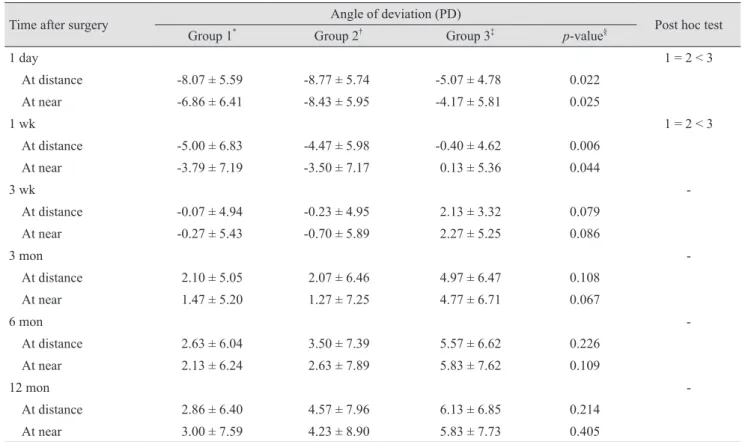
Postoperative angle of deviation for distant and near fix- ation of patients with intermittent X(T) in groups 1, 2 and 3 are shown in Table 2. The mean angle of deviation at post- operative one day and one week were significantly differ- ent at near and at distance among groups (one-way ANO- VA, p < 0.05). However, the mean angles of deviation at later postoperative checks were not statistically different (Table 2).
The mean preoperative near stereoacuities were 224.3 arcsec in group 1, 302.0 arcsec in group 2, and 1,107.3 arcsec in group 3. The mean postoperative 12 months near stereoacuities were 218.3 arcsec in group 1, 214.7 arcsec in group 2, and 743.0 arcsec in group 3. In groups 1, 2, and 3, near stereoacuities did not improve postoperatively (paired t-test; p = 0.858, p = 0.379, and p = 0.083, respectively).
Among the three groups, group 3 showed significantly dif- ferent preoperative and postopera tive near stereoacuities than the other two groups (one-way ANOVA; p = 0.001 and p = 0.001, respectively). A signifi cantly larger percent- age of patients in group 1 and group 2 exhibited superior preoperative near sensory fusional sta tus compared to group 3, as measured using the Worth 4-dot test (chi- square test, p = 0.024) (Table 3). In all groups, near sensory fusion improved postoperatively. In groups 1 and 3, distant
sensory fusion improved postoperatively. However, group 2 postoperative distant sensory fusion did not im prove.
The rate of surgical success was 80.0% in group 1, 73.3%
in group 2, and 73.3% in group 3. Between all three groups, the rates of surgical success were not statistically significant (chi-square test, p = 0.769). In addition, the un- dercorrection and overcorrection rates were not signifi- cantly different among the groups (Table 4).
Discussion
The surgical success rate of X(T) has been reported to range from 33% to 88% after unilateral LR recession and MR resection procedure [15-17]. A number of studies have analyzed the surgical success rates of X(T) with respect to angle deviation, and the surgical success rates have varied from 37.5% to 96.8% [4,18]. In our study, surgical success rate of the unilateral LR recession and MR resection was 80% in exodeviation under 20 PD, 73.3% in exodeviation between 20 to 40 PD, and 73.3% in exodeviation over 40 PD at one year postoperatively. Currie et al. [8] and Schwartz and Calhoun [9] previously reported that surgical success rates of large angle X(T) were 77%. Additionally, Li- vir-Rallatos et al. [7] reported that the surgical success rate of exodeviation over 35 PD was 62.0%. Some studies re- ported that the surgical success rate tended to decrease as Table 1. Characteristics of preoperative patients in intermittent exotropia who underwent unilateral lateral rectus recession and medial rectus resection
Group 1* Group 2† Group 3‡ p-value§
Sex
Male : female 12 : 18 13 : 17 18 : 12
Mean age at surgery (yr) 9.4 ± 5.8 9.4 ± 9.4 11.0 ± 6.5 0.621
Spherical equivalent, OD (diopter) -0.83 ± 1.85 -1.02 ± 2.25 -1.35 ± 3.10 0.630 Spherical equivalent, OS (diopter) -0.82 ± 1.88 -1.04 ± 2.26 -1.58 ± 3.05 0.277 Preoperative angle of X(T) at near primary position (PD) 18.4 ± 1.50 30.4 ± 8.89 44.4 ± 4.02 <0.001 Preoperative angle of X(T) at far primary position (PD) 18.8 ± 1.18 29.1 ± 8.10 43.4 ± 2.66 <0.001
Consistancy of deviation 16 (52.8) 14 (46.2) 18 (59.4) 0.508
Fixation preference 12 (39.6) 15 (49.5) 18 (59.4) 0.301
Lateral incomitancy 1 (3.3) 1 (3.3) 0 0.600
Superior or inferior oblique overaction 0 0 0
Follow-up period (mon) 15.5 ± 4.1 17.6 ± 5.8 18.9 ± 9.2 0.138
Values are presented as mean ± standard deviation or number (%).
OD = right eye; OS = left eye; X(T) = exotropia; PD = prism diopter.
*Group 1 ≤20 PD; †20 PD< group 2 <40 PD; ‡Group 3 ≥40 PD; §One-way analysis of variance.
the angle of deviation increased [4]. Jeong et al. [18] had reported that the surgical success rate of the unilateral LR
recession and MR resection was 67.5% in exodeviation less than 30 PD, 54.0% in exodeviation between 30 to 40 PD, Table 3. Preoperative and postoperative sensory status of patients with intermittent exotropia among groups classified by distance exodeviation
Group 1* Group 2† Group 3‡ p-value Post hoc test
Preoperative arcsec of stereopsis by
Titmus test 224.3 ± 544.3 302.0 ± 708.2 1,107.3 ± 1,304.3 0.001§ 1 = 2 < 3
Postoperative arcsec of stereopsis by
Titmus test 218.3 ± 548.6 214.7 ± 546.7 743.0 ± 1,172.9 0.001§ 1 = 2 < 3
Improvement : maintenance :
deterioration of stereopsis (standard:
2 octaves)
10 : 17 : 3 12 : 11 : 7 11 : 18 : 1 0.172Π -
Preoperative W4D at near (F : S : D) 22 : 8 : 0 22 : 5 : 3 13 : 15 : 2 0.024Π - Preoperative W4D at distance (F : S : D) 14 : 14 : 2 15 : 11 : 4 8 : 19 : 3 0.266Π - Postoperative W4D at near (F : S : D) 25 : 5 : 0 24 : 3 : 3 22 : 7 : 1 0.258Π - Postoperative W4D at distance (F : S : D) 21 : 8 : 1 15 : 12 : 3 19 : 8 : 3 0.508Π - Values are presented as mean ± standard deviation or number.
W4D = Worth 4-dot test; F = fusion; S = suppression; D = diplopia.
*Group 1 ≤20 prism diopter (PD); †20 PD< group 2 <40 PD; ‡Group 3 ≥40 PD; §One-way analysis of variance; ΠChi-square test.
Table 2. Postoperative angle of deviation for distant and near fixation of patients with intermittent exotropia among groups classi- fied by distance exodeviation
Time after surgery Angle of deviation (PD)
Post hoc test
Group 1* Group 2† Group 3‡ p-value§
1 day 1 = 2 < 3
At distance -8.07 ± 5.59 -8.77 ± 5.74 -5.07 ± 4.78 0.022
At near -6.86 ± 6.41 -8.43 ± 5.95 -4.17 ± 5.81 0.025
1 wk 1 = 2 < 3
At distance -5.00 ± 6.83 -4.47 ± 5.98 -0.40 ± 4.62 0.006
At near -3.79 ± 7.19 -3.50 ± 7.17 0.13 ± 5.36 0.044
3 wk -
At distance -0.07 ± 4.94 -0.23 ± 4.95 2.13 ± 3.32 0.079
At near -0.27 ± 5.43 -0.70 ± 5.89 2.27 ± 5.25 0.086
3 mon -
At distance 2.10 ± 5.05 2.07 ± 6.46 4.97 ± 6.47 0.108
At near 1.47 ± 5.20 1.27 ± 7.25 4.77 ± 6.71 0.067
6 mon -
At distance 2.63 ± 6.04 3.50 ± 7.39 5.57 ± 6.62 0.226
At near 2.13 ± 6.24 2.63 ± 7.89 5.83 ± 7.62 0.109
12 mon -
At distance 2.86 ± 6.40 4.57 ± 7.96 6.13 ± 6.85 0.214
At near 3.00 ± 7.59 4.23 ± 8.90 5.83 ± 7.73 0.405
Values are presented as mean ± standard deviation.
PD = prism diopter.
*Group 1 ≤20 PD; †20 PD< group 2 <40 PD; ‡Group 3 ≥40 PD; §One-way analysis of variance.
and 37.5% in exodeviation over 40 PD (p = 0.24). In our study, comparably good results were achieved for the uni- lateral LR recession and MR resection in children with large-angle X(T).
Although a number of studies have been conducted in patients with a large angle of exodeviation, they were fo- cused primarily on adults [5-9].Kim et al. [19] reported that the surgical success rate of unilateral LR recession and MR resection was 68.3% in exodeviation over 40 PD, at more than two-year follow-ups in children. However, they focused on large angle of exodeviation only. In our study, we evaluated the surgical success rate of unilateral LR recession and MR resection in both large angle and moderate angle X(T) in children. To the best of our knowl- edge, no study comparing surgical outcomes according to angle deviation in X(T) in children has been reported.
Near stereoacuity can be easily measured on an outpa- tient basis using simple equipment with Titmus stereotest (Stereo Optical) or by TNO stereotest (Lameris Tech., Utrecht, the Netherlands), which is cheap and readily available, and can be performed easily even in children.
Baker and Davies [20] reported that 87.1% of X(T) pa- tients’ near stereoacuities were not affected by surgical treatment. Simons [21]and Yildirim et al. [22]reported that there were no differences in near stereoacuities of normal eyes and X(T) eyes. In our study, the mean angle of devia- tion at near stereoacuity at one-year postoperative fol- low-up was significantly improved. However, near stereo- acuities by the Titmus stereotest showed no statistically significant change after surgery. Sharma et al. [11] reported improvement of both distant and near stereoacuity after strabismus surgery in intermittent X(T) patients. Adams et al. [12] reported improvement of distance stereoacuity af- ter surgery, but saw no difference in near stereoacuity out- come. Morrison et al. [13] reported that most cases showed no definite change of stereopsis after surgery in X(T). Our
results revealed that realignment at near did not improve near stereoacuities.
Previous studies have proposed that the patients with in- termittent X(T) have similar binocular sensory function at near as normal patients until significantly advanced stages [3,20]. Binocular sensory function at near is stable and sur- gery cannot be considered as an effective tool for patients with intermittent X(T) [23]. In this study, groups 1 and 2 showed good binocular sensory function at near and were minimally affected by strabismus surgery. However, group 3 showed a relatively deteriorated preoperative binocular sensory function at near which was improved by strabis- mus surgery. Since group 3 had a larger angle of exodevia- tion than groups 1 and 2, it could be considered to repre- sent advanced stage of disease. Feng et al. [24] reported that surgical intervention could restore central fusion and stereoacuity in patients with intermittent X(T). Our study showed that binocular sensory function at distance was improved in groups 1 and 3. When defining central fusion as a fusional status both at near and distance, surgical in- tervention restored central fusion in groups 1 and 3 in our study.
Early postoperative deviation is known to affect the sur- gical success rate. Keenan and Willshaw [25] reported that early postoperative overcorrection is the only factor that affected postoperative recurrence rate. Scott et al. [26]also reported that early postoperative deviation is important to surgical success rate. Further, Lee and Lee [17] insisted that one day postoperative deviation was a predictive fac- tor in surgical outcome of bilateral LR recession and uni- lateral LR recession and MR resection. A high surgical success rate has been reported for overcorrection on post- operative day 1 [27,28]. In our study, all three groups showed postoperative day 1 overcorrection at distance -8.07 ± 5.59 PD in group 1, -8.77 ± 5.74 PD in group 2, and -5.07 ± 4.78 PD in group 3. All three groups showed post- operative day one overcorrection at near as well. This re- sult might explain the relatively higher surgical success rate in our work.
This study has a few limitations. First, because this was a retrospective study, patients were not randomly assigned to each procedure, which might have caused a selection bias. Second, stereoacuity is variable over time in patients with X(T), which might have affected the validity of our conclusion [29,23]. Third, the mean follow-up period is rel- atively short. Finally, the sample size is small. Therefore, Table 4. Final surgical outcomes among groups of basic-type
intermittent exotropia
Group 1* Group 2† Group 3‡ p-value§ Overcorrection rate 1 (3.3) 1 (3.3) 0
Success rate 24 (80.0) 22 (73.3) 22 (73.3) 0.769 Undercorrection rate 5 (16.7) 7 (23.3) 8 (26.7) Values are presented as number (%).
*Group 1 ≤20 prism diopter (PD); †20 PD< group 2 <40 PD;
‡Group 3 ≥40 PD; §Chi-square test.
future studies with a larger sample size and longer period of follow-up are highly recommended.
In conclusion, the amount of angle deviation has no in- fluence on surgical outcomes in unilateral LR recession and MR resection in basic X(T) patients. The near stereo- acuities after LR recession and MR resection showed no statistically significant differences.
Conflict of Interest
No potential conflict of interest relevant to this article was reported.
References
1. Min EJ, Lee MK, Park BI. A clinical study on strabismus in children. J Korean Ophthalmol Soc 1991;32:319-28.
2. Wolff SM, Loupe DN. Binocularity after surgery for inter- mittent exotropia. In: Campos EC, editor. Strabismus and ocular motility disorders: proceedings of the sixth meeting of the International Strabismological Association, Surfers Paradise, Australia, 1990. Hampshire: Macmillan Press;
1991. p. 375.
3. Burian HM, Spivey BE. The surgical management of exo- deviations. Trans Am Ophthalmol Soc 1964;62:276-306.
4. Lee BH, Lee JW, Lee JH, et al. The accuracy of estimating postoperative deviation in exotropia with over 40 prism di- opters. J Korean Ophthalmol Soc 2010;51:1614-9.
5. Chang JH, Kim HD, Lee JB, Han SH. Supermaximal re- cession and resection in large-angle sensory exotropia. Ko- rean J Ophthalmol 2011;25:139-41.
6. Lau FH, Fan DS, Yip WW, et al. Surgical outcome of sin- gle-staged three horizontal muscles squint surgery for ex- tra-large angle exotropia. Eye (Lond) 2010;24:1171-6.
7. Livir-Rallatos G, Gunton KB, Calhoun JH. Surgical results in large-angle exotropia. J AAPOS 2002;6:77-80.
8. Currie ZI, Shipman T, Burke JP. Surgical correction of large-angle exotropia in adults. Eye (Lond) 2003;17:334-9.
9. Schwartz RL, Calhoun JH. Surgery of large angle exotrop- ia. J Pediatr Ophthalmol Strabismus 1980;17:359-63.
10. Abroms AD, Mohney BG, Rush DP, et al. Timely surgery in intermittent and constant exotropia for superior sensory outcome. Am J Ophthalmol 2001;131:111-6.
11. Sharma P, Saxena R, Narvekar M, et al. Evaluation of dis-
tance and near stereoacuity and fusional vergence in inter- mittent exotropia. Indian J Ophthalmol 2008;56:121-5.
12. Adams WE, Leske DA, Hatt SR, et al. Improvement in dis- tance stereoacuity following surgery for intermittent exo- tropia. J AAPOS 2008;12:141-4.
13. Morrison D, McSwain W, Donahue S. Comparison of sen- sory outcomes in patients with monofixation versus bifove- al fusion after surgery for intermittent exotropia. J AAPOS 2010;14:47-51.
14. Wright KW, Ryan SJ, editors. Color atlas of ophthalmic surgery: strabismus. Philadelphia: Lippincott; 1991. p. 261.
15. Jeoung JW, Lee MJ, Hwang JM. Bilateral lateral rectus re- cession versus unilateral recess-resect procedure for exo- tropia with a dominant eye. Am J Ophthalmol 2006;141:683- 8.
16. Chia A, Seenyen L, Long QB. Surgical experiences with two-muscle surgery for the treatment of intermittent exo- tropia. J AAPOS 2006;10:206-11.
17. Lee S, Lee YC. Relationship between motor alignment at postoperative day 1 and at year 1 after symmetric and asymmetric surgery in intermittent exotropia. Jpn J Oph- thalmol 2001;45:167-71.
18. Jeong TS, You IC, Park SW, Park YG. Factors of surgical success with unilateral recession and resection in intermit- tent exotropia. J Korean Ophthalmol Soc 2006;47:1987-92.
19. Kim KE, Yang HK, Hwang JM. Comparison of long-term surgical outcomes of 2-muscle surgery in children with large-angle exotropia: bilateral vs unilateral. Am J Oph- thalmol 2014;157:1214-20.e2.
20. Baker JD, Davies GT. Monofixational intermittent exotrop- ia. Arch Ophthalmol 1979;97:93-5.
21. Simons K. Stereoacuity norms in young children. Arch Ophthalmol 1981;99:439-45.
22. Yildirim C, Altinsoy HI, Yakut E. Distance stereoacuity norms for the mentor B-VAT II-SG video acuity tester in young children and young adults. J AAPOS 1998;2:26-32.
23. Holmes JM, Leske DA, Hatt SR, et al. Stability of near ste- reoacuity in childhood intermittent exotropia. J AAPOS 2011;15:462-7.
24. Feng X, Zhang X, Jia Y. Improvement in fusion and stere- opsis following surgery for intermittent exotropia. J Pediatr Ophthalmol Strabismus 2015;52:52-7.
25. Keenan JM, Willshaw HE. The outcome of strabismus sur- gery in childhood exotropia. Eye (Lond) 1994;8(Pt 6):632-7.
26. Scott WE, Keech R, Mash AJ. The postoperative results and stability of exodeviations. Arch Ophthalmol 1981;99:
1814-8.
27. Lee JY, Choi DG. The Clinical analysis of recurrence after surgical correction of intermittent exotropia. J Korean Ophthalmol Soc 2002;43:2220-6.
28. Ko KH, Min BM. Factors related to surgical results of in-
termittent exotropia. J Korean Ophthalmol Soc 1996;37:
179-84.
29. Hatt SR, Leske DA, Mohney BG, et al. Classification and misclassification of sensory monofixation in intermittent exotropia. Am J Ophthalmol 2010;150:16-22.