© 2016 The Korean Ophthalmological Society
This is an Open Access article distributed under the terms of the Creative Commons Attribution Non-Commercial License (http://creativecommons.org/licenses /by-nc/3.0/) which permits unrestricted non-commercial use, distribution, and reproduction in any medium, provided the original work is properly cited.
Original Article
Comparison of Postoperative Exodrift after First Unilateral and Second Contralateral Lateral Rectus Recession in Recurrent Exotropia
Eun Yeong Kim1, Hyun Kyung Kim2, Se Youp Lee3, Young Chun Lee1
1Department of Ophthalmology and Visual Science,Uijeongbu St. Mary’s Hospital, The Catholic University of Korea College of Medicine, Uijeongbu, Korea
2Department of Ophthalmology, St. Vincet’s Hospital, The Catholic University of Korea College of Medicine, Suwon, Korea
3Department of Ophthalmology, Dongsan Medical Center, Keimyung University School of Medicine, Daegu, Korea
Purpose: To compare postoperative exodrift of the first unilateral lateral rectus (ULR) muscle recession with the exodrift of the second contralateral ULR muscle recession in patients with recurrent small-angle exotropia (XT).
Methods: We evaluated the results of a second ULR muscle recession in 19 patients with recurrent XT with de- viation angles under 25 prism diopter (PD), following a first procedure of ULR muscle recession for small-angle XT. Recession of the lateral rectus muscle ranged from 8 to 9 mm. The postoperative motor alignment and degree of exodrift were investigated after the first ULR muscle recession and the second ULR muscle reces- sion in the same patients.
Results: Observed differences in postoperative ocular alignment between the first ULR muscle recession and the second ULR muscle recession were statistically significant at follow-up periods of six months (7.84 ± 4.43 vs. 3.89 ± 3.47 PD), one year (9.58 ± 4.97 vs. 5.21 ± 4.94 PD), and at a final follow-up (21.11 ± 2.98 vs. 7.52 ± 4.06 PD) after surgery (p = 0.006, 0.013, and 0.000). Postoperative exodrift was statistically different between the first and second ULR muscle recessions at three to six months (2.89 ±3.75 vs. 0.63 ± 3.45 PD) and one year to final follow-up (11.52 ± 5.50 vs. 2.32 ± 3.53 PD) (p = 0.034 and 0.000). All of the first ULR muscle recession patients showed XT with deviation angles of more than 15 PD at the final follow-up. Regardless, the surgical success rate (<8 PD) after the second ULR recession was 63.16% (12 patients) among the total amount of patients with recurrent XT.
Conclusions: This study shows that changes in exodrift after a second ULR muscle recession are less than changes after the first URL muscle recession among patients with recurrent XT. A second ULR muscle re- cession may be a useful surgery for small-angle XT patients with deviation angles of 25 PD or less after a first ULR muscle recession.
Key Words: Recurrent exotropia, Strabismus surgery, Unilateral rectus muscle recession
Exotropia (XT) has a strong tendency to drift outward after strabismus surgery, and frequently recurs over time [1]. Recently, it has become popular to elect one-muscle surgery over two-muscle surgery when a deviation angle is 25 prism diopter (PD) or less [2,3]. Because one-muscle surgery decreases the number of muscles to be operated
Received: February 27, 2015 Accepted: June 15, 2015
Corresponding Author: Young Chun Lee, MD, PhD. Department of Ophthalmology and Visual Science, Uijeongbu St. Mary’s Hospital, The Catholic University of Korea College of Medicine, #271 Cheonbo-ro, Ui- jeongbu 11765, Korea. Tel: 82-10-6327-5212, Fax: 82-31-847-3418, E-mail:
on, there are a number of advantages. One-muscle surgery is easier, faster, and leaves patients with a muscle that has not been operated on (and is therefore not in need of recov- ery), which allows many options for the next surgery and provides predictable results for follow-up procedures for small-angle to moderate-angle exodeviation [4,5]. Howev- er, some surgeons still avoid one-muscle procedures be- cause of undercorrection or ocular incomitance [6]. Ac- cordingly, surgeons often consider second surgery methods simultaneously with decisions about the first surgery.
The work of Sohn and Chang [7] reports the clinical results of patients who experienced repeated strabismus surgeries. All of the patients in their report underwent unilateral recession-resection procedures in a first operation.
In a second surgery, the other eyes of the patients were operated on with recession-resection procedures or unilateral lateral rectus (ULR) muscle recessions. In that particular case, the deviation angles changed less in the second surgery than in the first. However, there are no studies on patients who underwent ULR muscle recession in a first operation and contralateral lateral rectus recession in a second surgery. Accordingly, the present study observes and compares changes in deviation angles after first and second operations in patients with XT of 25 PD or less.
Materials and Methods
This study was approved by the ethics committee of Uijeongbu St. Mary’s Hospital, The Catholic University of Korea. We retrospectively reviewed the medical records of 63 patients who underwent ULR muscle recession for intermittent small-angle XT with exodeviation under 25 PD. One of the authors (YCL) performed surgeries between January 2002 and February 2012. We evaluated 19 of 63 patients who developed recurrent XT under 25 PD.
All surgeries were performed on patients under general anesthesia. Surgical dosage was based on distance deviation, as specified in Table 1. Surgery was performed based on the largest angle of deviation measured at the distance.
We evaluated the preoperative patient characteristics, including gender, age at time of surgery for XT, deviation angle of XT, extent of ULR recession, and interval between
recurrent XT and the second surgery. Prism and alternate cover tests with accommodative target fixation at a distance of 6 m (far) and 1/3 m (near) were conducted.
After the surgeries for XT and recurrent XT, we measured postoperative ocular alignment at postoperative week one, as well as at postoperative months one, three, six, 12, and at a final follow-up.
We also evaluated the postoperative exodrift for the first and second ULR recessions. Positive values indicate exodeviation, and negative values indicate esodeviation.
We compared the amount of exodrift between the first ULR muscle recession and the second ULR muscle recession of each patient.
Statistical analysis was performed using paired t-tests with SPSS ver. 15.0 (SPSS Inc., Chicago, IL, USA). Values of p < 0.05 indicate statistical significance.
Results
Table 2 summarizes the preoperative patient characteristics.
We evaluated 19 patients (11 males and 8 females). Patient mean age at the time of surgery was 67.53 ± 20.27 months at the first ULR recession, and 113.11 ± 27.05 months at the second ULR recession. Postoperative mean follow-up was 37.62 ± 10.04 months for the first ULR recession, and 49.76
± 12.83 months for the second ULR recession.
The preoperative mean deviation angle was 22.42 ± 2.85 PD in the first ULR recession, and 21.11 ± 2.98 PD in the second ULR recession. In the first ULR muscle recession group, 10 of 19 patients (52.63%) had XT with deviation angles of 20 to 25 PD preoperatively, whereas 13 of 19 patients (68.42%) had XT with deviation angles under 20 PD in the second ULR recession group.
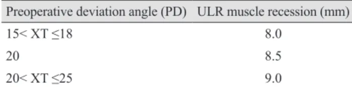
The postoperative changes in exodeviation following the first and second ULR muscle recessions for XT are Table 1. Surgical dosages of ULR muscle recession for pa- tients with XT and recurrent XT
Preoperative deviation angle (PD) ULR muscle recession (mm)
15< XT ≤18 8.0
20 8.5
20< XT ≤25 9.0
ULR = unilateral lateral rectus; XT = exotropia; PD = prism di- opter.
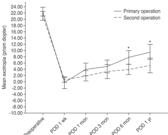
demonstrated and illustrated in Table 3 and Fig. 1. The postoperative exodeviation angles between the first ULR muscle recession and the second ULR muscle recession showed statistically significant differences at follow-up pe- riods of six months (7.84 ± 4.43 vs. 3.89 ± 3.47 PD), one year (9.58 ± 4.97 vs. 5.21 ± 4.94 PD), and at the last follow- up (21.11 ± 2.98 vs. 7.52 ± 4.06 PD) after surgery (p = 0.006, 0.013, and 0.000).
Postoperative exodrift was statistically different between the first and second ULR muscle recessions at three to six months (2.89 ± 3.75 vs. 0.63 ± 3.45 PD), and at one year to final follow-up (11.52 ± 5.50 vs. 2.32 ± 3.53 PD) (p = 0.034 and 0.000) (Table 4).
At the final postoperative follow-up, all of the first ULR muscle recession patients showed exodeviation of more
than 15 PD. Upon final follow-up, however, all of the second ULR muscle recession patients showed exodeviation of under 15 PD, with 12 patients (63.16%) showing exodeviation under 8 PD (Table 5).
Discussion
Corrective surgery for exodeviation should be considered if the binocular vision of a patient deteriorates [8], or if the patient demonstrates XT more than half of day, or if there is a problem with near fixation [9]. The results of surgery are affected by multiple factors, including patient age, preoperative deviation, the degree of stereopsis, and the surgical methods used to treat exodeviation [10].
Table 2. Preoperative patient characteristics in the first ULR muscle recession and second ULR muscle recession groups
Characteristics ULR muscle recession First (n = 19) Second (n = 19)
Male : female 11 : 8 11 : 8
Mean age at the time of
surgery (mon) 67.53 ± 20.27 113.11 ± 27.05 Mean follow-up period
(mon) 37.62 ± 10.04 49.76 ± 12.83
Preoperative deviation
angle at distance (PD) 22.42 ± 2.85 21.11 ± 2.98 ULR = unilateral lateral rectus; PD = prism diopter.
Table 3. Exodeviation after first ULR muscle recession and second ULR muscle recession
Period Exodeviation angle (PD) after
ULR muscle recession p-value
First Second
Preoperative 22.42 ± 2.85 21.11 ± 2.98 0.118 POD 1 wk -0.26 ± 3.91 0.53 ± 1.58 0.507 POD 1 mon 4.00 ± 4.11 1.89 ± 3.43 0.123 POD 3 mon 4.95 ± 4.13 3.26 ± 4.68 0.264 POD 6 mon 7.84 ± 4.43 3.89 ± 3.41 0.006* POD 1 yr 9.58 ± 4.97 5.21 ± 4.94 0.013* Last follow-up 21.11 ± 2.98 7.52 ± 4.06 0.000* Follow-up period
(mon) 37.62 ± 10.04 49.76 ± 12.83 - ULR = unilateral lateral rectus; PD = prism diopter; POD = post- operative day.
*p < 0.05.
24.00 22.00 20.00 18.00 16.00 14.00 12.00 10.00 8.006.00 4.002.00 -2.000.00 -4.00 -6.00 -8.00 -10.00
* *
Mean exotropia (prism diopter)
POD 1 yr POD 1 wk POD 1 mon POD 3 mon POD 6 mon Preoperative
Primary operation Second operation
Fig. 1. Average angle of exodeviation after first unilateral lateral rectus muscle recession and second unilateral lateral rectus mus- cle recession. I bars indicate 95% confidence intervals. POD = postoperative day. *p < 0.05.
Table 4. Exodrift after first ULR muscle recession and second ULR muscle recession
Follow-up period ULR muscle recession
p-value
First Second
1 wk-1 mon 4.26 ± 5.86 1.37 ± 3.45 0.139
1-3 mon 0.95 ± 2.78 1.36 ± 2.99 0.659
3-6 mon 2.89 ± 3.75 0.63 ± 3.45 0.034
6 mon-1 yr 1.74 ± 2.73 1.32 ± 2.91 0.619 1 yr-last follow-up 11.52 ± 5.50 2.32 ± 3.53 0.000 ULR = unilateral lateral rectus.
Jeong et al. [11]reported the success rate of unilateral recession and resection according to the degree of preoperative deviation in XT patients. The success rates of surgeries performed for patients with deviation angles under 30, 30 to 39, and over 40 PD were 67.5%, 54.0%, and 37.5%, respectively. Based on the findings of that study, the smaller the deviation angle is before surgery, the better the observed success rates are in patients with intermittent XT.
Wang and Nelson [12] demonstrated that the initial deviation angle is statistically related to the surgical success rate of ULR recession in XT patients with preoperative deviation angles of 15 to 35 PD.
Traditionally, XT surgery has been performed by bilateral lateral rectus recession or ULR recession and medial rectus resection. However, for exodeviation under 25 PD or less, ULR recession is often considered as a method of first surgery [4]. This is because ULR recession is effective and safe, while simultaneously preserving the other rectus muscle by only operating on one extraocular muscle. The one-muscle procedure also reduces surgery time and risks, such as sclera perforation and retinal detachment, which are possible during surgery. Among 56 patients with a moderate degree of exodeviation under 30 PD, 29 patients underwent ULR recession, and 27 underwent bilateral lateral rectus recession. There were no significant differences in the success rates between these two groups [5]. Weakley and Stager [2] suggested guidelines for ULR muscle recession. Comparing recurrent XT patients with exodeviation under 15 to 20 PD who underwent unilateral lateral recession as a first surgery with those who underwent it as second surgery, the authors reported that there was no significant difference in
the success rate between groups at 92.9% and 90.0%, respectively [13]. That study also recommends ULR surgery as a second surgery for patients with recurrent XT, but is different from our study in that its findings about the first surgeries for patients with recurrent XT are variable and inconsistent.
Despite the advantages of ULR recession, recurrence was frequently reported in the long-term observation of this study. Thus this study compares changes in deviation angles after first and second surgeries in patients who received ULR recession as a first surgery, and lateral rectus recession of the other eye as a second surgery. This study observes the tendency of exodeviation to recur after first and second surgeries over time, but the degree of recurrence and tendency of exodeviation to increase varies among patients. At three months after surgery, the characteristics of recurring exodeviation are apparently different, and after three, six, and 12 months, the degree of exodrift is significantly different between the first and second surgeries of patients. After the second surgery, any recurring exodeviation angles are smaller than the postoperative exodeviation angles from the first surgery, and the results are more stable. With an observation period of more than a year, we determined that 12 people out of 19 (63.16%) maintained exodeviation under 8 PD after their second surgeries.
The stability of ULR recession as a second surgery is attributable to several factors. The first factor is patient age at the time of surgery. At the time of the first and second surgeries, the mean patient age herein was 67.53 ± 20.27 and 113.11 ± 27.05 months, respectively, which are quite different from one another. Also, in the case of bilateral lateral rectus recession, recurrence rates are lower among older patients, which is clearly not related to any other factors [14]. Lower recurrence rates in older patients are similarly observed in ULR recession and medial rectus resection. After these surgeries (regarding both short-term and long-term results), recurrence tends to be more likely among patients with large angles of deviation [15]. There are no studies on the results of ULR recession recurrence, but based on the results of existing studies, we are able to attribute the lower recurrence rates to higher patient age.
The tendency of older patients to be more cooperative during preoperative examination may contribute to more accurate surgical outcomes, thereby impacting the lower recurrence rates. In addition, there is less likelihood among Table 5. Postoperative deviation angle of XT for first and sec-
ond ULR muscle recessions at last follow-up Postoperative deviation
angle (PD) ULR muscle recession
First Second
<8 0 12 (63.16)
8≤ XT <15 0 7 (36.84)
15≤ X <20 13 (68.42) 0
20≤ 6 (31.58) 0
Mean XT 21.11 ± 2.98 7.52 ± 4.06
Values are presented as number (%) or mean ± standard devia- tion.
XT = exotropia; ULR = unilateral lateral rectus; PD = prism di- opter; X = Exophoria.
older patients to return to exodeviation after a second surgery then after the first surgery. Second factor is because latent exodeviation appeared after the first surgery that doctors could examine the deviation angle more accurately before second surgery [7].
This study is limited insofar as it only studies patients who underwent ULR recession as a first surgery, and the same surgery in the other eye as a second surgery.
Consequently, we are unable to conclude that this surgery method is superior to other surgery methods as a second surgery. We recommend further research on ULR recession for a first surgery and medial rectus resection for a second surgery on the same eye in patients of comparable conditions. In terms of patient well-being, however, it is better to choose recession over resection, because the latter is more invasive and time consuming.
In conclusion, this study shows that exodrift changes after a second ULR muscle recession are less than exodrift changes after a first URL muscle recession in small-angle XT patients. Accordingly, a second ULR muscle recession may be a useful surgery for small-angle recurrent XT patients with deviation angles of 25 PD or less after a first ULR muscle recession. Therefore, when intermittent XT recurs after a patient’s first ULR recession, we recommend ULR recession for the patient’s other eye.
Conflict of Interest
No potential conflict of interest relevant to this article was reported.
References
1. Pukrushpan P, Isenberg SJ. Drift of ocular alignment fol- lowing strabismus surgery. Part 1: using fixed scleral su- tures. Br J Ophthalmol 2009;93:439-42.
2. Weakley DR Jr, Stager DR. Unilateral lateral rectus reces- sions in exotropia. Ophthalmic Surg 1993;24:458-60.
3. Olitsky SE. Early and late postoperative alignment follow- ing unilateral lateral rectus recession for intermittent exo-
tropia. J Pediatr Ophthalmol Strabismus 1998;35:146-8.
4. Wang L, Nelson LB. One muscle strabismus surgery. Curr Opin Ophthalmol 2010;21:335-40.
5. Spierer O, Spierer A, Glovinsky J, Ben-Simon GJ. Moder- ate-angle exotropia: a comparison of unilateral and bilater- al rectus muscle recession. Ophthalmic Surg Lasers Imag- ing 2010;41:355-9.
6. Deutsch JA, Nelson LB, Sheppard RW, Burke MJ. Unilat- eral lateral rectus recession for the treatment of exotropia.
Ann Ophthalmol 1992;24:111-3.
7. Sohn JH, Chang BL. Management for reoperation in recur- rent exotropia. J Korean Ophthalmol Soc 1994;35:580-4.
8. Wolff SM, Loupe DN. Binocularity after surgery for inter- mittent exotropia. In: Campos EC, editor. Strabismus and ocular motility disorder: proceedings of the 6th meeting of the International Strabismological Association; 1990;
Surfers Paradise, Australia. Hampshire: Macmillan Press;
1990. p. 375.
9. von Noorden GK, editors. Some aspects of exotropia. Pro- ceedings of Wilmer Resident's Association Meeting; 1966;
Baltimore, USA. Baltimore: Wilmer Resident's Association;
1966.
10. Kim SJ, Lee WS. Clinical analysis of surgical results in ex- odeviation. J Korean Ophthalmol Soc 1992;33:724-32.
11. Jeong TS, You IC, Park SW, Park YG. Factors of surgical success with unilateral recession and resection in intermit- tent exotropia. J Korean Ophthalmol Soc 2006;47:1987-92.
12. Wang L, Nelson LB. Outcome study of unilateral lateral rectus recession for small to moderate angle intermittent exotropia in children. J Pediatr Ophthalmol Strabismus 2010;47:242-7.
13. Lee K, Shin KS, Kim Y, Choi MY. Comparison of out- comes of unilateral lateral rectus recession for exotropia between first and second operations. Korean J Ophthalmol 2011;25:329-33.
14. Lim SH, Hwang BS, Kim MM. Prognostic factors for re- currence after bilateral rectus recession procedure in pa- tients with intermittent exotropia. Eye (Lond) 2012;26:846- 52.
15. Lim SH, Hong JS, Kim MM. Prognostic factors for recur- rence with unilateral recess-resect procedure in patients with intermittent exotropia. Eye (Lond) 2011;25:449-54.