Received: January 25, 2007 Accepted: August 17, 2007
Reprint requests to Young Suk Yu, MD. Department of Ophthal- mology, Seoul National University College of Medicine, 28 Yeongeon-dong, Jongno-gu, Seoul 110-744, Korea. Tel: 82-2-2072- 2438, Fax: 82-2-741-3187, E-mail: [email protected]
* The concept of this paper was reported at the 94th annual meeting of the Korean Ophthalmological Society, October 2005.
Visual Function after Primary Posterior Chamber Intraocular Lens Implantation in Pediatric Unilateral
Cataract: Stereopsis and Visual Acuity
Jung Hyun Park, MD,
1Young Suk Yu, MD,
1,2Jeong Hun Kim, MD,
1Seong Joon Kim, MD,
1,2Ho Kyung Choung, MD,
3Department of Ophthalmology, Seoul National University College of Medicine
1, Seoul, Korea Seoul Artificial Eye Center, Seoul National University Hospital Clinical Research Institute
2, Seoul, Korea
Seoul National University Boramae Hospital
3, Seoul, Korea
Purpose: To investigate the association between binocular function and vision after cataract removal and primary posterior chamber intraocular lens (PC-IOL) implantation in children with unilateral cataract and to identify visual function differences according cataract type.
Methods: Clinical records of 2- to 6-year-old patients with unilateral cataract removal and primary PC-IOL implantation were reviewed retrospectively. Visual acuity and ocular alignment were measured. Sensory fusion was assessed with the Worth 4-dot test, and stereoacuity with the Titmus stereo test. Cataracts were classified according to cause, lens opacity location, age at onset, and presence of strabismus. Clinical characteristics of patients who obtained good visual function were identified.
Results: Forty-seven patients were included. Among 22 (46.8%) with good vision (20/40 or better), only 6 (27.3%) achieved good binocular function (the presence of fusion and 100 seconds of arc or better of stereoacuity). Visual acuity was better in eyes with good binocular function (p=0.002). No other variables were significant for achieving good binocular function.
Conclusions: The removal of unilateral cataract in a visually immature child can result in a combination of good visual acuity and binocular function. Good binocular function is closely related to good visual acuity.
Korean Journal of Ophthalmology 21(4):195-200, 2007
Key Words: Cataract, Congenital anomaly, Intraocular lens, Visual acuity
Cataracts are a major cause of childhood blindness, with a prevalence of 1.2 to 6.0 cases per 10,000 births.
1Visual rehabilitation, especially in children with unilateral cataracts, remains challenging, and results are frequently disappointing.
Over the past several decades, visual outcomes after cataract extraction have dramatically improved due to earlier surgery, greater attention to aphakia optical correction with contact or intraocular lenses (IOL), and occlusion therapy.
2-6The development of useful binocularity has also been considered hard to achieve, but recent case reports and small case series describe patients with good central vision as well
as stereopsis or other evidence of binocularity.
7-10However, previous reports combined unilateral and bilateral cataract results,
10and studies on unilateral cataract only had small sample sizes.
7-8Furthermore, most studies included patients who underwent various treatment methods for aphakic eyes, such as contact lenses, epikeratophakia, and primary or secondary IOL implantation.
7-10The purpose of this study is to investigate the frequency of attaining good binocular function and vision after unilateral cataract removal and primary posterior chamber IOL implantation and to identify which children have the best opportunity for good visual function. The difference of visual function according to cataract type is also investigated.
Materials and Methods
A series of consecutive clinical records of 2- to 6-year-old
patients diagnosed with unilateral cataract who underwent
cataract removal and posterior chamber intraocular lens
(PC-IOL) implantation at Seoul National University Children’s
Congenital (n=14) Developmental (n=27) Traumatic (n=6)
Age at onset (mo) (median), (range) 43 (1-66) 48.4 (3-74) 56 (27-62)
BCVA (Snellen) (median), (range) 20/40 (CF-20/15) 20/50 (20/300-20/15) 20/30 (20/70-20/15)
Good VA (+/-) 7/7 11/16 4/2
Strabismus (+/-) 5/9 7/20 0/6
Good binocular function (+/-) 3/11 2/25 1/5
n = number, IOL = intraocular lens, BCVA = best corrected visual acuity, CF = counting fingers.
Good VA: 20/40 or better visual acuity on the Snellen chart.
Good binocular function: Fusion of Worth 4-dot test at near and distance and 100 sec of arc or better stereopsis.
Table 1. Characteristics according to cataract group Hospital between February 1995 and June 2004 were reviewed retrospectively. Patients with visual outcomes possibly predetermined by co-existing organic ocular defects were excluded; these defects included cataracts secondary to retinopathy of prematurity and those associated with dense corneal opacity and posterior-type persistent hyperplastic primary vitreous (PHPV).
Cataracts were classified by etiology as congenital, developmental, or traumatic. A congenital cataract was defined when the condition was detected before 3 months of age, with a consideration of opacity location, associated micro-ophthalmia or microcornea or the isolated presence of nuclear cataract. Cataracts were categorized by opacity location as nuclear, lamellar, posterior subcapsular, and mixed type. Age at onset was determined by pediatrician or ophthalmologist examination, parental observation, or date of ocular trauma. Ocular deviations were measured in prism diopters (PD). Children with visual axis deviation equal to or greater than 10PD and those with previous strabismus surgery were considered to have strabismus.
Sensory fusion was assessed by the Worth 4-dot test with red-green glasses. Near (1/3 meter) and distance (6 meters) fusion was measured. Stereoacuity was assessed by the Titmus stereo test. Near fusion and Titmus stereo test was performed with their near glasses or by adding +3.0 diopters trial lenses on the pseudophakic eyes. Binocular function was graded as “good” if there was distance and near fusion of the Worth 4-dot test, and if measured stereo acuity was 100 seconds of arc or better.
Visual acuity was measured with the fixation pattern or Snellen letters in preliterate and literate children, respectively.
The best-corrected visual acuity measured during binocular function test was noted. Visual acuity levels between 20/20 and 20/40 were considered “good.” During binocular function assessment, refractive error was also measured in manifest or cycloplegic manners. Patients with a refractive error difference of 2 diopters or larger were considered to have anisometropia.
Surgery was performed by a single surgeon (YS Yu). All procedures were performed under general anesthesia, including mechanical anterior capsulorrhexis and lens material aspiration by a mechanical irrigation-aspiration instrument. Posterior capsulectomy with anterior vitrectomy
was performed in children with posterior capsular opacities at the time of surgery. Children had simultaneous PMMA IOL implantation in the posterior chamber. IOL power was calculated using the SRK-II formula, with the goal of attaining emmetropia after the age of six. Between 2 and 5 years, the refractive goal was adjusted to allow 4.0D to 1.0D more of hyperopia.
Occlusion therapy was initiated when optics were sufficient to provide a clear retinal image. Recommended occlusion ranged from 1 to 6 hours a day and was prescribed according to patient age and the visual acuity level of the affected eye. Patching compliance was subjectively assessed as poor, moderate, or good by parental interviews on follow up.
Studied parameters were age at onset, sex, cataract type, opacity location, presence of strabismus pre- or postoperatively, anisometropia, and occlusion therapy compliance. The association of each independent variable with clinical outcome was estimated by chi-square test for discrete variables and student t test or Fisher’s exact test for continuous variables.
Results
Forty-seven patients were included in the study with 14, 27, and 6 congenital, developmental, and traumatic cataracts, respectively. Of the 27 eyes with developmental cataracts, 10 were posterior lenticonus and 2 were PHPV. Twenty-nine (61.7%) patients were male and 18 (38.3%) were female.
Mean age at onset was 47.7±18.9 months (3-71 months).
Mean age at cataract surgery was 53.2±15.6 months (24-72 months), with mean follow-up of 47.3±29.9 months (8-126 months).
Clinical characteristics of the different cataract groups are listed in Table 1. There were no significant differences in best corrected visual acuities, presence of strabismus, or good binocular function.
Overall, visual acuity was 20/40 or better in 22 (46.8%)
eyes, with 6 (27.3%) who achieved stereopsis of 100 seconds
of arc or less. Data from these 6 patients are listed in Table
2. One patient with good binocular function had exotropia,
but deviation was intermittent and measured 12PD at
distance.
Patient No
/Diagnosis Location of
opacity Age at onset
(months) Strabismus VA
operated eye VA
Fellow eye Stereopsis W4D (D/N)
1/congenital Nuclear 56 XT(12PD) 20/20 20/20 80 sec of arc 4/4
2/congenital Lamellar 64 none 20/40 20/15 100 sec of arc 4/4
3/congenital Nuclear 31 none 20/20 20/30 80 sec of arc 4/4
4/developmental Nuclear & PSC 22 none 20/25 20/20 100 sec of arc 4/4
5/developmental PSC 33 none 20/15 20/15 100 sec of arc 4/4
6/traumatic Nuclear 57 none 20/30 20/20 80 sec of arc 4/4
VA = visual acuity, W4D = worth 4 dots, D/N = distant/near, PSC = posterior subcapsular cataract, ASC = anterior subcapsular cataract, XT = exotropia, PD = prism diopters.
Table 2. Clinical characteristics of six patients who achieved good binocular function
Eyes with good VA
(n=22) (45.7 %) Eyes without good VA
(n=25) (54.3 %) P-value
Age at onset (mo) (mean±SD) 46.4±18.3 48.2±19.7 0.707
*Age at operation 52.0±15.9 53.9±15.6 0.692
*Age at examination 101.6±29.4 97.0±36.8 0.383
*Sex 1.000
†Male/female 14/8 15/10
Type of cataract 0.432
†Congenital 7 (31.8%) 7 (28.0%)
Developmental 11 (60%) 16 (64.0%)
Traumatic 4 (18.2%) 2 (8.0%)
Location of opacity 0.158
†Nuclear 6 (27.3%) 1 (4.0%)
Lamellar 2 (9.1%) 3 (12.0%)
Posterior subcapsular 9 (40.9%) 15 (60.0%)
Mixed 5 (22.7%) 6 (24.0%)
Anisometropia 0.754
†Anisometropia >2 diopters 6 9
Anisometropia ≤2 diopters 16 16
Strabismus, preoperatively 0.749
†Strabismus (+) 6 6
Strabismus (-) 16 19
Strabismus, postoperatively 0.747
†Strabismus (+) 5 (22.7%) 7 (28.0%)
Strabismus (-) 17 (77.3%) 18 (72.0%)
Compliance with Occlusion Therapy 1.000
†Good 8 13
Moderate or poor 4 8
*
independent t-test,
†chi-square test.
VA = visual acuity, IOL = intraocular lens, CL = contact lens.
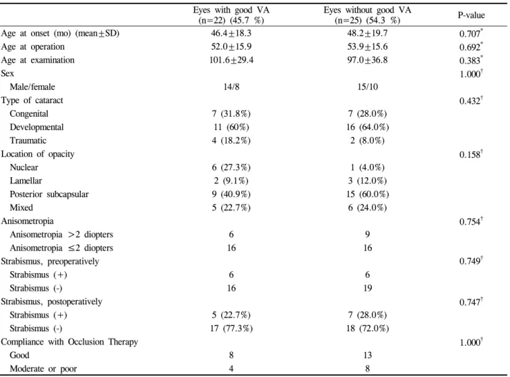
Table 3. Clinical and Ocular Variables in Patients with and without Good Visual Acuity Table 3 compares the characteristics of patients who
achieved or did not achieve good visual acuity, with univariate analysis results. Age at onset, cataract type, opacity location, presence of pre- or postoperative strabismus, and occlusion therapy compliance was not different between patients with or without good visual acuity.
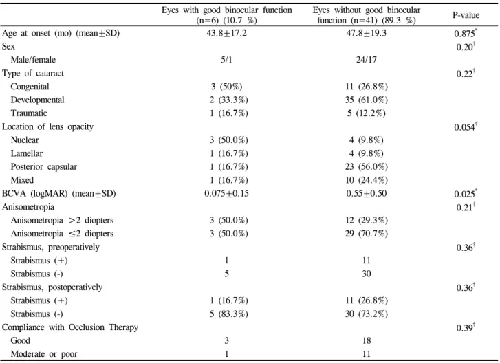
Table 4 compares characteristics of patients with or
without good binocular function with univariate analysis
results. In eyes with good binocular function, visual acuity
was better (p=0.025) compared with eyes without good
binocular function. No other variables were significant for
achieving good binocular function.
Eyes with good binocular function
(n=6) (10.7 %) Eyes without good binocular
function (n=41) (89.3 %) P-value
Age at onset (mo) (mean±SD) 43.8±17.2 47.8±19.3 0.875
*Sex 0.20
†Male/female 5/1 24/17
Type of cataract 0.22
†Congenital 3 (50%) 11 (26.8%)
Developmental 2 (33.3%) 35 (61.0%)
Traumatic 1 (16.7%) 5 (12.2%)
Location of lens opacity 0.054
†Nuclear 3 (50.0%) 4 (9.8%)
Lamellar 1 (16.7%) 4 (9.8%)
Posterior capsular 1 (16.7%) 23 (56.0%)
Mixed 1 (16.7%) 10 (24.4%)
BCVA (logMAR) (mean±SD) 0.075±0.15 0.55±0.50 0.025
*Anisometropia 0.21
†Anisometropia >2 diopters 3 (50.0%) 12 (29.3%)
Anisometropia ≤2 diopters 3 (50.0%) 29 (70.7%)
Strabismus, preoperatively 0.36
†Strabismus (+) 1 11
Strabismus (-) 5 30
Strabismus, postoperatively 0.36
†Strabismus (+) 1 (16.7%) 11 (26.8%)
Strabismus (-) 5 (83.3%) 30 (73.2%)
Compliance with Occlusion Therapy 0.39
†Good 3 18
Moderate or poor 1 11
*