대한치주과학회지 : Vol. 35, No. 4, 2005
마그네슘 티타네이트 표면의 조골세포 부착도와 분화
최승민1ㆍ이재관1ㆍ고성희2ㆍ엄흥식1ㆍ장범석1
1강릉대학교 치과대학 치주과학교실
2강릉대학교 치과대학 약리학교실
Ⅰ. 서론
Brånemark에 의해 개발된 골유착 임플란트1)는 장기적으로 높은 성공률을 보였으나, 골질이 나쁜 경우 기계절삭형 임플란트는 낮은 성공률을 보였고, 이를 보완하기 위해 다양한 표면처리 방법에 대한 연구들이 진행되었다.1)
일반적으로 티타늄 금속은 실온의 공기 중에서 자 연적으로 1.5~10nm의 표면 산화막으로 덮히게 된 다.2)기계절삭형 티타늄 임플란트는 이 산화막이 대 부분 TiO2로 구성된다.3-4) 이러한 얇은 표면 산화막 은 전기전도성이 낮고5), 열역학적 안정성이 높으며
6-7), 수성환경에서 이온형성 경향이 낮아8), 부식에 대한 저항성이 높다. 이러한 산화막의 성질로 인하여 티타늄 임플란트는 다른 금속에 비해 생체친화성이 우수하다.9)
하지만 티타늄 임플란트에 자연적으로 형성되는 얇은 산화막은 우수한 생체적합성에도 불구하고 좀 처럼 골조직과 직접적인 화학적 결합을 형성하지 않 아 종종 비활성의 세라믹 생체적합물질로 정의되기
도 한다.10)
기계절삭형 임플란트는 골질이 나쁜 부위에서 성 공률이 현저하게 떨어지기 때문에 티타늄 임플란트의 골 반응을 개선하기 위해 다양한 표면처리 기술들이 개발되었다. 최근에 개발되어 임상적으로 사용되는 치과 임플란트들은 표면의 기하학적 개선에 초점을 맞추어왔다.11-13) 이러한 시도는 임플란트의 표면이 거칠어지면 골조직과 임플란트 사이에 기계적 결합이 이루어질 수 있다는 가정에 그 기초를 두고 있다.
표면 조도의 증가는 조골세포의 부착을 향상시키 며14), 골세포가 성장하였을 때, 보다 성숙한 표현형 을 나타낼 수 있도록 해준다.15)표면 조도가 골모세 포의 증식과 분화, 그리고 골 형성에 관여하는 국소 인자들의 생산에 영향을 주며17), 동물실험에서도 표 면 조도를 높였을 경우 더 우수한 골 반응을 나타낸 다는 것이 조직학적으로 입증되었다.17-19)
다른 한편으로는 티타늄 임플란트의 표면을 유리, 인산칼슘, 세라믹, 수산화인회석 등으로 처리하여 임플란트의 화학적 성질을 개선하여 생활성 물질로 만들려는 다양한 표면 처리 기술들이 개발되었다
*이 논문은 2005년도 강릉대학교 학술연구조성비 지원에 의하여 수행되었음
*교신저자 : 장범석, 강원 강릉시 강릉대학로 120번지 강릉대학교 치과대학 치주과학교실, 우편번호 : 210-320 E-mail : [email protected], 전화번호 : 033-640-3150
21-27). 하지만 임플란트 표면에 생활성 물질을 코팅하 는 경우 이러한 코팅 재료가 골조직과 결합하는 과정 에서 생리적 반응에 의해 탈락하는 문제가 발생하게 된다.31) 몇몇 동물실험과 임상실험의 결과에 의하면, 코팅 재료의 박리현상과 생 흡수 현상은 코팅물질 내부 혹은 계면층에서 일어나고 골조직과 결합하는 생리적 반응에 의해 발생하였다.29-31)
최근에 발표된 몇몇 생체 실험은 티타늄 임플란트 의 산화막 성질 변형이 골조직 반응을 향상시킬 수 있다는 결과를 보여주었다.32-48) 표면의 산화막 두께 가 600 ㎚ 이상인 다공성 표면구조를 가진 양극산화 티타늄 임플란트의 경우 기계절삭형 티타늄 임플란 트에 비해 동물실험에서 매우 좋은 결과를 보여 주 었다.44-46)
양극산화법은 티타늄 표면에 존재하는 산화막을 보다 균일하고, 두껍고, 치밀하게 만들어주며, 높은 전압을 가했을 경우에는 표면에 분화구 형태의 다공 성 구조를 형성해 준다. 양극산화에 의해 형성된 다 공성 구조는 임플란트 식립 시 골과의 접촉면적을 증가시켜줄 뿐 아니라 골 단백질과 같은 골 재생에 관여하는 인자들을 골 내에 장기간 전달할 수 있는 전송수단으로 이용할 수 있다. 티타늄에 대한 양극 산화는 전해질의 종류, 전해질의 농도, 전해질의 온 도, 상대 음극의 종류, 전류의 양 등 여러 가지 요소 들에 의해 다른 결과를 나타낸다.
Sul 등은 이러한 양극산화 티타늄 임플란트의 긍 정적인 골 반응에 기초하여 티타늄 표면 성분을 다 양하게 변형하여 비교하였다. 양극산화 시 각각 다 른 이온들(Ca, S, P)을 포함한 전해질 용액을 사용하 여 산화층의 성질을 변형한 임플란트에 대한 골조직 반응을 비교한 이 연구에서 Ca와 P 이온이 포함된 임플란트의 경우 골과 티타늄 임플란트 사이의 생화 학적 결합이 이루어졌을 가능성이 제시되었다.49)
이 연구의 목적은 마그네슘 이온을 첨가한 전해질 용액으로 양극산화를 시행하여 제작한 마그네슘 티 타네이트 표면과 기계절삭형 티타늄 시편에 조골세 포양 세포를 배양하여 세포 부착, 증식 및 분화를 비 교, 평가하기 위한 것이다.
Ⅱ. 실험재료 및 방법
1. 실험재료
직경 10 mm, 두께 1 mm의 티타늄 디스크를 300번과 800번 실리콘 카바이드 사포를 사용하여 균일한 표면을 갖도록 만들었다. 그리고 세정용액에 넣고 초음파 세척한 후 50℃ 오븐에서 24시간 동안 건조 시켰다. 표본들을 각각 유리 튜브에 넣고 trichlorethylene과 순수 에탄올을 각각 사용한 초 음파 세척을 15분간 2회 시행하고 50℃ 오븐에서 24시간 동안 건조한 후 멸균였다. 실험군은 멸균 전 마그네슘 이온이 포함된 전해질 시스템에서 높은 전 압과 전류의 galvanostatic mode로 양극산화시킨 후 멸균시킨 시편으로 하였고, 양극산화 과정을 거 치지 않은 시편을 대조군으로 설정하였다.
2. 세포배양방법
세포는 사람 골육종에서 부터 확립된 조골세포양 세포주인 Saos-2 세포를 한국세포주은행(서울, 대한 민국)으로부터 구입하여 사용하였다. Saos -2 세포 를 100mm 크기의 세포배양접시(Coring Co. Coring, NY, USA)에 10% fetal bovine serum (Hyclone Laboratories, Inc., Logan, UT, USA)과 100 U/㎖의 penicillin, 100 ㎍/㎖의 streptomycin이 함유된
⍺-MEM(Invitrogen, Gibco brand, Grand Island, NY, USA)을 이용하여, 공기혼합배양기(37℃, 100%
습도, 5% CO2)에서 배양하였다.
3. 세포부착 및 세포증식능 측정
배양 1일, 6일째에 0.25% trypsin, 4 mM EDTA 혼합용액(Invitrogen, Gibco brand)을 넣고 37℃에서 5분간 배양하여 부착된 세포를 시편으로 부터 떼어낸 후 tryphan blue(Invitrogen, Gibco
brand)로 염색하고 혈구계수기로 염색되지 않은 세
포의 수를 측정하였다.
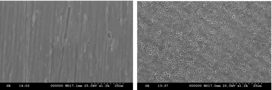
Figure 1. SEM pictures, machined surface(left), magnesium titanate surface at
×1200(right)
4. 알칼리성인산분해효소의 활성도 측정
배양 1일과 6일째에 0.1% Triton X-100이 포함 된 10mM Tris-HCl 용액(pH7.4)과 cell scra- per 를 사용하여 세포를 모으고 얼음 위에서 초음파 분쇄 기를 이용하여 1분간 초음파 처리하여 일부는 알카 리성인산분해효소활성 측정에 일부는, 단백질 정량에 사용하였다. 알카리성인산분해효소활성은 0.05 M glycin-NaOH buffer(pH10.3)를 완충액으로 사용하 고, 15mM
p
-nitrophenol phosphate(p-NPP)(Sig- ma-Aldrich, Saint Louis, Mo, USA)를 효소 기질로 첨가하여 37℃에서 30분 반응시킨 후 0.1 N NaOH 로 반응을 중지시키고, 효소작용에 의해서 기질로부 터 유리된p
-nitrophenol을 410nm에서 spectro- photometer(amersham pharmacia biotech, Cam- bridge, England)로 비색정량하였다. 총 단백질량은 bovine serum albumin을 표준용액으로 사용하여 BCA protein assay reagent(Pierce Inc, Rockford, IL)로 정량하였다. 알칼리성인산분해효소의 활성도는 nmol of pNP/min/㎍ of protein으로 표기하였다.5. 통계학적 분석
초기부착, 세포증식능 그리고 알칼리성인산분해효 소의 활성을 SPSS 12.0을 사용하여 통계처리 하였 다. 실험군과 대조군 간의 세포증식능 및 알칼리성 인산분해효소 활성도를 t-test를 이용하여 검증하였 다(p < 0.05).
Ⅲ. 결과
1. 주사전자현미경 관찰
주사전자현미경 관찰에서 마그네슘 티타네이트 시 편의 표면은 특징적인 다공성 구조로 이루어져 있는 것을 확인할 수 있었다(Figure. 1 right).
2. 세포부착 및 증식
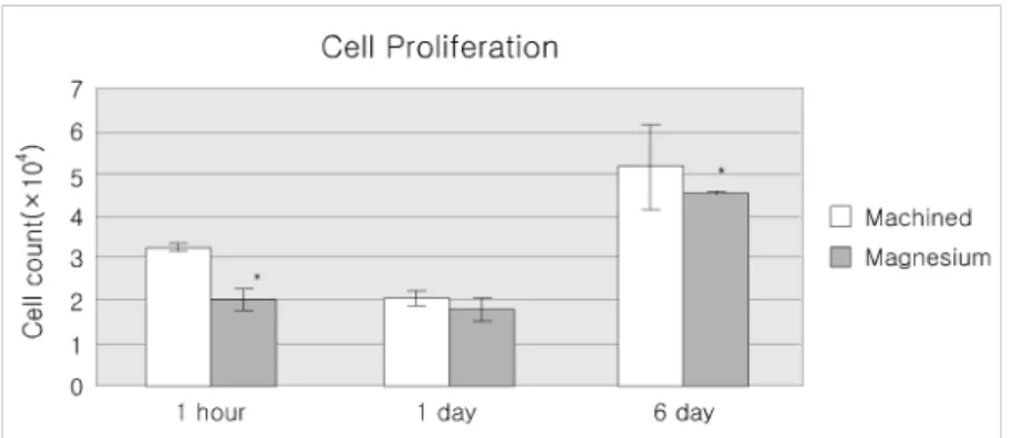
배양 1시간, 1일 및 6일째 세포를 떼어낸 후 혈구 계수기로 세포수를 측정하였다. 배양 1시간째 대조군 인 기계절삭형 시편에서 실험군인 마그네슘 티타네이 트에 비해 통계적으로 유의하게 많은 수의 세포를 관 찰할 수 있었고(p < 0.05), 배양 1일째에는 대조군과 실험군이 통계적으로 유의한 차이를 보이지 않았으 며, 배양 6일째에는 대조군의 세포 수가 실험군에 비 해 통계적으로 유의하게 많았다(p < 0.05)(Table 1, Figure 2).
3. 알칼리성인산분해효소의 활성도
배양 1일째의 활성도는 대조군에서 다소 높게 나 타났으나 통계적으로 유의한 수준은 아니었다. 배양 6일째의 활성도는 대조군에 비해 실험군인 마그네슘 티타네이트에서 통계적으로 유의하게 높은 수치를 보였다(p < 0.05)(Table 2, Figure 3).
Table 1. Cell adhesion and proliferation (×104 cell count)
1 hour 1 day 6 day
Machined 3.241 ± 0.080 2.058±0.162 5.133±0.973
Magnesium 2.027 ± 0.240* 1.790±0.267 4.519±0.026*
Values are Mean ±S.D (n=4)
* Statistically significant difference at the p = 0.05
Figure 2. Cell adhesion and proliferation(×104 cell count)
Table 2. Alkaline phosphatase activity (nmole/㎍/min)
1 day 6day
Machined 9.222±0.706 13.324±0.704
Magnesium 7.566±0.606 16.499±1.241*
Values are Mean ±S.D (n=4)
* Statistically significant difference at the p = 0.05
Figure 3 Alkaline phosphatase activity (nmole/μg/min)
Ⅳ. 총괄 및 고찰
이 연구에서 양극산화법을 이용하여 제작한 마그네 슘 티타네이트에서 배양한 조골세포양 세포는 기계절 삭형 티타늄에서 배양한 세포와 증식과 분화에 차이 가 있다는 것을 확인하였다.
초기에 세포 부착을 평가하기 위해 배양 후 1시간 째에 세포수를 측정하였으며, 이후 세포의 증식을 평가하기 위해 배양 1일과 6일째에 세포수를 측정하 였다. 초기에는 상대적으로 매끈한 표면을 가진 기 계절삭 표면에서 더 많은 수의 세포가 관찰되었으나, 1일째의 결과에서는 통계적으로 유의한 차이를 보이 지 않았는데 이는 초기에 부착되었던 세포들 중에서 일부는 살아남지 못하고 증식과정 중 탈락했기 때문 인 것으로 생각된다. 그리고 배양 6일째는 마그네슘 티타네이트에 비해 기계절삭 표면에서 조골세포의 증식이 더 많이 일어난 것으로 관찰되었다.
조골세포의 분화 정도를 평가하기 위해 1일과 6 일째 알칼리성인산분해효소의 활성을 평가하였다.
세포의 초기 부착이나 증식의 결과와는 달리 배양 6 일째 마그네슘 티타네이트 시편의 세포에서 더 높은 수준의 알칼리성인산분해효소 활성도를 나타내고 있 었다. 즉 마그네슘 티타네이트 시편에서 성장한 Saos-2 세포가 더 높은 수준으로 분화하였다는 것 을 의미한다.
표면의 화학적인 특성과 더불어 골과의 반응에서 중요한 것이 표면의 물리적 특성, 즉 표면 조도이다.
여러 가지 방법을 이용하여 표면 조도에 변화를 주 는 이유는 골과 임플란트 사이의 기계적인 상호고정 효과를 증진시키기 위해서이고, 또한 임플란트 표면 에서 골이 형성되는 과정에 표면 조도가 크게 영향 을 주기 때문이다.
티타늄 임플란트의 골 반응을 향상시키기 위해 표 면의 기하학적 개선 방법들이 소개되었고 동물 실험 에서도 우수한 결과를 얻었다17-19). 평균 표면 조도 (Sa)가 1.5 μm 일때 가장 강력한 골 반응이 나타났 으며, 더 매끄럽거나 거친 경우에는 더 낮은 골 반응 이 나타났다.50)
현재까지 표면 조도에 변화를 주기 위해 티타늄 피복법, 티타늄 분사법, 산부식법, 분사-산부식법, 양극산화법 등의 방법들이 이용되고 있다. 그러나 피복법의 경우에는 임플란트를 식립하는 도중 피복 된 입자들이 골과의 밀착에 의해 마모되어 떨어져 나가거나 시간경과에 따라 임플란트 표면으로부터 분리되는 현상이 발생하였고29-31), 분사법의 경우에 는 분사에 사용한 금속 입자가 표면에 잔류하거나 임플란트 표면 성질에 변화를 야기하므로 골반응에 문제를 야기할 수 있었다.51,52)
임플란트 표면의 형태 및 화학적 성질을 개선하는 데 있어 양극산화법은 간단하고, 조절가능하며 비용 대비 효과적인 방법이다. 또한 양극산화법을 이용하 면 복잡한 형태의 임플란트에서 가장 이상적인 형태 의 산화막을 만들 수 있다. 산화막의 두께, 미세 구 조, 조도 그리고 칼슘이나 마그네슘과 같은 이온의 농도는 양극산화 시 전압, 전류, 반응시간 그리고 전 해질의 종류 및 농도를 변화시킴으로써 조절할 수 있다.42,43)
이전의 연구결과 최적의 생체 골조직 반응은 티타 늄 임플란트의 표면 산화막 두께가 600~1500 nm 이며, 다공성 구조로 12~24% 공극률을 가지며, 순 수한 티타늄 옥사이드(TiO2) 보다는 칼슘 티타네이 트(Ti2CaO3) 혹은 마그네슘 티타네이트(MgTiO3)와 같은 생활성 화합물의 경우에서 더 우수한 것으로 알려져 있다.44-49)
마그네슘 티타네이트 임플란트의 표면 화학적 성 질은 임플란트 안정성과 결합강도에 있어 중요한 향 상을 가져올 수 있다. 마그네슘은 2가의 양이온상태 에서 가장 안정적이다. Mg2+, Ca2+는 세포 표면 수 용기의 integrin superfamily와 fibronectin, vitronectin, fibrinogen 등의 ligand protein의 결합 작용에 있어 중요한 역할을 하는 것으로 알려 져 있다.50-53)이런 integrin-ligand 상호작용이 세 포의 부착, 이주, 증식 그리고 분화를 조절하게 되는 것이다.
마그네슘 이온의 이동과 이온 교환 반응이 inte- grin-ligand 상호작용을 촉진하는 중요한 추진력이
되고 그 결과 향상된 골 반응으로 마그네슘 티타네 이트 임플란트의 생화학적 결합이 가능할 수 있게 되는 것이다. 또한 다공성의 표면을 지니고 있어 표 면의 미세한 공간 내부로 골조직의 성장을 유도하게 된다.
골세포 배양 모델은 골과 생체적합물질의 상호작 용을 연구하는데 있어 그 사용이 증가하고 있다. 대 부분의 배양은 조골세포를 이용해 왔다.54) 이러한 시험관 모델은 생체 내의 환경을 그대로 재현할 수 는 없으나 다양한 생체적합물질의 계면에서 조골세 포의 부착과 세포 간질의 합성에 대해 형태학적, 생 화학적, 분자적인 정보를 제공하여 초기의 골과 임 플란트 계면에서 발생하는 골유착 현상을 이해하는 데 도움을 준다.55) 인간의 골육종으로부터 얻어진 Saos-2 세포는 골 형성을 유발할 수 있는 고분화된 세포주로서 골세포의 반응을 연구하는데 있어 좋은 모델인 것으로 알려져 있다.
알칼리성인산분해효소는 골 생성을 위한 세포 분 화에 있어 초기의 지표가 된다. 이 효소의 활성도는 모든 세포에 존재하지만 골모세포와 같은 주위 기질 을 광화시키려는 세포에서 높은 수준으로 나타나게 된다.56) 조골세포가 성숙해 질수록 그들이 생산하는 세포외 기질 소포에는 알칼리성인산분해효소의 활성 도가 높아지게 된다. 이러한 특징적인 증가 양상으 로 인해 알칼리성인산분해효소는 세포외 기질의 지 표효소가 된 것이다.57) 기질 소포는 광화과정의 시 작과 관련이 있고, 그 내부에는 기질에서 결정의 침 착과 성장에 필요한 효소들이 포함되어 있다.58)
Ⅴ. 결론
마그네슘 티타네이트 표면이 조골세포의 증식 및 분화에 미치는 영향을 알아보기 위하여 기계절삭형 표면 시편을 대조군으로 하여 마그네슘 티타네이트 시편에서 조골세포양세포인 Saos-2 세포의 부착, 증식 및 알칼리성인산분해효소의 활성도를 관찰하여 다음과 같은 결과를 얻었다.
1. 배양 후 1시간에 측정한 초기 세포 부착은 대 조군에 비하여 마그네슘 티타네이트 표면에서 유의하게 낮았다.
2. 1일째와 6일째의 세포증식도는 대조군에 비하 여 마그네슘 티타네이트 표면에서 유의하게 낮 았다.
3. 6일째 알칼리성인산분해효소의 활성도는 마그 네슘 티타네이트 표면에서 유의하게 높게 나타 났다.
이 실험의 결과에서 마그네슘 티타네이트 표면이 기계절삭형 티타늄 표면에 비해 조골세포의 분화를 촉진시키는 것으로 나타났으나, 대조군으로 기계절 삭형 표면을 가진 티타늄만을 비교하였기 때문에 아 직 마그네슘 티타네이트의 조골세포에 대한 효과를 확신할 수는 없다. 최근 생산되는 임플란트들에서 사용되고 있는 여러 표면처리 방법들과의 비교 연구 그리고 동물 실험을 통한 연구가 추가적으로 필요할 것이다.
Ⅵ. 참고문헌
1. Brånemark PI, Hansson BO, Adell R, Breine U, Lindström U, Hallén O, Öman A. Osseointegrated implants in the treat- ment of the edentulous jaw. Experience from a 10-year period. Scand J Plast Reconstr Surg 1977;16(Suppl. 1):7-127.
2. Kasemo B, Lausmaa J. Aspect of surface physics on titanium implants. Swed Dent J 1983;28(Suppl.):19-36.
3. Lausmaa J, Kasemo B. Surface spectros- copic characterization of titanium implant materials. Appl Surf Sci 1990;45:133-46.
4. Olefjord I, Hansson S. Surface analysis of four dental implant systems. Int J Oral Maxillofac Impl 1993;8:32-40.
5. Zitter H, Plenk HJ. The electrochemical
behaviour of metallic implant materials as indicator of their biocompatibility. J Biomed Mater Res 1987;21:881-96.
6. Williams DF. Corrosion of implant mate- rials. Ann Rev Mater Sci 1976;6: 237-65.
7. Solar RJ, Pollack SR, Korostoff E. In vitro corrosion testing of titanium surgical implant alloys: an approach to understan- ding titanium release from implants. J Biomed Mater Res 1979;13:217-50.
8. Tengvall P, Lundström I. Physicoche- mical considerations of titanium as a biomaterial. Clin Mater 1992;9:115-34.
9. Johansson CB. On tissue reactions to metal implants. Thesis, University of Göteborg, Sweden, 1991.
10. L J, Hastings GW. Oxide ceramics:
inert ceramic materials in medicine and dentistry. In: Black J, Hastings G, editors. Handbook of biomaterial pro- perties. London: Chapman & Hall;
1998. p. 340-354.
11. Deportet D, Watson P, Pharoah M, Levy D, Todescan R. Five to six-year results of a prospective clinical trial using the ENDOPORE dental implant and a man- dibular overdenture. Clin Oral Implants Res 1999;10:95-102.
12. Buser D, Nydegger T, Oxland T, Co- chran DL, Schenk RK, Hirt HP, Snetivy D, Nolte LP. Interface shear strength of titanium implants with a sandblasted and acid-etched surface: a biomecha- nical study in the maxilla of miniature pigs. J Biomed Mater Res 1999;45:
75-83.
13. Palmer RM, Palmer PJ, Smith BJ. A 5-year prospective study of astra single
tooth implants Clin Oral Implants Res 2000;11:179-182.
14. Testori T, Wiseman L, Woolfe S, Porter SS. A prospective multicenter clinical study of the osseotite implant: four-year interim report. Int J Oral Maxillofac Implants 2001;16:193-200.
15. Bowers, K.T., Keller, J.C. Randolph, B.A., Wick, D.G., Michaels, C.M. Opti- mization of surface micromorphology for enhanced psteoblast response in vitro.
Int J Oral Maxillofac Implants 1992;
7(3):302-310.
16. Kieswetter, K, Schwartz, Z, Dean, D.D., Boyan, B.D.:The role of implant surface characteristics in the healing of bone.
Cret. Rev. oral Biol. Med. 1996;7(4):
329-345.
17. Lohmann, C.H., Sagun, R. Jr., Sylvia, V.L., Cochran, D.L., Dean, D.D., Boyan B.D., Schwartz, Z. Surface roughness modulates the response of MG63 osteo- blast-like cells to 1,25-(OH)(2)D(3) through regulation of phospholipase A (2) activity and activation of protein kinase A.J. Biomed. Mater. Res., 1999;
47(2):139-151.
18. Ericsson, I., Johansson, C.B., Bystedt, H., Norton, M.R. A histomorphometric evaluation of bone-to-implant contact on machine prepared and roughened tita- nium dental implants. A pilot study in the dog. Clin. Oral Implants Res. 1994;
5(4):202-206.
19. Larsson, C., Thomsen, P., Lausmaa, J., Rodahl, M., Kasemo, B., Ericson, L.E.
Bone response to surface modified tita- nium implants : studies on electropo-
lished imlants with different oxide thicknesses and morphology. Bioma- terials 1994;15(13):1062- 1074.
20. Wennerberg, A., Ektessabi, A., Albrekt- sson, T., Johansson, C., Andersson, B.
A 1-year follow up of implants of differing surface roughness placed in rabbit bone. Int. J. Oral Maxillofac.
Implants 1997;12(4):486-494.
21. Jansen JA, Wolke JG, Swann S, Van der Waerden JP. Application of magnet- ron sputtering for producing ceramic coatings on implant materials. Clin Oral Implants Res 1993;4:28-34.
22. Ishizawa H, Ogino M. Formation and characterization of anodic titanium oxide films. J Biomed Mater Res 1995;
29:65-72.
23. Ektessabi AM. Ion beam processing of bio-ceramics. Nucl Instrum Methods Phys Res B 1995;99:610-3.
24. Campbell PA, Gledhill HC, Brown SR, Turner IG. Vacuum plasma sprayed hydroxyapatite coatings on titanium alloy substrates: surface characteriza- tion and observation of dissolution pro- cesses using atomic force microscopy. J Vac Sci Technol B 1996;14:1167-72.
25. Haddow DB, James PF, Van Noort R.
Sol.gel derived calcium phosphate coa- tings for biomedical applications. J Sol.
Gel Sci Technol 1998;13:261-5.
26. Burgess AV, Story BJ, La D, Wagner WR, LeGeros JP. Highly crystalline Mp-1(TM) hydroxylapatite coating Part I: in vitro characterization and com- parison to other plasma-sprayed hydro- xylapatite coatings. Clin Oral Implants
Res 1999;10:245-56.
27. Lo WJ, Grant DM, Ball MD, Welsh BS, Howdle SM, Antonov EN, Bagratashvili VN, Popov VK. Physical, chemical, and biological characterization of pulsed laser deposited and plasma sputtered hydroxyapatite thin films on titanium alloy. J Biomed Mater Res 2000;50:
536-45.
28. Hench LL, Kokubo T. Properties of bioactive glasses and glassceramics.
Handbook of biomaterial properties. In:
Black J, Hastings G, editors. Handbook of biomaterial properties. London:Chap- man & Hall; 1998. p. 355-63.
29. Gottlander M. On hard tissue reactions to hydroxyapatite-coated titanium im- plants. Ph.D. thesis, Department of Biomaterials/Handicap Research, Uni- versity of G. oteborg, Sweden, 1994.
30. Albrektsson T. Hydroxyapatite-coated implants: a case against their use. J Oral Maxillofac Surg 1998;56:1312-26.
31. Ong JL, Chan DCN. Hydroxyapatite and their use as coatings in dental im- plants: a review. Crit Rev Biomed Eng 2000;28:667-707.
32. Ishizawa H, Fujino M, Ogino M. Mecha- nical and histological investigation of hydrothermally treated and untreated anodic titanium oxide .lms containing Ca and P. J Biomed Mater Res 1995;
29:1459-68.
33. Larsson C, Emanuelsson L, Thomsen P, Ericson L, Aronsson B, Rodahl M, Kasemo B, Lausmaa J. Bone response to surfacemodi .ed titanium implants:
studies on the tissue response after one
year to machined and electropolished implants with different oxide thick- nesses. J Mater Sci Mater Med 1997;
8:721-9.
34. Hanawa T, Kamiura Y, Yamamoto S, Kohgo T, Amemiya A, Ukai H, Mura- kami K, Asaoka K. Early bone formation around calcium-ion-implanted titanium inserted into rat tibiae. J Biomed Mater Res 1997;36:131-6.
35. Ichikawa T, Hanawa T, Ukai H, Murakami K. Three-dimensional bone response to commercially pure titanium, hydroxyapatite, and calcium-ion-mixing titanium in rabbits. Int J Oral Maxillofac Implants 2000;15:231-8.
36. Yan WQ, Nakamura T, Kobayashi M, Kim HM, Miyaji F, Kokubo T. Bonding of chemically treated titanium implants to bone. J Biomed Mater Res 1997;
37:267-75.
37. SkripitzR, Aspenberg P. Tensile bond between bone and titanium. Acta Or- thop Scand 1998;69:2-6.
38. Fini M, Cigada A, Rondelli G, Chiesa R, Giardino R, Giavaresi G, Aldini N, Torricelli P, Vicentini B. In vitro and vivo behaviour of Ca and P-enriched anodized titanium. Biomaterials 1999;20 :1587-94.
39. Nishiguchi S, Nakamura T, Kobayashi M, Kim HY, Miyaji F, Kokubo T. The effect of heat treatment on bone- bon- ding ability of alkali-treated titani- um.
Biomaterials 1999;20:491-500.
40. Henry P, Tan AE, Allan BP. Removal torque comparison of TiUnite and turned implants in the Greyhound dog
mandible. Appl Osseointegration Res 2000;1:15-17.
41. Albrektsson T, Johansson C, Lundgren AK, Sul YT, Gottlow J. Experimental studies on oxidized implants. A histo- morphometrical and biomechanical analysis. Appl Osseointegration Res 2000;1:21-24.
42. Sul YT, Johansson CB, Jeong Y, Albrektsson T. The electrochemical oxide growth behaviour on titanium in acid and alkaline electrolytes. Med Eng Phys 2001;23:329-46.
43. Sul YT, Johansson CB, Petronis S, Krozer A, Jeong Y, Wennerberg A, Albrektsson T. Characteristics of the surface oxides on turned and electro- chemically oxidized pure titanium im- plants up to dielectric breakdown: the oxide thickness, micropore con.gura- tions, surface roughness, crystal struc- ture and chemical composition. Bioma- terials 2002;23:491-501.
44. Sul YT, Johansson CB, Jeong Y, Wennerberg A, Albrektsson T. Reso- nance frequency and and removal torque analysis of implants with turned and anodized surface oxide. Clin Oral Im- plants Res 2002;13:252-259.
45. Sul YT, Johansson CB, R.oser K, Al- brektsson T. Qualitative and quan- titative observations of bone tissue reactions to anodized implants. Bioma0 terials 2002;23:1809-19.
46. Sul YT, Johansson CB, Jeong Y, R. oser K, Wennerberg A, Albrektsson T. Oxi- dised implants and their influence on the bone response. J Mater Sci Mater
Med 2001;12:1025-31.
47. Sul YT, Johansson CB, Kang YM, Jeon DG, Albrektsson T. Bone reactions to oxidized titanium implants with electro- chemically anion S and P incorporation.
Clin Implant Dent Relat Res 2002;
4:478-87.
48. Sul YT, Johansson CB, Albrektsson T.
Oxidized titanium screws coated with calcium ions and their performance in rabbit bone. Int J Oral Maxillofac Implants 2002;17:625-634.
49. Sul YT. The significance of the surface properties of oxidized titanium to the bone response: special emphasis on potential biochemical bonding of oxi- dized titanium implant. Biomaterials 2003;24:3893-3907.
50. Kwon, S.Y., Takei, H., Pioletti, D.P., Lin, T., Ma, Q.J., Akeson, W.H., Wood, D.J., Sung, K.L. Titanium particles inhibit osteoblast adhesion to fibronec- tin-coated substrates. J. Orthop. Res.
2000; 18(2):203-211.
51. Gailit J and Ruoslahti E. Regulation of the fibronectin receptor affinity by diva- lent cations. J Biol Chem 1988;263:
12927-12932.
52. Mould AP, Akiyama SK, and Humphries MJ. Regulation of integrin alpha 5 beta 1-fibronectin interactions by divalent cations. Evidence for distinct classes of binding sites for Mn2+, Mg2+, and Ca2+. J Biol Chem 1995:270;26270-26277.
53. Krause A, Cowles EA, Gronowicz G.
Integrin-mediated signaling in osteo-
blasts on titanium implant materials. J Biomed Mater Res 2000:52; 738-747.
54. Cooper LF, Masuda T, Yliheikkila PK, Felton DA. Generalizations regarding the process and phenomenon of osseo- integration. Part II. In vitro studies. Int J Oral Maxillofac Implants 1998;13:
163-74.
55. Davies JE. In vitro modeling of the bone /implant interface. Anat Rec 1996;245:
426-45.
56. Owen TA, Aronow M, Shalhoub V, Barone LM, Wilming L, Tassinari MS, Kennedy MB, Pockwinse S, Lian JB, Stein GS. Progressive development of the rat osteoblast phenotype in vitro:
reciprocal relationships in expression of genes associated with osteoblast proli- feration and differentiation during for- mation of the bone extracellular matrix.
J Cell Physiol 1990;143:420- 30.
57. Ali SY. Mechanisms of calcification. In : Owen R, Goodfellow J, Bollough P, editors. Scientific foundation of ortho- paedics and traumatology. London:
Heinemann, 1984:175-95.
58. Dean DD, Schwartz Z, Bonewald LF, Muniz OE, Morales SM, Gomez R, Brooks BP, Qiao M, Howell DS, Boyan BD. Matrix vesicles produced by osteo- blast-like cells in culture become signi- ficantly enriched in proteoglycandeg- rading metallo-proteinases after addi- tion of β-glycerophosphate and as- corbic acid. Calcif Tissue Int 1994;54:
399-408.
-Abstract-
Osteoblast adhesion and differentiation on magnesium titanate surface
Seung-min Choi1, Jae-kwan Lee1, Sung-hee Ko2, Heung-sik Um1, Beom-seok Chang1
1Department of Periodontics, College of Dentistry, Kangnung National University
2Department of Phamacology, College of Dentistry, Kangnung National University
The nature of the implant surface can directly influence cellular response, ultimately affecting the rate and quality of new bone tissue formation. The aim of this
in vitro
study was to investigate if human osteoblast-like cells, Saos-2, would respond differently when plated on disks of magnesium titanate and machined titanium.Magnesium titanate disks were prepared using Micro Arc Oxidation(MAO) methods. Control samples were machined commercially pure titanium disks.The cell adhesion, proliferation and differentiation were evaluated by measuring cell number, and alkaline phosphatase(ALPase) activity at 1 day and 6 day after plating on the titanium disks.Measurement of cell number and ALPase activity in Saos-2 cells at 1 day did not demonstrate any difference between machined titanium and magnesium titanate. When compared to machined titanium disks, the number of cells was reduced on the magnesium titanate disks at 6 day, while ALPase activity was more pronounced on the magnesium titanate.
Enhanced differentiation of cells grown on magnesium titanate samples was indicated by decreased cell proliferation and increased ALPase activity.2)
Key words : Magnesium titanate; Osteoblast cell differentiation; Alkaline phosphatase activity