골종은 골막에서 형성된 비정상적으로 치밀한 돌출성 종괴 로 정상적인 골조직을 가지며, 계속 성장하는 양성 골종양이다.
대부분 두개골이나 안면골에 호발하며 외이도에 생기는 경우 는 드물다. 외이도에 생기는 골종의 경우, 두통, 외이염, 전도성 청력 손실 등 유사한 임상증상을 보이며 비교적 외이도에 흔히 발생하는 외골종(exostoses)과의 감별이 중요하다. 외골종은 보존적 치료에 반응이 없는 경우를 제외하곤 수술적 처치가 필 요치 않은 반면에 외이도 골종은 계속 성장하기 때문에 대부분 은 수술적 처치가 필요하다. 그러나 임상 증상과 이학적 소견 만으론 외이도 골종과 외골종의 감별이 어려우며, Graham(1) 과 Kemink 등 (2)은 조직학적으로 두 질환을 섬유혈관 (fibrovascular) 통로의 유무로 감별할 수 있다고 하였으나, Fenton 등(3)에 의하면 두 질환 모두에서 섬유혈관 통로가 관 찰되어 섬유혈관 통로의 존재 유무로 두 개의 질환을 조직학적 으로 감별할 수 없다고 주장하였다. 그러므로 조직병리소견과 더불어 전산화 단층 촬영을 이용한 두 질환의 방사선학적 감별 진단은 중요하다. 그러나 외이도 골종은 대부분 이과의
(otologist)에 의해 알려져 왔으며 외이도 골종의 전산화 단층 촬영 소견에 대한 보고는 드물다. 이에 저자들은 저자들이 경 험한 외이도 골종의 전산화단층촬영 소견을 문헌 고찰과 함께 보고하고자 한다.
대상과 방법
1994년 6월부터 2002년 2월까지 수술로 외이도 골종으로 확진되고 전산화 단층 촬영(Computed tomography; 이하 CT 로 약함)을 시행한 8명의 환자를 대상으로 하였다. 성별은 남 자가 4명, 여자가 4명이었으며 연령분포는 8세부터 41세까지 로 평균 연령은 21.4세였다. 환자의 증상으로는 청력 장애, 이 충만감(aural fullness), 외이염 등이 있었다.
CT는 GE CT/i와 GE high speed(GE Medical System, Milwaukee, Wisconsin, U.S.A.) 기기를 이용하였으며, 절편 두 께는 1 mm였고, 골 알고리즘(bone algorism)을 이용한 관상 면과 축상면 영상을 촬영하였다.
CT상 병변의 크기, 모양, 분포, 형태, 발생 부위 등을 분석하 였으며, 이중 형태는 부분적인 골화가 진행된 경우나 골화 소
외이도에 생긴 골종의 전산화 단층 촬영 소견
1김하영・송창준・윤충대・박미현・신병석
목적: 외이도에서 발생한 외이도 골종의 전산화단층촬영 소견을 보고하고자 한다.
대상과 방법: 수술 후 조직병리학적으로 확진된 8명의 환자(남: 여=4:4,8-41세)를 대상으로 외
이도 골종의 전산화 단층 촬영 소견을 후향적으로 분석하였다. 측두골 CT에서 외이도 골종의 크기, 모양, 분포, 발생부위와 함께 병변과 고실유양돌기 봉합선 혹은 고실인상 봉합선과의 관 계, 추적검사를 시행한 환자에서 CT상 병변의 변화를 분석하였다.
결과: 외이도 골종의 모든 예에서 작은 각을 가진 단일성 병변으로 보였다. 왼쪽에서 발생한 경 우가 5예였으며, 오른쪽은 3예였다. 외골종의 크기는 가장 작은 것이 0.5 cm, 가장 큰 것이 1.2 cm으로 평균 0.6 cm이었다. 병변의 위치는 모두 골성 외이도의 외 측 말단부, 골연골 접합부에 서 발생하였다. 경이 발생하는 곳은 측두골 고실부의 전하벽이 5예(63%)로 가장 많았고, 전상 벽 즉 고실인상 봉합선이 2예(25%), 전벽이 1예였다. 외골종의 형태는 치밀형이 5예, 해면상 형이 3예였다. 1예의 해면상형 외골종은 35개월 후 추적 검사에서 골화가 진행되어 치밀형 외 골종으로 변화하였다.
결론: 외이도 골종은 외이도에 일측성, 단발성으로 발생하며 경에 의해 외이도에 부착하는 특징 적인 영상소견을 보였다. 또한, 외이도 골종은 일반적으로 알려진 것과는 달리 고실유양돌기 봉 합선이나 고실인상 봉합선과 관련 없이 대부분 고실 벽에서 발생하였다.
1충남대학교 의과대학 진단방사선학교실
이 논문은 2005년 8월 3일 접수하여 2006년 1월 6일에 채택되었음.
견 없이 균질성의 저밀도 종괴로 보이는 경우를 해면상 (cancellous form)으로, 분엽(lobulation)을 보이는 치밀한 (dense) 종괴는 치밀형(compact form)으로 방사선학적 소견 에 의해 분류하였다. 또한 골종이 고실유양돌기 봉합선 혹은 고 실인상 봉합선을 따라 발생하였는가와 추적검사를 시행한 환 자에서 병변의 변화 등을 후향적으로 분석하였다.
결 과
외이도 골종은 전 예에서 작은 각(peduncle)을 가진 단발성 병변으로 우측과 좌측이 각각 3예와 5예였다.
병변은 모두 골성 외이도의 외측 말단부인 골연골접합부 (osteochondral junction)에서 발생하였다. 각성 외이도 골종의 경(stalk)이 부착된 부위는 외이도의 전하벽(antero-inferior wall)이 5예(63%)로 가장 많았고, 전상벽(antero-superior wall), 즉 고실인상 봉합선이 2예(25%), 전벽(anterior wall)
이 1예였다.
외골종의 형태는 고밀도의 치밀형이 5예, 해면상형이 3예였 다. 해면상 외골종은 2예에서 각각 저밀도의 종괴 내부에 층판 형 골화와 점상형 골화가 관찰되었으며 1예에서는 골화 소견 없이 균질성의 저밀도 종괴였다. 층판형 골화를 보인 1예의 해 면상형 외골종은 35개월 후 추적 검사에서 골화가 진행되어 치 밀형 외골종으로 변화하였다.
1예에서 좌측의 외이도 골종을 경외이도 접근법으로 절제술 을 시행한 뒤, 34개월 뒤에 동측에 치밀형의 골종이 재발하여 재수술을 시행하였다.
외골종의 크기는 가장 작은 것이 0.5 cm, 가장 큰 것이 1.2 cm으로 평균 0.6 cm였다.
고 찰
골종은 단발성의 양성종양으로 대부분 이과의에 의해 알려
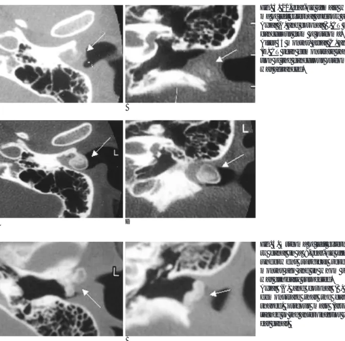
Fig. 2. Osteoma of the left external au- ditory canal in a 15-year-old woman with external otitis.
A. Axial CT scan of the left temporal bone demonstrates the string-like thin stalk (black arrow) connecting the mass (white arrow) to the anterior wall of the ear canal.
B. Coronal CT scan shows a cancellous form of oseteoma (arrow) located at the lateral part of the external auditory
A B
Fig. 1. Osteoma of the right external auditory canal in a 22-year-old man with external otitis.
A. Axial CT scan of the right temporal bone shows a pedunculated bony mass (arrow) located at the lateral part of the external auditory canal.
B. Coronal CT scan demonstrates the stalk connecting the mass (arrow) to the anterosuperior portion of the ear canal.
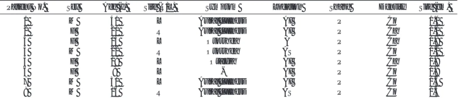
Table 1. Summary of Clinical and Radiologic Findings in 8 Patients with Osteoma of External Auditory Canal
Patient No. Sex Age (y) Site (R/L) Symptom Location Shape Density Size (cm)
1 M 41 L Aural fullness AI P Co 1.0
2 F 20 R Aural fullness AI P Ca 1.0
3 F 15 L Otorrhea A P Ca 0.8
4 M 22 R Otorrhea AS P Co 1.2
5 F 19 L Otalgia AI P Ca 0.9
6 F 8 L - AI P Co 0.9
7 M 30 L Aural fullness AI P Co 0.6
8 M 16 R Aural fullness AS P Co 0.5
R: Right, L: Left, AI: Anteroinferior, A: Anterior, AS: Anterosuperior, P: Pedunculated, Co: Compact form, Ca: Cancellous form
진 질환이다(4). 측두골의 모든 부위에서 생길 수 있으나 외이 도 외측에서 발생하는 경우가 가장 많으며, 대부분 일측에서 발 생하는 양성 질환으로 크기가 지속적으로 증가한다. 대부분의 골종이 후천적으로 발생하지만(5), Smelt(6)는 내이도에 생긴 선천성 골종을 기술한 바 있다.
외이도에 생기는 골종은 외골종과의 감별이 중요한데 임상 적, 조직병리학적으로 두 질환은 다른 별개의 질환이다(1). 그 러나 이들 두 질환은 모두 외이도의 협착을 가져와 전도성 청 력손실, 외이도염, 이루, 이통 등 유사한 임상증상을 유발할 수 있다(7).
외골종은 외이도에 생기는 가장 흔한 골 종양으로 사람에게 만 발생한다. 보통 찬물에 20년 이상 장기간 노출된 기왕력이 있는 사람에게서 흔히 발견되는 것으로 알려졌다. 지리적 분포 상 해변에서 주로 발생하며, 임상적으로 파도타기꾼 귀(Surfur’
s ear) 또는 오스트레일리아 사람 귀(Australian ear)라고 불 리기도 한다. 외골종은 양측성, 다발성 종괴로 골종에 비해 넓
은 기저부를 갖고, 20:1로 남자에서 호발하며 10대에서는 흔히 발견되지 않는다. 반면에 골종은 원인이 확실히 알려져 있지 않 은 일측성, 단발성 종괴로 외골종 보다는 드문 질환으로 남자 에서 3배 이상 많이 생기며, 전 연령층에서 발생할 수 있다.
Sheehy(8)에 따르면 수술로 제거된 16예의 외이도 골종 환자 중 3명(18.8%)이 15세 이하였다. 7예(44%)가 30세 이하였 으며, 6예(38%)의 환자는 50세 이상이었다. 본 연구에선 전체 8 예 중 5예가 20세 이하에서 발생하였으며, 평균 연령은 21.4 세였다. 다발성 골종은 대부분 거의 가드너 증후군(Gardner’s syndrome)과 관련이 있는 것으로 알려져 있는데 반해, 외골종 은 이 증후군과 연관이 없다(3). 일반적으로 외골종 환자들은 대부분 증상이 없어 치료가 필요하지 않으나, 외이도의 폐쇄로 증상이 있을 경우 대증요법을 시행한다. 귀지와 부스러기 (debris) 등이 외이도에 정체되어 폐쇄가 초래되면 청력 장애 등이 발생하게 된다. 반면에 골종은 계속해서 성장해서 외이도 의 폐쇄를 초래하기 때문에 대부분은 수술적 제거가 치료 원칙
A B
Fig. 3. 22-year-old female with osteo- ma of left external auditory anal.
Axial (A) and coronal (B) CT scan show cancellous form of osteoma.
After 35 months, axial (C) and coronal (D) CT scan demonstrate that ossifica- tion of the cancellous osteoma (arrow) was advanced.
C D
A B
Fig. 4. Osteoma of left external audito- ry canal in a 8-year-old female who underwent surgical resection 34 months ago and in whom recurrence was clinically suspected.
Axial (A) and coronal (B) CT scan demonstrate that the cauliflower- shaped, osseous mass (arrow) is at- tached to the anteroinferior wall of the ear canal.
이다. 그러나 외골종은 보존적 치료에 반응을 보이지 않는 경 우를 제외하고는 수술적 처치가 필요치 않기 때문에 두 질환의 감별 진단이 중요하다.
Graham(1)과 Kemink 등(2)에 의하면 조직학적으로 외이 도 골종은 기저 골막에 편평상피 세포로 덮여 있으며, 판상골 (lamellated bone)로 둘러싸인 풍부한 섬유혈관 통로를 보이는 것이 특징이며, 풍부한 섬유성조직과 굴모양혈관(sinusoidal- like blood vessels)을 가지며, 불규칙한 형태를 지닌 다른 통 로를 보이는 경우도 있다. 외골종 역시 기저 골막에 편평상피 세포로 덮여 있으며, 풍부한 골세포를 가지고 있는 골막하골의 동심성층을 보이나 섬유혈관 통로는 볼 수 없다는 것이 외이도 골종과 다른 점이라고 주장하였다. 그러나 Fenton 등(3)에 의 하면 외골종과 골종 모두에서 섬유혈관 통로가 관찰되어 섬유 혈관 통로의 존재 유무로 두 개의 질환을 조직학적으로 감별할 수 없다고 주장하였다.
외골종과 골종 모두 임상증상으로 분류할 때 시기에 따라 3 단계로 나눈다. 1단계는 검사자의 눈에는 병변이 명확하나 환 자는 증상이 없는 경우이고, 2단계는 증상이 있으나 대증적 요 법으로 치료할 수 있는, 3단계는 증상이 있고 수술이 필요한 경 우이다(9).
골종의 형태는 치밀형과 해면상형의 두 가지로 나누어진다.
방사선학적으로 치밀형은 분엽을 보이는 치밀한 골종괴이고, 해 면상형은 부분적으로 골화를 보이는 골종괴이다(7). 본 연구에 서는 치밀형이 5예였고, 해면상형이 3예로 치밀형 골종이 해면 상형 골종보다 더 많았다.
Hsiao 등(9)과 박 등(10)은 외이도 골종이 조영증강 전 CT 소견상 관상면과 축상면에서 골성의, 단일성의 작은 각을 가진 병변으로 외이도에서 특징적으로 관찰되는 것으로 기술하였으 며, 본 연구에서도 외이도 바깥쪽의 단일성의 각을 가진 골성 병변들로 관찰되었다.
외이도 골종은 대부분 고실인상 봉합선 또는 고실유양돌기 봉합선을 따라서 발생하는 것으로 알려졌다(8). 그러나 본 연 구에서는 지금까지 보고된 것과는 달리 봉합선을 따라 발생한 골종이 전체 8예 중 2예(25%)에 불과해 골종과 봉합선과의 관 계에 대해 더 많은 증례를 바탕으로 새롭게 규명해볼 필요가 있을 것으로 생각되었다.
외이도 골종의 경우 골종의 완전한 절제가 이루어진 경우 재
발을 하지 않는 것으로 알려져 있으나(11), 본 연구에서는 1 예에서 좌측의 외이도 골종을 경외이도 접근법으로 절제술을 시행한 뒤, 34개월 뒤에 재발하여 재수술을 시행하였다. 이후 추적 검사는 시행되지 않았다.
이 연구의 제한점으로 첫 번째, 대상 환자 수가 많지 않아 통 계 처리를 제대로 할 수 없었고, 두 번째 영상과 임상 경과의 충분한 추적검사가 이루어지지 못했다는 점이다.
결론적으로 외이도 골종은 외이도의 바깥쪽에서 발생하는 양 성 종양으로 CT소견상 경에 의해 외이도의 골조직과 연결되어 있는 단일성의 골성 종괴로 관찰되었다. 일반적으로 고실유양 돌기 봉합선이나 고실인상 봉합선에서 발생하는 것으로 알려 졌으나, 본 연구에선 대부분의 경우에 CT와 수술 소견 상 모 두에서 봉합선과 관계없이 발생하여 이에 대한 추가 연구가 필 요할 것으로 생각한다.
참 고 문 헌
1. Graham MD. Osteomas and exostosis of the external auditory canal. A clinical, histopathological and scanning electron micro- scopic study. Ann Otol Rhinol Laryngol 1979:88:566-572
2. Kemink JL, Graham MD. Osteomas and exostoses of the external auditory canal medical and surgical management. J Otolaryngol 1982;11:101-106
3. Fenton J, Turner J, Fagan PA. A histopathologic review of tempo- ral bone exostoses and osteoma. Laryngoscope 1996;106:624-628 4. Friedmann I. Pathological lesions of the external auditory meatus:
a review. J R Soc Med 1990;83:34-37
5. Di Bartolomeo JR. Exostoses of external auditory canal. Ann Otol Rhinol Laryngol Suppl 1979;61:2-20
6. Smelt GJ. Exostoses of the internal auditory canal. J Laryngol Otol 1984;98:347-350
7. Valvassori GE, Mafee MF, Carter BL. Imaging of the Head and Neck. New York: Thieme Medical Publishes Inc 1995;110-131 8. Sheehy JL. Diffuse exostoses and osteomata of the external audito-
ry canal: a report of 100 operations. Otolaryngol Head Neck Surg 1982;90:337-342
9. Hsiao SH, Liu TC. Osteoma of the external ear canal. Otol Neurotol 2003;24:960
10. 박만수, 방진현, 정재걸, 이덕희, 정승문, 류대식. 외이도에 생긴 골 종: 1예 보고. 대한방사선의학회지 2001;45:143-145
11. Noordzij JP, Ariaga MA, Stone AB. Differentiating bony lesions of the external auditory canal. Ear Nose Throat J 1995;74:49-51
J Korean Radiol Soc 2006;55:33-37
Address reprint requests to : Chang Joon, Song, M.D., Department of Diagnostic Radiology, Chungnam National University School of Medicine, 640 Daesa-dong, Jung-ku, Taejeon 301-721, Korea.
Tel. 82-42-220-7834 Fax. 82-42-253-0061 E-mail: [email protected]
CT Findings of the Osteoma of the External Auditory Canal
1Ha-young Kim, M.D., Chang Joon Song, M.D., Chung Dae Yoon, M.D., Mi Hyun Park, M.D., Byung Seok Shin, M.D.
1Department of Diagnostic Radiology, Chungnam National University, School of Medicine
Purpose: We wanted to report the CT image findings of the osteoma of the external auditory canal.
Materials and Methods: Temporal bone CT scanning was performed on eight patients (4 males and 4 females aged between 8 and 41 years) with pathologically proven osteoma of the external auditory canal after opera- tion, and the findings of the CT scanning were retrospectively reviewed. Not only did we analyze the size, shape, distribution and location of the osteomas, we also analyzed the relationship between the lesion and the tympanosqumaous or tympanomastoid suture line, and the changes seen on the CT scan images for the pa- tients who were able to undergo follow-up.
Results: All the lesions of the osteoma of the external auditory canal were unilateral, solitary, pedunculated bony masses. In five patients, the osteomas occurred on the left side and for the other three patients, the osteo- mas occurred on the right side. The average size of the osteoma was 0.6 cm with the smallest being 0.5 cm and the largest being 1.2 cm. Each of the lesions was located at the osteochondral junction in the terminal part of the osseous external ear canal. The stalk of the osteoma of the external auditory canal was found to have oc- curred in the anteroinferior wall in five cases (63%), in the anterosuperior wall (the tympanosqumaous suture line) in two cases (25%), and in the anterior wall in one case. The osteoma of the external auditory canal was a compact form in five cases and it was a cancellous form in three cases. One case of the cancellous form was changed into a compact form 35 months later due to the advanced ossification.
Conclusion: Osteoma of the external auditory canal developed in a unilateral and solitary fashion. The charac- teristic image findings show that it is attached to the external auditory canal by its stalk. Unlike our common knowledge about its occurrence, osteoma mostly occurred in the tympanic wall, and this is regardless of the tympanosquamous or tympanomastoid suture line.
Index words :Osteoma Ear, CT