INTRODUCTION
With advances in imaging modalities, perioperative care, surgi- cal techniques, and indications for liver resection have been ex- panded. Both right and extended right hepatic lobectomy are oc- casionally associated with postoperative liver failure (1) because it involves a resection of a large portion of the liver. Therefore, choosing which procedure to perform must be done with care for each individual. Removing more than 70% of liver parenchyma has been known to increase the risk of postoperative mortality (1). Previous clinical and experimental reports have shown that
when portal veins are occluded or stenotic, it leads to atrophy of the affected lobe or segment along with compensatory hypertro- phy of the unaffected hepatic lobe (2, 3). Based on this result, pre- operative portal vein embolization (PVE) has become a well-es- tablished means to upsize the future remnant liver as well as to prevent postoperative liver insufficiency in patients considered for extensive liver resection (4-6). Additional hepatic artery emboli- zation combined with PVE has been reported to be beneficial for compensatory hypertrophy compared with PVE alone (7, 8).
Our study has been performed to compare the volume change and the regenerative capacity between portal vein ligation (PVL)
J Korean Soc Radiol 2013;68(4):289-296 http://dx.doi.org/10.3348/jksr.2013.68.4.289
Received November 9, 2012; Accepted January 8, 2013 Corresponding author: Gyeong Sik Jeon, MD Department of Radiology, CHA Bundang Medical Center, College of Medicine, CHA University, 59 Yatap-ro, Bundang-gu, Seongnam 463-712, Korea.
Tel. 82-31-780-5382 Fax. 82-31-780-5381 E-mail: [email protected]
This is an Open Access article distributed under the terms of the Creative Commons Attribution Non-Commercial License (http://creativecommons.org/licenses/by-nc/3.0) which permits unrestricted non-commercial use, distri- bution, and reproduction in any medium, provided the original work is properly cited.
Purpose: To compare the volume change and the regenerative capacity between portal vein ligation (embolization) (PVL) and heterochronous PVL with hepatic artery ligation (HAL) in a rodent model.
Materials and Methods: The animals were separated into three groups: group I, li- gation of the left lateral and median portal vein branches; group II, completion of PVL, followed by ligation of the same branches of the hepatic artery after 48 h;
control group, laparotomy without ligation was performed. Five rats from each group were sacrificed on 1, 3, 5, and 7 days after the operation. Volume change measurement, liver function tests and immunohistochemical analysis were per- formed.
Results: The volume of the nonligated lobe between groups I and II was not signifi- cantly different by day 5 and day 7. Mean alanine aminotransferase and total biliru- bin levels were significantly higher in group II, while the albumin level was higher in group I. Both c-kit- and MIB-5-positive cells used in the activity detection of regen- eration were more prevalent in group I on day 1, 3, and 5, with statistical signifi- cance. There was no operation related mortality.
Conclusion: PVL alone is safe and effective in compensatory liver regeneration.
Performing both PVL and HAL does not confer any additional benefits.
Index terms Portal Vein Hepatic Artery Ligation
Liver Regeneration
Comparative Study of Compensatory Liver Regeneration in a Rat Model: Portal Vein Ligation Only versus Sequential Ligation of the Portal Vein and Hepatic Artery
1백서에서 단독 간문맥 결찰과 간문맥과 간동맥의 순차적 결찰 후 간재생의 비교 연구1
Soo Young Chung, MD
1, Gyeong Sik Jeon, MD
2, Byungmo Lee, MD
31Department of Pathology, Dongnam Institute of Radiological & Medical Sciences, Busan, Korea
2Department of Radiology, CHA Bundang Medical Center, College of Medicine, CHA University, Seongnam, Korea
3Department of Surgery, Seoul Paik Hospital, Inje University College of Medicine, Seoul, Korea
Immunohistochemistry
Whole liver was dissected and postfixed in 10% neutral-buff- ered formalin for 24 hours. Primary antibodies used for immu- nostaining were MIB-5 (a novel antibody reactive with the equiv- alent Ki-67 protein, 1 : 800, LabVision, Cheshire, UK), and c-kit (1 : 400, DAKO, Glostrup, Denmark). MIB-5 is used for the de- tection of cells in the active phases of the cell cycle and c-kit is used for the detection of stem cell expression. The liver was di- luted in normal goat serum buffer. We used the Zymed nonbio- tin amplification system (Zymed Laboratories Inc., South San Francisco, CA, USA). Only nuclear-stained cells in Ki-67 and cytoplasmic-stained cells in c-kit were considered meaningful.
The number of Ki-67-positive cells and c-kit-positive cells were counted using × 400 magnification in 10 randomly selected vi- sual fields, using samples from five animals per group from each of the three experimental groups. Two pathologists evaluated the staining independently, and any discrepancy was resolved by a consensus review.
Assessment of Liver Proliferation and Function
The ratio of the weight of the nonligated lobe to the body weight was assessed using the following equation: R = weight of nonligated lobes/body weight (nLW/BW). Plasma was separat- ed by centrifuge using blood samples collected by a direct heart puncture. The collected plasma was used for the standard liver function tests. The alanine aminotransferase (ALT), aspartate aminotransferase (AST), total bilirubin, alkaline phosphatase, gamma glutamyl transpeptidase, albumin, prothrombin time, and the international normalized ratio (INR) of the blood sam- ples were determined. All of the parameters were measured us- ing the standard laboratory methods.
Statistical Analysis
The values for the results were expressed as means ± standard error of the mean. Student’s t-test and Kruskal-Wallis analyses were used for the statistical analysis. The probability values low- er than 0.05 were considered significant.
RESULTS
Two animals were lost in group II following the operation.
During the initial stage of the experiments, rats that had under- and heterochronous PVL with hepatic artery ligation (HAL) in
a rodent model.
MATERIALS AND METHODS
Experimental Design and Surgical Procedures
This study was approved by Dankook University’s “Animal Care and Use Committee” (South Korea). All procedures were conducted in accordance with the ‘Guide for Care and Use of Laboratory Animals’ published by the National Institutes of Health and the ethical guidance of the International Association for the Study of Pain.
Male Sprague-Dawley rats (Samtako Co, Seoul, Korea) aged 13 weeks and weighing between 350 and 450 g were used for all of the experiments. All the animals were kept in identical hous- ing units with an alternating 12 h light-dark cycle. Food and wa- ter were available ad libitum.
After an acclimation period of at least 7 days, the animals were separated into three groups as follows: group I, performed only ligation of the left lateral and median portal vein branches (which supplies approximately 70% of total liver volume); group II, first completed ligation of the portal vein, followed by ligation of the same branches of the hepatic artery after 48 hours; control group, conducted laparotomy without ligation.
Prior to the operations, all of the animals were anesthetized with an intraperitoneal injection of zolazepam mixed with xyla- zine and normal saline at room temperature, using a non-sterile, clean technique. In group I, via midline incision, the branch of the portal vein supplying the median and the left lateral lobes of the liver was ligated using 6-0 silk. The abdominal wall and skin were closed with 4-0 silk. In group II, the second operation was performed after 48 hours, by performing ligation of the same branch of the hepatic artery. Abdominal cavity irrigation using a mixture of normal saline and antibiotics was performed as the last step of each operation.
Postoperatively, the animals had free access to water and food.
Rats from each group were sacrificed using the same anesthesia method described above on 1, 3, 5, and 7 days after the opera- tion. At least five animals per group were employed for every study time point. Blood was collected by the heart puncture method just before sacrifice. The liver was removed, and the weight of the body and non-ligated lobes were weighed.
gone the 1st operation were accidentally not separated, injured at the operation site, and eventually died. Thereafter, all rats were separated and remained alive until the end of the study.
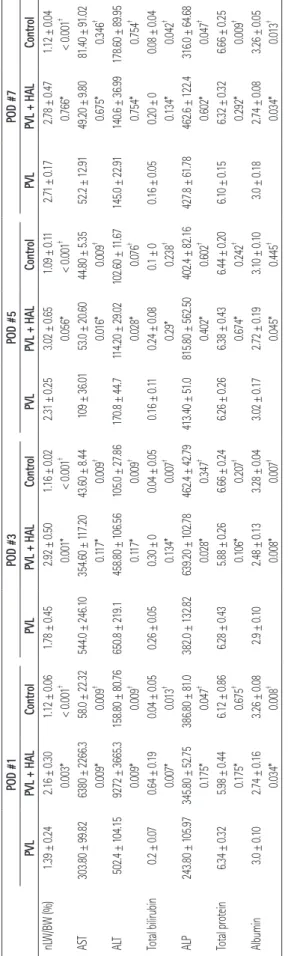
Weight of Nonligated Lobes/Body Weight (nLW/BW) and Liver Function Test
In group I, the nLW/BW gradually increased and was highest at 7 days after surgery. The nLW/BW was higher in group II un- til the 5th day after surgery. However, there were no significant differences at days 5 and 7 (Fig. 1).
Serum AST and ALT values was markedly increased in group II compared with group I on day 1 after surgery; the AST and ALT values was highest at day 3 in group I. The total bilirubin level was also higher in group II on day 1 after surgery; however, this difference was not statistically clear. In contrast, the albumin level was significantly higher in group I than in group II through- out all time periods. The INR was significantly lower in group I on days 5 and 7. These results are summarized in Table 1.
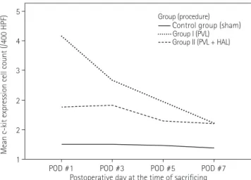
Expression of C-Kit
There was an overall increase in the number of c-kit-positive cells in group I (PVL) compared to group II (PVL + HAL) (p <
0.05). However, there were no statistically significant differences 7 days after surgery (p = 1.00). The peak levels of c-kit-positive hepatocytes in group I was detected on day 1; yet, the peak level
Fig. 1. Results of nonligated liver weight/body weight. At postopera- tive days 1 and 3, significant increase of nonligated liver weight is seen in group II. However, eventually there are no significant differ- ences on day 7 in both groups.
Note.-HAL = hepatic artery ligation, POD = postoperative day, PVL =
portal vein ligation jurofiles of Liver Inryic andunction Ta P Fogede 1. Regenblerolat Liver Weight and Ser #7D PO#5D #3POPO#1D POD PVL + HALControl HLPVL +ALControlLPVlPVrolPVLPV HALContL +LPVL + HALContPVro ± ±.09 165 0. 0..02 3251 ±2.311 0.47 2. 0. 0.04 0. ±.12 1 ±7178 2. 17 0. ±021.1 ±30W/BW (%) 1.39 ± 0..16 2.16 ± 0.24 nL50 0. ±92 2. 145 0.8 ±1.706 0.2 ± †††† 0.0*66010.7010.0< 6*< 0.050.0010.003*< 010.001*< 0.0 53 44.6020± .0 .01 36 ±09 1 ±44.80.91 5.80 ±.02 910 ±1.4 80 ±359.2 4 12 ±2.2 5 8. 9..60 .3 66 22 ±80 63AS 5.82 990 ±3.830T 43 8.00 6.1 110 ±4.635 ±240 ± .0 44 5.32 227.2 †††† .00.009096.34 0*750.6*90.0 0 06*0.117*.009 0.01 0 4.22.610.02 290 ±.8 11.7 44± 1170 1.860 ±.91.67.99± .95 890 ±8.617 3614 ±0.6 14 22 ±5.027 ±.0 .3 65 36 ±72 925 054.1 102.450 T AL 158.80 ± 16 6.5 100 ±8.8 45 9.1 21 ±0.865.76 80 †††† .00 090.04.75 0*0.7096.07548* 0*9.00 0*170.1.02 0 0 0. ±.240511 0.6 ±0.1 0.108 0. ± 0 0.16 ± 0.0.20 ± 0 0.08 ± ±04 0.05.04 ± 0Total bilirubin 0.2 0.07 0.64 ± 0.6 ± 0 190.305 0.0 ±0.2 0.4 ±0.0 05 †††† 08 00.134*.0429*.230.2.0170.007 0*30.134* 0.00 56± .4 02 40 2.5 51 ±.8015 8.0 .160 ±3.482.79422.4.6864± .0 16 3 127.8 ±2.6 46 .78 61 ±410 ±425.96.838.755.8347 10 81 ±.8043 2P AL± 0 ± 52.0 9.2.4 62 38 2.7 100 ± 4 63 2 2.813± .0 82 †††† 7 0*020.62*2.60 0.04.34.407 075* 0.040.10.0* 0728 0. ±.44 643 6 ±.3826 0.6 ±20 6.2 6.6.10 ± 0.15 32 ± 0.32 6.66 ± 0.25 0.24 0. ± 0..66Total protein 6.34 ± 0.32 5.98 ±44 626 0. ±88 5.43 0.8 ±6.286 0.2 ±6.1 †††† .67.2450.1759.00 0920.2*2* 0* 00.14*06 0.207 0.67 0. ±.10 3 0.1917 ±.72 2 0.2 ± 33.010.26.0 ± 0.18 2.74 ± 0.08 3 ± 0.0504 0. ±Albumin 3.0 ± 0.10 2.74 ± 0.16 3.26 ± 0.08.28 313 0. ±48 2. 0 0.1± .9 2 †††† .00 080.03.01*340.0345.44 05**.04 00.008 0.007* 0 esPVt between + L and PVL HAL.e t-t th- vithNote.*The palue estimated w † L, + L PVsisPVg onmL, HAupand the control gro. akaly wnaTh value estimatede pithW thlisal al–usKre orae th =W/BLWtioat nn,ioligy errt aicheatf t lo ws ht gheiwy od btobeeiedatligon nhef tt oghepph =asame inanal= T ALe, atotosALe linkaal= P ALinphra ans, HseraferaotinmnsteSTfetarase, A = aspar PValL = portat vein ligioday, cie tivrapetoos p =OD, Pngificr saatn 0.0000
0.0300
0.0100 0.0200 0.0400
POD #3 POD #7
POD #1 POD #5
Postoperative day at the time of sacrificing
Mean non-ligated liver weight/body weight
Group (procedure) Control group (sham) Group I (PVL) Group II (PVL + HAL)
in group II was not detected until 3 days after surgery, as shown in Figs. 2, 3.
Expression of MIB-5
There was an overall increase in the nuclear expression of MIB- 5 in group I compared to group II. The number of MIB-5-positive hepatocytes was significantly higher in group I than in group II from day 1 to 5 (p < 0.05). Unlike c-kit, the peak level of MIB-5- positive hepatocytes was noted on day 3 in both groups I and II.
There were little differences in the expression of MIB-5 between groups I and II on day 7 (p = 0.80), as presented in Figs. 4, 5.
DISCUSSION
Liver regeneration following PVL has been studied in depth Fig. 2. Results of c-kit expression. Between group I and II, there are
statistically significant differences in expression of c-kit from postop- erative days 1 to 5 (p < 0.05).
Note.-HAL = hepatic artery ligation, HPF = high power field, POD = postoperative day, PVL = portal vein ligation
Fig. 3. Microscopic images of nonligated lobes with c-kit staining (× 100). Brown-colored, stained cells are more apparent in group I (A) than in II (B) on day 1. On day 7, there are no significant differences between group I (C) and II (D).
C A
D B 1
4
2 2 3 5
POD #3 POD #7
POD #1 POD #5
Postoperative day at the time of sacrificing
Mean c-kit expression cell count (/400 HPF)
Group (procedure) Control group (sham) Group I (PVL) Group II (PVL + HAL)
for several decades (9). Many studies have reported that PVE is an effective method for liver regeneration prior to liver resection (4-6). However, other studies have reported that adding hepatic artery embolization yields better results (6, 7, 10, 11). Kong et al.
(10) reported that compensatory hypertrophy of the unaffected lobes was evident. Data from their study revealed that when PVL and HAL are performed simultaneously, the ligated liver tissues undergo massive necrosis (10). Nagino et al. (7) reported that additional hepatic artery embolization was effective in in- ducing compensatory hypertrophy; however, they also reported adverse events, such as necrosis and abscess formation. Like the results of Nagino et al., in our study, necrosis of the ligated lobe was grossly present in group II in which both PVL and HAL were performed at an interval of 48 h. As the evaluation of the ligated lobe was not the major point of our study, we did not
Fig. 4. Results of MIB-5 expression. Between group I and II, there are statistically significant differences in the expression of MIB-5 from postoperative days 1 to 5 (p < 0.05).
Note.-HAL = hepatic artery ligation, HPF = high power field, POD = postoperative day, PVL = portal vein ligation
Fig. 5. Microscopic images of nonligated lobes with MIB-5 staining (× 200). Stained cells are more apparent in group I (A) than II (B) on day 3.
On day 7, there are no significant differences between group I (C) and II (D).
C A
D B
0 80
20 40 60 100
POD #3 POD #7
POD #1 POD #5
Postoperative day at the time of sacrificing
Mean MIB-5 expression cell count (/400 HPF)
Group (procedure) Control group (sham) Group I (PVL) Group II (PVL + HAL)
types of cells to grow. Signaling through c-kit protein plays a role in cell survival, proliferation, and differentiation (16). In con- trast to the results of Kong et al. (10), the MIB-5-labeling index showed higher levels in group I in our study. The levels of c-kit- positive hepatocytes were also higher in group I. Considering the increase in liver enzyme levels and bilirubin, acute damage to the hepatocytes in group II may have interfered with liver re- generation of the nonligated lobe. These results are similar to a previous report by Rozga et al. (9), who reported the highest values of DNA-synthetic activity in rats in a 70% ligation group at 24 h. In these rats, the increased DNA-synthetic activity per- sisted at least until 2 weeks after the procedure. The most vigor- ous mitotic activity in PVL in rats was noted at 48 h and persist- ed for 2 weeks (12). In group I of our study, the expression of c-kit and Ki-67 cells was highest on day 1 and day 3, respective- ly. On day 7, the levels of both markers were very low, possibly due to the markedly decreased regeneration activity. As a result, it is difficult to determine whether significant volume hypertro- phy occurred after 7 days or at the time interval of the two mark- ers has any correlation with liver regeneration.
There are several limitations to our study. First, portal vein li- gation and portal vein embolization is not the same procedure.
Broering et al. (17) and Iida et al. (18) reported that PVE was more efficient than PVL for the induction of hypertrophy of the contralateral lobe. They explained that one of the reasons for the larger liver volumes is the portoportal collateral vessels. Howev- er, as in other several studies, fortnight normalization of in- creased portal blood flow induced by portal vein occlusion in humans and the early peak of hepatocytes proliferation after portal occlusion in rodents suggests that liver hypertrophy after portal occlusion is induced earlier than the formation of porto- portal collateral formation (19-21). Hence, this may have little impact on liver hypertrophy. Therefore we considered that our results can be applicable to PVE. Second, this study was con- ducted over a short period of time. Third, we did not perform a microscopic evaluation of the ligated lobe. Without this, the re- lationship between ligated and nonligated lobes was not re- vealed from our results. Fourth, we had not confined the c-kit- positive expression cells to the stem cells of the liver; therefore, the relevance of the stem cell expression cannot be concluded.
In conclusion, PVL alone was found to be safe and effective to induce compensatory liver regeneration. Performing both PVL comment on this.
Kong et al. (10) also reported that the ratio of the weight of the nonligated lobe to the body weight was significantly higher at 48 h in a group that underwent PVL plus the HAL heteroch- ronous procedure than in a PVL-only group and a simultaneous PVL plus the HAL group. The time of the peak level of hyper- trophy (168 h) was similar to our results. At first, hypertrophy of the nonligated lobe seemed more prominent in group II (PVL + HAL); however, there were no significant differences on day 7 in the two groups due to the delayed progressive hypertrophy in group I. Also, group II showed a slightly decreased volume ratio on day 7. This is in common with the reports by Veteläinen et al.
(12) who noted that the abrupt increase in the volume followed by a subsequent decrease in the volume can be explained by a massive, progressive apoptosis. According to the report by Kong et al. (10), massive necrosis of the ligated lobe found in group II may have interfered with the regeneration of the nonligated lobe.
However, further investigation regarding this was not performed in this study since apoptosis could not be proven due to the rela- tively short follow-up time of 7 days.
Rozga et al. (9) reported almost total resorption of the necro- ses in the portal vein of the ligated lobes, which was seen in 4 days after ligation. However, in our study, grossly necrotic tissue was found in all rats in group II at all time points. In our opinion, this may have been due to the fact that there was not enough time for the healing process from PVL because of the subse- quent HAL.
Liver function tests revealed significantly higher levels of AST/ALT and total bilirubin in group II. Severe liver injury was also confirmed in group II with additional HAL. Albumin levels were also lower in group II, and INR was increased in group II.
These results imply a decreased synthetic function of the liver in group II. Nakao et al. (13) also reported similar results.
In 1975, it was established that portal venous blood flow pro- moted hepatic cell regeneration (14). For the evaluation of liver regeneration, immunohistochemical staining for MIB-5 and c- kit were used in this study. MIB-5 is a novel antibody reactive with the rat equivalent to Ki-67 protein. The Ki-67 reacts exclu- sively with the nuclei of the proliferating cells and is expressed during all active parts of the cell division cycle (G1, S, G2, and M); however, it is absent in the resting cells (G0) (15). C-kit pro- tein binds to the stem cell factor, a substance that causes certain
11. Aoki T, Imamura H, Hasegawa K, Matsukura A, Sano K, Sugawara Y, et al. Sequential preoperative arterial and portal venous embolizations in patients with hepatocellu- lar carcinoma. Arch Surg 2004;139:766-774
12. Veteläinen R, Dinant S, van Vliet A, van Gulik TM. Portal vein ligation is as effective as sequential portal vein and hepatic artery ligation in inducing contralateral liver hy- pertrophy in a rat model. J Vasc Interv Radiol 2006;17:
1181-1188
13. Nakao N, Miura K, Takahashi H, Ohnishi M, Miura T, Oka- moto E, et al. Hepatocellular carcinoma: combined hepat- ic, arterial, and portal venous embolization. Radiology 1986;161:303-307
14. Bucher ML, Swaffield MN. Regulation of hepatic regener- ation in rats by synergistic action of insulin and glucagon.
Proc Natl Acad Sci U S A 1975;72:1157-1160
15. Gerlach C, Sakkab DY, Scholzen T, Dassler R, Alison MR, Gerdes J. Ki-67 expression during rat liver regeneration after partial hepatectomy. Hepatology 1997;26:573-578 16. Bin WT, Ma LM, Xu Q, Shi XL. Embryonic hepatocyte trans-
plantation for hepatic cirrhosis: efficacy and mechanism of action. World J Gastroenterol 2012;18:309-322
17. Broering DC, Hillert C, Krupski G, Fischer L, Mueller L, Achil- les EG, et al. Portal vein embolization vs. portal vein ligation for induction of hypertrophy of the future liver remnant. J Gastrointest Surg 2002;6:905-913; discussion 913
18. Iida H, Aihara T, Ikuta S, Yoshie H, Yamanaka N. Compari- son of percutaneous transhepatic portal vein embolization and unilateral portal vein ligation. World J Gastroenterol 2012;18:2371-2376
19. Goto Y, Nagino M, Nimura Y. Doppler estimation of portal blood flow after percutaneous transhepatic portal vein embolization. Ann Surg 1998;228:209-213
20. Yachida S, Ikeda K, Kaneda K, Goda F, Maeba T, Maeta H.
Preventive effect of preoperative portal vein ligation on endotoxin-induced hepatic failure in hepatectomized rats is associated with reduced tumour necrosis factor alpha production. Br J Surg 2000;87:1382-1390
21. Heinrich S, Jochum W, Graf R, Clavien PA. Portal vein liga- tion and partial hepatectomy differentially influence growth of intrahepatic metastasis and liver regeneration in mice.
J Hepatol 2006;45:35-42 and HAL does not confer additional benefits.
REFERENCES
1. Thompson HH, Tompkins RK, Longmire WP Jr. Major he- patic resection. A 25-year experience. Ann Surg 1983;197:
375-388
2. Rous P, Larimore LD. Relation of the portal blood to liver maintenance: a demonstration of liver atrophy condition- al on compensation. J Exp Med 1920;31:609-632
3. Takayasu K, Muramatsu Y, Shima Y, Moriyama N, Yamada T, Makuuchi M. Hepatic lobar atrophy following obstruction of the ipsilateral portal vein from hilar cholangiocarcino- ma. Radiology 1986;160:389-393
4. Kubota K, Makuuchi M, Kusaka K, Kobayashi T, Miki K, Hasegawa K, et al. Measurement of liver volume and he- patic functional reserve as a guide to decision-making in resectional surgery for hepatic tumors. Hepatology 1997;
26:1176-1181
5. Abulkhir A, Limongelli P, Healey AJ, Damrah O, Tait P, Jackson J, et al. Preoperative portal vein embolization for major liver resection: a meta-analysis. Ann Surg 2008;247:
49-57
6. Imamura H, Shimada R, Kubota M, Matsuyama Y, Nakayama A, Miyagawa S, et al. Preoperative portal vein embolization:
an audit of 84 patients. Hepatology 1999;29:1099-1105 7. Nagino M, Kanai M, Morioka A, Yamamoto H, Kawabata Y,
Hayakawa N, et al. Portal and arterial embolization before extensive liver resection in patients with markedly poor functional reserve. J Vasc Interv Radiol 2000;11:1063-1068 8. Inaba S, Takada T, Amano H, Yoshida M, Yamakawa Y, Yas-
uda H, et al. Combination of preoperative embolization of the right portal vein and hepatic artery prior to major hepatectomy in high-risk patients: a preliminary report.
Hepatogastroenterology 2000;47:1077-1081
9. Rozga J, Jeppsson B, Bengmark S. Portal branch ligation in the rat. Reevaluation of a model. Am J Pathol 1986;125:
300-308
10. Kong D, Kusano M, Arase T, Nishino N, Jin Z, Kameyama S, et al. Liver regeneration after portal vein plus hepatic ar- tery ligation performed heterochronously in rats. J Hepa- tobiliary Pancreat Surg 2002;9:86-92
백서에서 단독 간문맥 결찰과 간문맥과 간동맥의 순차적 결찰 후 간재생의 비교 연구1
정수영
1· 전경식
2· 이병모
3목적: 백서에서 간문맥 단독결찰과 간문맥과 간동맥의 순차적 결찰 후 반대측 간의 재생에 대하여 비교하고자 한다.
대상과 방법: 동물들은 세 그룹으로 분류하였다. 1) 좌측 간문맥 결찰만 시행한 그룹, 2) 좌측 간문맥 결찰 후 48시간 이 후에 동일한 간동맥 결찰을 시행한 그룹, 3) 혈관 결찰 없이 개복술만 시행한 경우. 술 후 1, 3, 5, 7일째 되는 날 각 그룹 에서 최소 5마리씩을 희생시켜 간 부피 변화와 간 기능 검사 결과, 간 재생에 관한 면역염색화학 결과를 얻었다.
결과: 결찰하지 않은 반대측 간의 부피 변화는 5일과 7일째 첫 번째와 두 번째 그룹에서 유의한 차이는 없었다. Alanine aminotransferase와 총 빌리루빈 값은 두 번째 그룹에서 유의하게 높았으며, 알부민값은 첫 번째 그룹에서 유의하게 높았 다. 간 재생의 과정에 있는 세포 확인을 위한 c-kit와 MIB-5 면역세포화학 검사 양성세포는 술 후 1, 3, 5일에 첫 번째 그룹에서 통계적으로 유의하게 많은 분포를 보였다. 수술의 직접적인 결과로 사망한 경우는 없었다.
결론: 단독 간문맥 결찰은 대상성 간재생에 비교적 안전하고 유의한 방법이다. 본 연구에서는 추가적인 간동맥 결찰은 부 가적인 이득이 없을 것으로 보인다.
1동남권원자력의학원 병리과, 2CHA의과학대학교 분당차병원 영상의학과, 3인제대학교 의과대학 서울백병원 외과