Brief Report
Vol. 29, No. 5, 2017 663
Received June 16, 2016, Revised August 20, 2016, Accepted for publication October 19, 2016
Corresponding author: Ohsang Kwon, Department of Dermatology, Seoul National University College of Medicine, 101 Daehak-ro, Jongno-gu, Seoul 03080, Korea. Tel: 82-2-2072-2417, Fax: 82-2-742-7344, E-mail: [email protected]
This is an Open Access article distributed under the terms of the Creative Commons Attribution Non-Commercial License (http://creativecommons.org/
licenses/by-nc/4.0) which permits unrestricted non-commercial use, distribution, and reproduction in any medium, provided the original work is properly cited.
Copyright © The Korean Dermatological Association and The Korean Society for Investigative Dermatology
thosis, extended rete ridges, and various degrees of hyper- keratosis in the epidermis4. The origins of the mucin and the mechanism of its development are not known, but pre- vious studies suggested that mucin formation might in- crease as a result of fibroblast upregulation5. Because the present case showed a decrease in fat composition and an increase in fibrosis in the NLCS lesion over time, it is con- sidered to be an interesting case supporting this correla- tion. Apart from fibroblast upregulation, external stim- ulation by constant friction, and coincidental co-occur- rence of mucinous nevus and NLCS should be regarded as possible hypotheses, and more cases and study will be needed to better understand the relation between two dis- eases and mechanism of mucin deposition.
The clinical features of the present case were similar to those of soft fibromas or neurofibromas, so histopatho- logical examination and special staining were required for differential diagnosis. In conclusion, we report a rare case of acquired mucinous nevus in NLCS. Our case provides additional support for the hypothesis that mucin deposi- tion may develop in association with fibroblast.
CONFLICTS OF INTEREST
The authors have nothing to disclose.
REFERENCES
1. Hoffmann E, Zurhelle E. Über einen naevus lipomatosus cu- taneus superficialis der linken glutaalgegend. Arch Dermatol Syphilol 1921;130:327-333.
2. Perez-Crespo M, Lopez-Navarro N, Betlloch I, Herrera E, Niveiro M, Gallego E. Acquired and familial mucinous nevus. Int J Dermatol 2011;50:1283-1285.
3. Redondo Bellón P, Vázquez-Doval J, Idoate M, Quintanilla E. Mucinous nevus. J Am Acad Dermatol 1993;28:797-798.
4. Song BH, Park S, Park EJ, Kwon IH, Kim KH, Kim KJ.
Mucinous nevus with fat: an unusual case report and liter- ature review. Am J Dermatopathol 2012;34:e146-e148.
5. Carapeto FJ, Chárlez L, Marrón J, Grasa MP, Marrón SE.
Infantile and progressive papular mucinosis. Med Cutan Ibero Lat Am 1985;13:525-530.
https://doi.org/10.5021/ad.2017.29.5.663
A Familial Case of Aplasia Cutis Congenita in Two Korean Siblings: A Review of Genetic Aspects
Dong Young Kim1,2,3, Jong Seo Park1,2,3, Ohsang Kwon1,2,3
1Department of Dermatology, Seoul National University College of Medicine, 2Laboratory of Cutaneous Aging and Hair Research, Biomedical Research Institute, Seoul National University Hospital, and 3Institute of Human-Environment Interface Biology, Medical Research Center, Seoul National University, Seoul, Korea
Dear Editor:
Aplasia cutis congenita (ACC) is a rare congenital malfor- mation, characterized by a localized defect of epidermis, dermis, and sometimes subcutaneous tissues1-3. Although
it has been suggested that ACC results from disrupted de- velopment or degeneration of skin in utero, the patho- genesis remains unclear. There are five familial cases of ACC reported in Korea (Table 1)1-5. Three cases were type
Brief Report
664 Ann Dermatol
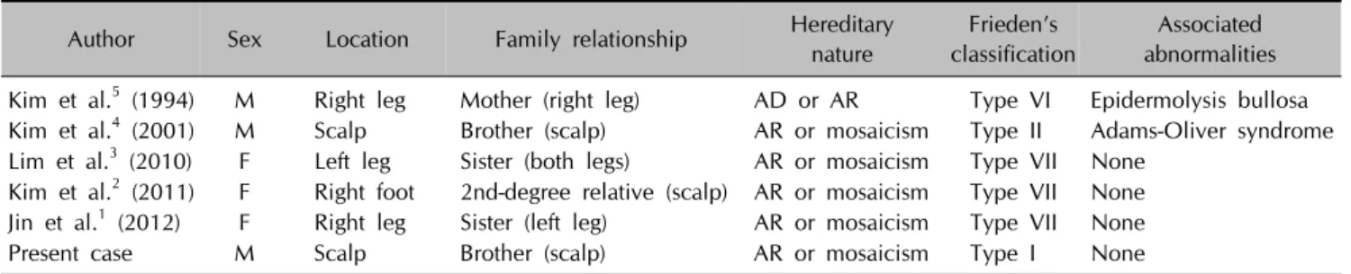
Table 1. Summary of reported cases of familial ACC in Korea
Author Sex Location Family relationship Hereditary
nature
Frieden’s classification
Associated abnormalities Kim et al.5 (1994) M Right leg Mother (right leg) AD or AR Type VI Epidermolysis bullosa Kim et al.4 (2001) M Scalp Brother (scalp) AR or mosaicism Type II Adams-Oliver syndrome Lim et al.3 (2010) F Left leg Sister (both legs) AR or mosaicism Type VII None
Kim et al.2 (2011) F Right foot 2nd-degree relative (scalp) AR or mosaicism Type VII None Jin et al.1 (2012) F Right leg Sister (left leg) AR or mosaicism Type VII None
Present case M Scalp Brother (scalp) AR or mosaicism Type I None
ACC: aplasia cutis congenita, M: male, F: female, AD: autosomal dominant, AR: autosomal recessive.
Fig. 1. (A) Clinical manifestation of irregular crusted skin defects at one month after birth. (B) Clinical features with non-hairy large shiny scar at 18 months after birth. (C) His 8-year-old brother also presented with scalp aplasia cutis congenita (ACC). (D) Histopathological findings revealed dermal fibrosis with absence of adnexal structure, consistent with ACC (H&E, ×200).
VII ACC localized to legs1-3. The other cases were com- bined with syndromic conditions such as epidermolysis bullosa and Adams-Oliver syndrome4,5. Herein, we report 18-month-old male and his 8-year-old brother with ACC on their scalps, which represent type I ACC in siblings without associated congenital anomalies (Fig. 1A∼C).
A 13-day-old infant was transferred to us for evaluation of a large ulcerated-crusted skin defect on the scalp, which was present since birth (Fig. 1A). The infant was born at term (38 weeks, birth weight 2,580 g) by caesarean section. The newborn was otherwise healthy with Apgar score 10 at birth. His parents were healthy without con- sanguinity, known medical problems, history of drug in- take, infection or trauma during pregnancy.
He presented with a well-demarcated irregular ulcerated scalp defect covering an area 10×10 cm. Outside brain computed tomography and neurosonography showed no skull defects or abnormalities. There was no chromosomal abnormality such as trisomy 13 or 4p-syndrome. Clinical diagnosis was ACC, and he was treated conservatively by simple dressing with antibiotic ointment. After 18 months, he returned to discuss advanced treatment (Fig. 1B). We referred him to plastic surgeon for staged repair. A histo- logical study confirmed the diagnosis of ACC, and there were no remnant viable hair follicles on the biopsy speci- men (Fig. 1D).
ACC is a rare congenital disorder with an underlying mechanism that remains unclear. It is characterized by lo- calized or widespread areas of affected skin most com- monly on the scalp, and usually present at birth3. ACC may be associated with defects of the underlying skull, es- pecially when the skin defect is larger than 10 cm2. Our patient had no underlying skull abnormalities, although he had a relatively large scalp lesion. According to Frieden’s classification of ACC, type I (ACC of the scalp without multiple abnormalities) is most common with autosomal dominant or sporadic inheritance1. In our case, the parents had no evidence of ACC, indicating the inheritance pat- tern could be autosomal recessive or represent genetic mosaicism, both of which differ from the typical heredi- tary pattern of type I ACC.
In conclusion, we report a rare case of familial scalp ACC in Korea which follows atypical inheritance patterns. Our case could provide additional support for the diverse he- reditary characteristics of type I ACC.
CONFLICTS OF INTEREST
The authors have nothing to disclose.
REFERENCES
1. Jin SY, Kim DH, Lim WS, Choi YS, Lee AY, Lee SH. Aplasia
Brief Report
Vol. 29, No. 5, 2017 665 cutis congenita in two siblings. Korean J Dermatol 2012;
50:714-717.
2. Kim JY, Lee YK, Ko SY, Kim KA, Shin SM. Clinical course of aplasia cutis congenita. J Korean Soc Neonatol 2011;18:
359-364.
3. Lim JH, Park H, Kim JW, Yu DS. A case of aplasia cutis con- genita in two siblings. Korean J Dermatol 2010;48:517-520.
4. Kim YJ, Jee YH, Lee CH, Kim YC, Cinn YW, Kim EJ, et al. A case of Adams-Oliver syndrome which was observed in two brothers. J Korean Soc Neonatol 2001;8:171-174.
5. Kim HS, Hwang SJ, Kim CH, Kim DC, Park HR. Aplasia cutis congenita: a case report. J Korean Soc Plast Reconstr Surg 1994;21:365.