https://doi.org/10.4174/astr.2017.92.2.82 Annals of Surgical Treatment and Research
Surgical resection of synchronous and metachronous lung and liver metastases of colorectal cancers
Shinseok Jeong, Jin Seok Heo, Jin Young Park, Dong Wook Choi, Seong Ho Choi
Department of General Surgery, Samsung Medical Center, Sungkyunkwan University School of Medicine, Seoul, Korea
INTRODUCTION
Colorectal cancer (CRC) is one of the most common cancers.
About half of all CRC patients will develop hepatic metastases and 20% will develop pulmonary metastases during the course of their disease [1]. For metastatic CRC, prognosis without any treatment is poor. The 5year survival rate of colon cancer with distant metastasis is about 13% in the United States [2,3]. With isolated hepatic metastasis, metastasectomy is an established treatment for resectable metastasis. Previous studies on patients with hepatic metastases treated with surgical treatment re
ported a 58% of 5year survival rate [4,5]. Surgical treatment of pulmonary metastases is common for CRC patients and studies have demonstrated 5year survival rates up to 56% [6,7].
Encouraged by these results, many centers contemporarily offer pulmonary and hepatic metastasectomy to selected patients with both pulmonary and hepatic metastases.
However, such an aggressive approach is controversial [8].
The role of surgery in the management of patients who have both liver and lung metastases is not well defined. There were 12 articles reporting survival after hepatic and pulmonary metastasectomy following CRC surgery (Table 1) [920]. The Purpose: Surgical resection of isolated hepatic or pulmonary metastases of colorectal cancer is an established procedure, with a 5-year survival rate of about 50%. However, the role of surgical resections in patients with both hepatic and pul- monary metastases is not well established. We aimed to analyze overall survival of these patients and associated factors.
Methods: Data retrospectively collected from 66 patients who underwent both hepatic and pulmonary metastasectomy after colorectal cancer surgery from August 2002 through August 2013 were analyzed. In univariate analysis, the log-rank test compared patient survival between groups. P < 0.1 was considered indicative of significance. Multivariate analysis of the significance data using a Cox proportional hazard model identified factors associated with overall survival. The synchronous group (n = 57) was defined as patients who had metastasectomy within 3 months from primary colorectal cancer surgery. The remaining nine patients constituted the metachronous group.
Results: Median follow-up was 126 months from the primary colorectal cancer surgery. The 5-year survival was 73.4%.
There was no difference in overall survival between the synchronous and metachronous groups, consistent with previous studies. Distribution (involving one hemiliver or both, P = 0.010 in multivariate analysis) of liver metastases and multiplicity of the pulmonary metastasis (P = 0.039) were predictors of poor prognosis.
Conclusion: Sequential or simultaneous resection of both hepatic and pulmonary metastasis of colorectal cancer resulted in good long-term survival in selected patients. Thus, an aggressive surgical approach and multidisciplinary decision making with surgeons seems to be justified.
[Ann Surg Treat Res 2017;92(2):82-89]
Key Words: Colorectal cancer, Liver metastasis, Lung metastasis, Synchronous metastasis
Received September 5, 2016, Reviewed September 22, 2016, Accepted October 17, 2016
Corresponding Author: Jin Seok Heo
Department of General Surgery, Samsung Medical Center, Sungkyunkwan University School of Medicine, 81 Irwon-ro, Gangnam-gu, Seoul 06351, Korea
Tel: +82-2-3410-0926, Fax: +82-2-3410-6982 E-mail: [email protected]
Copyright ⓒ 2017, the Korean Surgical Society
cc Annals of Surgical Treatment and Research is an Open Access Journal. All articles are distributed under the terms of the Creative Commons Attribution Non- Commercial License (http://creativecommons.org/licenses/by-nc/4.0/) which permits unrestricted non-commercial use, distribution, and reproduction in any medium, provided the original work is properly cited.
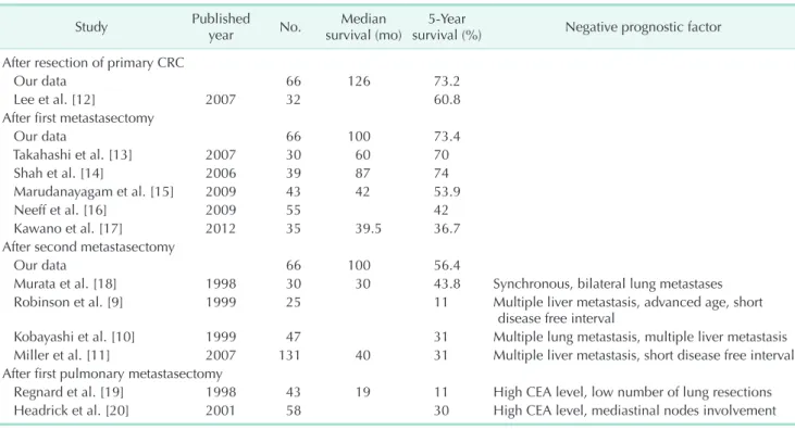
conditions and out comes varied, and factors associated with prolonged survival were debatable. Fiveyear survival rates as high as 11% from the second metastasectomy was reported [9], with 5year survival rate as high as 70% reporting from the first metastasectomy [13].
In this study, clinical, pathologic, and treatmentrelated features of 66 patients who underwent hepatic and pulmonary meta stasectomies at a single institution were prospectively collected over 11 years. Our goal was to compare the outcomes bet ween synchronous and metachronous metastasectomy groups, and to define prognostic factors for survival in this pa
tient group. The data should be useful in patient selection.
METHODS
Patients
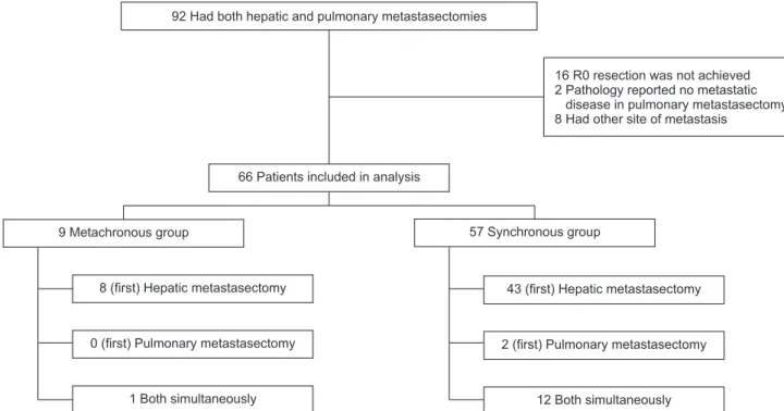
Data collected retrospectively from 92 patients who under
went both hepatic and pulmonary metastasectomy after CRC surgery from August 2002 to August 2013 at a single institution, Samsung Medical Center, in Seoul, Korea. Through March 2015, the patients were offered hepatic and pulmonary metastasectomy only if oncologically curative resection was possible. There were no wellestablished criteria used in selection of pa tients. Selec tion of patients for resection of multiple site meta static disease was at the discretion of the surgeon. Patient information included tumor, treatment, recurrence, and deathrelated in forma tion. Twentysix patients
were excluded; 16 patients failed to achieve R0 resection in metastasectomy and required pallia tive surgery, 2 patients confirmed to have no lung metastasis after lung resection, and 8 patients had other site of metastasis. Finally, 66 patients were included in this analysis.
Definitions
Synchronous metastasis was defined as metastasis to the liver or lung at the time of resection of the primary CRC or the first metastasectomy done within 3 months from primary CRC surgery. The metachronous group was defined as patients who had the first metastasectomy later than 3 months from the primary colorectal tumor resection (Fig. 1). The occurrence of bilateral lung metastases represented one episode of metastasis regardless of whether the disease was resected during one operation or staged thoracotomies. Radiofrequency ablation (RFA) was also counted as metastasectomy of hepatic metastasis if the RFA procedure was done according to the radiologists’
reports. Diseasefree intervals (DFI) were calculated as the time between surgeries. The primary DFI was between primary CRC surgery and first site metastasectomy as well as the secondary DFI was between the first site metastasectomy and second site metastasectomy. Primary DFI was 0 to 3 months in the synchronous metastasectomy group. Secondary DFI was 0 in patients presenting with simultaneous liver and lung metastases.
Table 1. Previous reports about survival of sequential hepatic and pulmonary metastasectomy following CRC surgery
Study Published
year No. Median
survival (mo) 5Year
survival (%) Negative prognostic factor After resection of primary CRC
Our data 66 126 73.2
Lee et al. [12] 2007 32 60.8
After first metastasectomy
Our data 66 100 73.4
Takahashi et al. [13] 2007 30 60 70
Shah et al. [14] 2006 39 87 74
Marudanayagam et al. [15] 2009 43 42 53.9
Neeff et al. [16] 2009 55 42
Kawano et al. [17] 2012 35 39.5 36.7
After second metastasectomy
Our data 66 100 56.4
Murata et al. [18] 1998 30 30 43.8 Synchronous, bilateral lung metastases
Robinson et al. [9] 1999 25 11 Multiple liver metastasis, advanced age, short
disease free interval
Kobayashi et al. [10] 1999 47 31 Multiple lung metastasis, multiple liver metastasis Miller et al. [11] 2007 131 40 31 Multiple liver metastasis, short disease free interval After first pulmonary metastasectomy
Regnard et al. [19] 1998 43 19 11 High CEA level, low number of lung resections
Headrick et al. [20] 2001 58 30 High CEA level, mediastinal nodes involvement
CRC, colorectal cancer.
Statistical analysis
Survival and its associated factors were analyzed by univa
riate and multivariate analyses. Some continuous or categorical variables were converted into dichotomous variables, such as age (<60 years vs. ≥60 years), T stage (0-2 vs. 3, 4), N stage (0 vs.
1, 2), and DFI (<12 months vs. ≥12 months). Survival analysis used IBM SPSS Statistics ver. 22.0 (IBM Co., Armonk, NY, USA).
Actuarial survival was determined by KaplanMeier analysis.
Relationships of patient, tumor, and treatment characteristics to outcomes were tested by logrank test. Multivariate analysis was done with the Cox proportional hazards model explaining the overall survival after first metastasectomy. In univariate analysis, factors whose Pvalues were <0.1 were selected for multivariate analysis. All factors whose Pvalues were <0.05 in multivariate analysis were considered statistically significant.
RESULTS
Sixtysix patients were included in the analysis. They had both liver and lung metastasectomies after CRC surgery during the period of study. Table 2 shows the clinicopathologic and operative characteristics. Median age was 59 years (range, 25 to 79 years) at first diagnosis. Fortyfive were male patients and 21 were female. Nine patients were in the metachronous group and 57 patients were in the synchronous group (M1 stage).
Twentyfive patients had CRC originating from the colon, while 41 patients had CRC originating from the rectosigmoid junction or rectum. The majority of the subjects was T3 (47, 71.2%). Four
patients were less than T2 and 15 subjects were T4. In nodal status, 25 (37.9%) was negative. Twentyone patients (31.8%) were N2 and 20 (30.3%) were diagnosed with N1. 1 of them has proven to achieve pathologic complete remission (ypT0N0) in primary surgery (colon and liver). Ten patients received
92 Had both hepatic and pulmonary metastasectomies
66 Patients included in analysis 9 Metachronous group
16 R0 resection was not achieved 2 Pathology reported no metastatic
disease in pulmonary metastasectomy 8 Had other site of metastasis
57 Synchronous group 8 (first) Hepatic metastasectomy
0 (first) Pulmonary metastasectomy 1 Both simultaneously
2 (first)Pulmonarymetastasectomy 43 (first) Hepatic metastasectomy
12 Both simultaneously
Fig. 1. Patient grouping according to initial presentation and sequence of metastasis site.
Table 2. Patient demographics
Variable Value
Sex
Male 45 (68.2)
Female 21 (31.8)
Age (yr), median (range) 59 (25–79)
Chronology
Metachronous (M0) 9 (13.6)
Synchronous (M1) 57 (86.4)
Colorectan cancer site
Colon 25 (37.9)
Rectum or rectosigmoid junction 41 (62.1) T
T2 or less 4 (6.1)
T3 47 (71.2)
T4 15 (22.7)
N
N0 25 (37.9)
N1 20 (30.3)
N2 21 (31.8)
Values are presented as number (%) unless otherwise indicated.
neoadjuvant chemotherapy before primary CRC surgery.
Survival
Fortysix of 66 patients (69.6%) survived. Median survival was 100 months (95% confidence interval [CI], 55.3 to 196.7) from primary CRC surgery, 100 months (95% CI not available) from first metastasectomy, and 100 months (95% CI, 40.5 to 159.5 months) from second site metastasectomy. The 5year survival rate was 73.2% from primary CRC surgery, 73.4%
from first metastasectomy, and 56.4% from the second site metastasectomy. Survival curves are shown in Fig. 2.
Synchronous versus metachronous metastasis
Synchronous group (n = 57) was defined as patients who had first metastasectomy within 3 months from primary CRC surgery, the others (who had first metastasectomy after
3 months from the primary CRC surgery) comprised the metachronous group (n = 9). Median survival for synchronous metastasis was 126 months, while the median survival was not available for metachronous metastasis group because of the lack of mortality cases. In synchronous metastases patients, the majority (n = 43, 65%) presented with liver metastasis (liver first group) compared to 8 subjects (12.1%) in the metachronous metastases group. Two patients (3.0%) were in the lung first group in the synchronous metastasis group. Twelve of those with synchronous metastases (18.2%) presented with lung and liver metastases simultaneously, compared with 1 patient (1.5%) in the metachronous metastases group.
There was no difference in longterm survival between the synchronous and metachronous group, and this result was consistent with previous studies [911]. The 5year survival rate after primary CRC surgery was 77.8% in the synchronous group,
Survivalprobability
Observation from the first colorectal cancer surgery (mo)
A B
C D
0 150
0 1.0 0.8 0.6 0.4 0.2
Synchronous group versus metachronous group
100 50
Metachronous group
Metachronous group (censored) Synchronous group
Synchronous group (censored)
Survivalprobability
Observation from the first metastasectomy (mo)
0 140
0 1.0 0.8 0.6 0.4 0.2
Synchronous group versus metachronous group
Metachronous group
Metachronous group (censored) Synchronous group
Synchronous group (censored) 120 100 80 60 40 20
Survivalprobability
Observation from the first metastasectomy (mo)
0 140
0 1.0 0.8 0.6 0.4
0.2 Solitary lung metastasis
Solitary lung metastasis (censored) Multiple lung metastasis
Multiple lung metastasis (censored) 120 100 80 60 20 40
Survivalprobability
Observation from the first metastasectomy (mo)
0 140
0 1.0 0.8 0.6 0.4 0.2
Solitary versus multiple liver metastases
Solitary liver metastasis
Solitary liver metastasis (censored) Multiple liver metastasis
Multiple liver metastasis (censored) 120 100 80 60 20 40
Solitary versus multiple lung metastases
Fig. 2. Survival results. (A) Synchronous versus metachronous survival analysis, by logrank test (P = 0.327, by logrank) (1, meta chronous; 2, synchronous). (B) Survival from first metastatetomy by logrank test (P = 0.813, by logrank). (C) Comparison bet ween liver metastases involving single lobe or both lobe in survival from first site metastasectomy (P = 0.031, by logrank). (D) Com parison between single and multiple lung metastasis in survival from first site metastasectomy (P = 0.784, by logrank).
similar to the metachronous group (73.1%). The 5year survival rate of the synchronous and metachronous group following first metastasectomy was 74.1% and 73.2%, respectively. In logrank test, P was 0.327 after first CRC surgery and 0.819 after first metastasectomy.
In the synchronous metastasis group, 5 patients received primary colectomy, hepatic metastasectomy, and pulmonary metastasectomy within a month. Three patients received those surgeries in the same day, and the others received colectomy and hepatic metastasectomy first and then pulmonary meta
stasectomy within 7 to 14 days. The aforementioned 5 patients displayed no 30day mortality and surgeryrelated morbidities.
Liver metastasis
Fiftyfive patients (83.3%) presented with single lobe involve
ment no matter, while only 11 patients (16.7%) had multiple metastasis involving both hepatic lobes. Fiftythree (65.2%) patients had single metastasis and 23 patients (34.8) had multiple metastastases at first presentation of liver metastasis.
Fiftyseven of patients (86.3%) received hepatic metastasectomy only once, 7 had 2 hepatic metastasectomies, and 2 had 3 hepatic metastasectomies. Median survival for single hepatic lobe involvement group was 125 months (95% CI, 33 to 212) and 42 months (95% CI, 33 to 52) for both hepatic lobe involvement group. While multiple liver metastasis versus single liver metastasis did not differ (P = 0.661; hazard ratio [HR], 1.123;
95% CI, 0.483 to 3.151) in logrank test, bilobar involvement of liver metastasis did show poor prognosis compared to single lobar involvement in first liver metastasis (P = 0.022; HR, 3.36;
95% CI, 1.192 to 9.474). Fig. 2 shows cumulative survival curve from primary CRC surgery depending on the multiplicity and bilobar distribution of liver metastases.
Lung metastasis
Concerning lung metastasis, 51 patients (77.3%) had lung lesions on one side, while 15 (22.7%) had both lungs involved at first pulmonary metastases. Fortyfour patients (66.7%) had single metastasis and 22 (33.3%) had multiple lung metastases at first presentation of lung metastases. Single lung metastasis group was compared with the multiple lung metastasis group;
no difference was evident (P = 0.078). Both lung involvement groups were compared with the single lung (right or left only) metastasis group; concerning survival, no difference was evident (P = 0.086). Fig. 2 shows cumulative survival curve from primary CRC surgery depending on the multiplicity and lobar distribution of lung metastases.
Disease-free interval
Median primary DFI was 0 months (range, 0 to 31 months) and median secondary DFI was 16 months (range, 0 to 78 months). In primary DFI, 59 patients were categorized as
shorter primary DFI (<1 year) and the other 7 patients as longer pri mary DFI. Twentytwo patients were categorized as shorter secondary DFI, and the other 44 patients as longer secondary DFI. Comparision between shorter DFI groups and longer DFI groups; the difference was not significant for primary DFI (P = 0.89), but was for secondary DFI (P = 0.013).
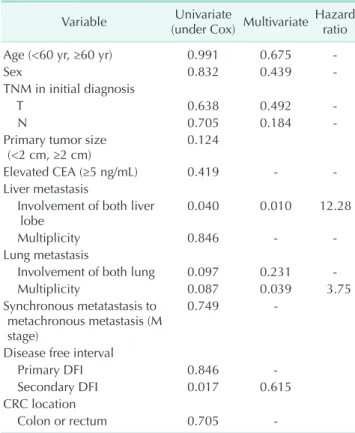
Prognostic factors
Analysis of potential prognostic factors for survival from the time of resection of the first metastasis is shown in Table 3.
Involvement of both liver lobe and lung, multiplicity of lung metastasis, and longer secondary DFI were counted possible prognostic factors and included in multivariate analysis. In multivariate analysis with Coxproportional hazard model, involvement of both liver lobe (P = 0.010) and multiplicity of lung metastasis (P = 0.039) were inversely associated with prolonged survival.
DISCUSSION
In this analysis, as many previous studies have shown, metastasectomies of CRC in selected patients showed accept
Table 3. Univariate (Cox regression) and multivariate an
alysis (Cox proportional hazard model) for overall survival after synchronous or sequential pulmonary and hepatic meta stasectomy
Variable Univariate
(under Cox) Multivariate Hazard ratio
Age (<60 yr, ≥60 yr) 0.991 0.675
Sex 0.832 0.439
TNM in initial diagnosis
T 0.638 0.492
N 0.705 0.184
Primary tumor size
(<2 cm, ≥2 cm) 0.124
Elevated CEA (≥5 ng/mL) 0.419
Liver metastasis
Involvement of both liver
lobe 0.040 0.010 12.28
Multiplicity 0.846
Lung metastasis
Involvement of both lung 0.097 0.231
Multiplicity 0.087 0.039 3.75
Synchronous metatastasis to metachronous metastasis (M stage)
0.749
Disease free interval
Primary DFI 0.846
Secondary DFI 0.017 0.615
CRC location
Colon or rectum 0.705
DFI, diseasefree interval; CRC, colorectal cancer.
able and good survival prognosis. This result is comparable with 5year survival of pulmonary metastasectomy of CRC [21]. During the time of this study, our institution offered CRC surgery with curative intent for 7,531 patients. Of these, 1,032 presented with metastatic disease (M1). Regardless of the cause of the death, the overall 7,531 patients had a 5year survival rate of 49.1%, while those with metastatic disease had a rate of 49.3%. Sixtysix patients with both pulmonary and hepatic metastasectomies had a 5year survival rate from CRC surgery of 73.4% and 56.4% from the first metastasectomy. This result indicates that patients with both pulmonary and hepatic metastasectomies may be strictly selected for resectability.
Median followup was 48 months (ranging from 14 to 147 months) from the first metastasectomy, and 31 (ranging from 1 to 107 months) from the second site metastasectomy. At the end of the period of observation, 20 patients had died and
46 patients were still alive. Among the survivors, disease had progressed in 13, partial response or stable disease was evident in 3, and the remaining 30 patients showed no evidence of disease recurrence. Table 4 summarizes longterm outcomes of several recent studies on metastatic CRC. Survival after sequential and simultaneous metastatectomies of liver and lung for metastatic CRC was reported 12 articles including more than 25 patients. Survival varied widely and could be partly due to differences in patient selection and study method.
Presently, synchronous and metachronous metastasis seem
ed not to affect prognosis (P = 0.327 by logrank test), similar to other studies [911]. Multiplicity of liver metastasis has been associated with poor prognosis [911]. This was not evi
dent in the present analysis. In univariate analysis, multiple liver metastasis had a similar survival outcome with single liver metastasis group (P = 0.659 in logrank test). However,
Table 4. Site and chronology of metastases
Variable No. (%) Overall survival (%)
Median survival Pvalue
3Year 5Year
Over all 66 (100) 89.8 73.4 100 0.141
Liver first 51 (77.3) 91.6 78.7 N/A
Lung first 2 (3.0) 100 50.0 39
Both simultaneously 13 (19.7) 80.0 54.9 N/A
Chronology group 0.814
Synchronous metastasesa) 57 (86.4) 89.9 73.2 100
Metachronous metastasesb) 9 (13.6) 88.9 74.1 N/A
Synchronous metastasesa) 57 (86.4) 89.9 73.2 100 0.120
Liver first 43 (65.2) 92.4 79.8 N/A
Lung first 2 (3.0) 100.0 50.0 69.5
Both simultaneously 12 (18.2) 77.8 48.6 47
Metachronous metastasesb) 9 (13.6) 88.9 77.8 N/A 0.723
Liver first 8 (12.1) 87.5 N/A N/A
Lung first 0 (0) N/A N/A N/A
Both simultaneously 1 (1.5) 100.0 100.0 N/A
Liver metastasis N/A
Single lobe 55 (83.3) 93.8 79.2 N/A 0.031
Both lobe 11 (16.7) 70.0 42.0 43
Single metastasis 53 (65.2) 91.9 78.3 100 0.843
Multiple metastasis 23 (34.8) 86.4 65.7 N/A
Lung metastasis
Single lung 51 (77.3) 93.3 78.0 N/A 0.086
Both lung 15 (22.7) 76.9 54.9 68
Single metastasis 44 (66.7) 92.4 78.2 N/A 0.078
Multiple metastasis 22 (33.3) 84.7 62.1 68
Disease free interval
Primary DFI < 12 mo 59 (89.4) 90.3 74.3 100 0.89
Primary DFI ≥ 12 mo 7 (10.6) 85.7 68.6 N/A
Secondary DFI < 12 mo 22 (33.3) 72.7 56.9 64 0.013
Secondary DFI ≥ 12 mo 44 (66.7) 97.6 80.4 N/A
DFI, diseasefree interval; N/A, not available.
a)First metastasectomy within 3 months from primary cancer surgery. b)First metastasectomy after 3 months from primary cancer surgery.
involve ment of both lobes in hepatic metastasis was associated with poor prognosis (P = 0.015, by logrank test). This may be because prior studies did not consider the distribution of the liver metastasis rather than multiplicity of the liver metastasis as a prognostic factor. In univariate analysis, distribution of liver metastasis, multiplicity of lung metastasis, and secondary DFI had P < 0.1. In multivariate analysis, only the distribution of the liver metastasis and multiplicity of lung metastasis showed P < 0.05 (HR, 12.28 and 3.75, respectively).
In this analysis, as many previous studies have done, the data have a limitation that it does not represent wall metastatic CRC patients. Prior survival data (Table 4) involved different and subjective criteria, so the results were not amenable to statistical analysis.
Sequential resection of both hepatic and pulmonary colorec
tal metastases resulted in good longterm survival in the pre
sent subjects. Moreover, distribution of the liver metastases rather than multiplicity was a significantly poor prognosis factor for patients with both hepatic and pulmonary metastases.
Thus, an aggressive surgical approach and multidisciplinary decision making with surgeons may be justified with this data.
For defining detailed surgical criteria for both hepatic and pulmonary metastases, further studies that include more pa
tients are needed.
CONFLICTS OF INTERESTS
No potential conflict of interest relevant to this article was reported.
1. Liu LX, Zhang WH, Jiang HC. Current treatment for liver metastases from colorectal cancer. World J Gastroenterol 2003;9:193200.
2. Simmonds PC. Palliative chemotherapy for advanced colorectal cancer: systematic review and metaanalysis. Colorectal Cancer Collaborative Group. BMJ 2000;
321:5315.
3. American Cancer Society. Cancer facts &
figures. Atlanta: American Cancer Society;
2015.
4. Fong Y, Fortner J, Sun RL, Brennan MF, Blumgart LH. Clinical score for predicting recurrence after hepatic resection for metastatic colorectal cancer: analysis of 1001 consecutive cases. Ann Surg 1999;230:30918.
5. Shah SA, Bromberg R, Coates A, Rempel E, Simunovic M, Gallinger S. Survival after liver resection for metastatic colorectal carcinoma in a large population. J Am Coll Surg 2007;205:67683.
6. Zabaleta J, Aguinagalde B, Fuentes MG, Bazterargui N, Izquierdo JM, Hernández CJ, et al. Survival after lung metastasectomy for colorectal cancer:
importance of previous liver metastasis as a prognostic factor. Eur J Surg Oncol 2011;37:78690.
7. Hornbech K, Ravn J, Steinbruchel DA.
Outcome after pulmonary metastasec
tomy: analysis of 5 years consecutive surgical resections 20022006. J Thorac Oncol 2011;6:173340.
8. Kneuertz PJ, Chang GJ, Hu CY, Rodriguez
Bigas MA, Eng C, Vilar E, et al. Overtreat
ment of young adults with colon cancer:
more intense treatments with unmatched survival gains. JAMA Surg 2015;150:4029.
9. Robinson BJ, Rice T W, Strong SA, Rybicki LA, Blackstone EH. Is resection of pulmonary and hepatic metastases warranted in patients with colorectal cancer? J Thorac Cardiovasc Surg 1999;117:
6675.
10. Kobayashi K, Kawamura M, Ishihara T.
Surgical treatment for both pulmonary and hepatic metastases from colorectal cancer. J Thorac Cardiovasc Surg 1999;
118:10906.
11. Miller G, Biernacki P, Kemeny NE, Gonen M, Downey R, Jarnagin WR, et al.
Outcomes after resection of synchronous or metachronous hepatic and pulmonary colorectal metastases. J Am Coll Surg 2007;205:2318.
12. Lee WS, Yun HR, Yun SH, Chun HK, Lee WY, Kim SJ, et al. Treatment outcomes of hepatic and pulmonary metastases from
colorectal carcinoma. J Gastroenterol Hepatol 2008;23(8 Pt 2):e36772.
13. Takahashi S, Nagai K, Saito N, Konishi M, Nakagohri T, Gotohda N, et al. Multiple resections for hepatic and pulmonary metastases of colorectal carcinoma. Jpn J Clin Oncol 2007;37:18692.
14. Shah SA, Haddad R, AlSukhni W, Kim RD, Greig PD, Grant DR, et al. Surgical resection of hepatic and pulmonary metastases from colorectal carcinoma. J Am Coll Surg 2006;202:46875.
15. Marudanayagam R, R amkumar K, Shanmugam V, Langman G, Rajesh P, Coldham C, et al. Longterm outcome after sequential resections of liver and lung metastases from colorectal carcinoma. HPB (Oxford) 2009;11:6716.
16. Neeff H, Horth W, Makowiec F, Fischer E, Imdahl A, Hopt UT, et al. Outcome after re section of hepatic and pulmonary meta
stases of colorectal cancer. J Gastrointest Surg 2009;13:181320.
17. Kawano D, Takeo S, Tsukamoto S, Katsura M, Masuyama E, Nakaji Y. Prediction of the prognosis and surgical indications for pulmonary metastectomy from colorectal car ci noma in patients with combined hepa tic metastases. Lung Cancer 2012;75:
20912.
REFERENCES
18. Murata S, Moriya Y, Akasu T, Fujita S, Sugihara K. Resection of both hepatic and pulmonary metastases in patients with colo rectal carcinoma. Cancer 19985;83:
108693.
19. Regnard JF, Grunenwald D, Spaggiari L, Girard P, Elias D, Ducreux M, et al. Sur
gical treatment of hepatic and pul mo nary meta stases from colorectal cancers. Ann Thorac Surg 1998;66:2148.
20. Headrick JR, Miller DL, Nagorney DM, Allen MS, Deschamps C, Trastek VF, et al.
Surgical treatment of hepatic and pul mo
nary metastases from colon cancer. Ann
Thorac Surg 2001;71:9759.
21. Pfannschmidt J, Dienemann H, Hoffmann H. Surgical resection of pulmonary metastases from colorectal cancer: a systematic review of published series.
Ann Thorac Surg 2007;84:32438.