• 검색 결과가 없습니다.

Identification and Characterization of Protease-Resistant Proteins from Adzuki Beans

N/A
N/A
Protected

Academic year: 2021

Share "Identification and Characterization of Protease-Resistant Proteins from Adzuki Beans"

Copied!
6
0
0

로드 중.... (전체 텍스트 보기)

전체 글

(1)

Original Article

Identification and Characterization of Protease-Resistant Proteins from Adzuki Beans

Eun-Jung Song 1 , Sun-Min Park 2 , Qun Wang 1 , and Jinkyu Lim 1 *

1 School of Food Science & Biotechnology, Major in Life and Food Sciences, Kyungpook National University, Daegu , Korea

2 Gyeongbuk Technopark, Pohang, Gyeongsangbuk-do, Korea

소화 효소 저항성을 지니는 팥 단백질의 성질 규명

송은정 1 ·박선민 2 ·왕 췬 1 ·임진규 1 *

1 경북대학교 응용생명과학부 생명식품공학전공, 경북대학교 식품공학부 , 2 경북 테크노파크, 포항, 경상북도

Received: September 15 2014 / Revised: September 26 2014 / Accepted: September 26 2014

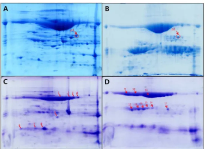
Abstract It is already known that adzuki beans (Vigna angularis) are able to control appetite. Therefore, this study tested the proteins isolated from adzuki beans for their protease resistance and interaction with the intestinal muco- sa. The major proteins from adzuki beans were found to be resistant to the digestive enzymes pepsin and pancreatin, and were identified using 2D-SDS-polyacrylamide gel elec- trophoresis and mass spectrometry. The major adzuki proteins were easily fractionated by treating the soluble protein extract with 10 mM CaCl 2 , and were found to contain lacto- transferrin, a homologous protein to the dynein light chain domain, proteinase inhibitor, and proteins with unknown func- tions. From a tissue binding assay using mouse intestinal tissue sections, the major protein fraction showed weak, yet significant and specific binding to the mucosa layer of the small intestine. Thus, the current results suggest that adzuki proteins are resistant to digestive enzymes, which enables them to survive protease digestion in the intestinal tract, plus they may interact with the intestinal mucosa layer.

Therefore, the molecules responsible for controlling appetite in adzuki beans are presumably protease-resistant proteins that interact with the intestinal mucosa or delay digestion in the digestive tract.

Keywords: adzuki bean, protease resistant protein, pepsin, pancreatin, tissue binding

서 론

팥은 떡고물, 양갱, 팥빙수용으로 많이 쓰이는 곡류로 잡곡 등 혼식용으로도 이용되고 있다. 팥에는 녹말 등의 탄수화물이 약 50% 함유되어 있으며, 그 밖에 단백질이 약 20% 함유되 어 있다. 이 단백질 함량은 콩류 안에서는 중간 정도의 함량 이고 이중 80%는 글로불린이다. 글로불린 단백질 중 대부분 이 glycine이며 valine을 제외한 필수 아미노산이 풍부하다 (Hwang et al., 2005). 특히 lysine 함량이 높아 lysine이 부족 한 쌀과 함께 혼식하면 단백질 보충에 매우 효과적이다. 이외 에도 비타민 B1이 현미보다도 더 많이 함유되어 있어 각기병 치료에 효과적이다. 이뇨작용, 피로회복에도 도움이 되며 비타 민 A, B2, 칼슘, 인, 철분 등이 함유되어 있다. 또한 팥의 외 피에는 사포닌과 콜린이라는 성분이 들어있어 항암효과와 성 인병 예방에도 도움이 된다고 알려져 있으며, 포함된 사포닌 의 거품을 내는 특성 때문에 피부 미용에도 이용된다(Kang and Han, 2012). 팥은 열량도 낮아 다이어트 식품으로도 각광 받고 있으며(Thomson and Winham, 2013) 팥의 식이섬유 (5 g/100 g) 는 장운동을 도와 변비해소에도 좋다(Kim et al., 2003). 이처럼 팥은 콩에 비해서는 이용이 적지만 최근 웰빙

*Corresponding author: Jinkyu Lim Tel: 82-53-950-5755; Fax: 82-53-950-6750 E-mail: [email protected]

Song EJ and Park SM contributed egually in this study.

This is an Open Access article distributed under the terms of the Creative Commons Attribution Non-Commercial License (http://creativecommons.

org/licenses/by-nc/3.0/) which permits unrestricted non-commercial use, distribution, and reproduction in any medium, provided the original work is properly cited.

© 2014 Institute of Agricultural Science and Technology, Kyungpook

National University

(2)

한 단백질들을 단백질 분해효소 저항성 단백질이라 보고 팥 또한 이러한 콩 단백질처럼 완전히 분해되지 않고 소장 점막 과 어떠한 상호작용을 하는 팥 단백질이 있을 가능성이 있으 므로 팥 단백질 중의 소화효소 저항성 단백질에 대한 연구를 중점으로 실험을 진행하였다.

재료 및 방법

Defatted adzuki bean 샘플용액 제조

팥을 4 o C 증류수에 18 시간 동안 침지시켜 외피를 모두 제거 한 후 전체의 반은 10분 동안 100 o C로 열처리를 하고 나머지 반은 열처리 없이 각각 동결건조하여 완전히 수분을 제거하 였다. 그 후 건조된 열처리 및 무열처리 팥을 각각 분쇄기에 갈아서 고운 분말로 만들고 분말 30 g 당 0.1 × PBS 150 mL 를 첨가 후 1 시간 동안 실온에서 200 rpm으로 교반하여 단 백질을 추출하고 12000 rpm, 4 o C 에서 30분간 원심분리하여 수 용성 단백질 상층액을 얻었다.

Simulated gastric/intestinal fluid 에서 팥 단백질 분해 수용성 팥 단백질 용액을 9 mL씩 취하고 pepsin을 최종 3.2 mg/mL되도록 첨가하여 pH 2로 맞추어 주고(Galia et al., 1998) 37 o C water bath 에서 반응시키며 0, 1, 2 h에 각각의 반 응물을 채취하고 1 M NaOH을 가하여 pH 7로 맞추어 반응 을 종료 시킨 후 0 o C 에서 보관하였다. 채취하고 남은 팥 단백 질 반응물에 1 M NaOH 용액을 가하여 pH 6.8로 맞춘 후 pancreatin 의 최종 농도가 10 mg/mL 되도록 첨가하고(Galia et al., 1998; Stippler et al., 2004) water bath incubator(37 o C) 에 서 반응시키며 1, 2, 3, 4, 18 h에서 각각 반응물을 채취하고 5분간 100 o C 물에서 중탕 열처리하여 반응을 종결 시킨 후 0 o C 에서 보관하였다.

단백질분해효소 저항성 단백질의 분리

열처리구와 무열처리구 팥 단백질을 pepsin 및 pancreatin으로 소 화시킨 후 소화효소에 저항성을 보이는 단백질분석을 위해 12%

Sodium dodecylsulfate-polyacrylamide gel electrophoresis(SDS- PAGE) 로 100 V에서 전기영동 후 gel을 Coomassie Brilliant Blue G-250으로 overnight staining, 10% ethanol 용액으로

무열처리 및 열처리 팥 단백질을 pancreatin으로 4 h 처리한 것, 그리고 CaCl 2 처리하여 얻은 major protein을 simulated intestinal fluid 용액에 녹이고 이 용액을 pancreatin으로 4 h 처리한 시료 3 mL 에 1 : 1 비율로 phenol을 넣은 후 vortexing 하고 5분간 3000 rpm, 37 o C 에서 원심분리하였다. 그 뒤 단백질이 들어 있 는 아래쪽 phenol층을 취하여 5 × 부피의 0.1 M ammonium acetate/methanol 을 넣어 단백질을 침전시켰다. 침전된 단백질 을 70% ethanol을 넣고 vortexing하여 washing한 뒤 5분간 12000 rpm, 37 o C 에서 원심분리하고 pellet을 세척하는 과정을 3 번 반복하였다. 세척된 단백질 시료를 동결건조하고 그 단백질 시료 450 µg을 사용하여 pH 4.0-7.0 구간의 IPG-strip(Biorad, Herculus, CA, USA)을 이용하여 36000 V-h로 isoelectric focusing 하였다. 1차원 전기영동이 이루어진 strip을 equilibration buffer 에서 30분간 처리 후 2차원 전기영동을 12% SDS-PAGE로 실시하였다. 전기영동이 끝난 후 gel을 Coomassie Brilliant Blue G-250으로 staining(overnight)한 후 10% ethanol로 destaining하고 증류수로 세척한 후 gel 싱에 분리된 단백질 image를 scan하였다.

MALDI-TOF 측정

2-D gel에서 선발한 단백질 spot들을 50% acetonitrile/0.1 M NaHCO 3 , pH 8.0 로 destaining한 후 냉동건조 하여 보관하였다.

Matrix Assisted Laser Desorption Ionization-Time of Flight (MALDI-TOF) 질량분석 할 spot을 정하고 동결건조한 gel에 1 mg/mL 0.1 M NaHCO 3 , pH 8.0, Trypsin(Promega, MI, USA) 을 15 µL씩 넣고 얼음에서 30분 동안 incubation 한 뒤 37 o C water bath 에 16시간 반응시켰다. Trypsin을 처리한 tube에 서 15 µL 용액을 취하여 새 tube에 담고 얼린 후에 동결 건조 하여 MALDI-TOF를 위한 샘플을 만든다. MALDI-TOF 분석에 서 얻어진 mass spectra를 M-over-Z 프로그램으로 calibration하 고 그 값들을 가지고 NCBI-database를 검색하여 단백질을 동 정하였다(Webster, 2012). MASCOT을 검색엔진으로 사용하여 얻은 score로부터 가장 신뢰도가 높은 것들을 동정된 단백질로 결정하였다.

CaCl 2 처리로 얻은 major 팥 단백질을 이용한 tissue binding 냉동 보관하였던 major 팥 단백질인 CaCl 2 pellet 을 biotinylation 하기 위해 1 × PBS 로 10 mg/mL 농도로 맞춰주고 10 mg biotin/

1 mL dimethyl sulfoxide 0.1 mL 과 1 h 실온에서 반응 시켜주고

(3)

얼음 위에서 냉장보관하였다. 쥐의 소장을 paraffin으로 고정하 여 4 µm 절편으로 lateral 하게 자른 slide를 이용하여 tissue binding 을 실행하였다. 먼저 paraffin 제거를 위해 xylene에 10분 간 slide를 담그고 차례대로 100% ethanol에 3분, 95% ethanol 에 3분, 80% ethanol에 3분, 70% ethanol에 3분 동안 담근다.

그 뒤 물에 5분, 3% H 2 O 2 용액에 13분 처리 후 물에 15분간 담근다. Antigen retrieving을 위해 slide를 0.01 M citric acid, pH 6.0에 넣고 마이크로웨이브 오븐에서 10분간 가열시켜준다.

30 분간 열을 식힌 후 물에 10분, 1 × PBS 에 10분 처리한다.

Slide 조직 바깥 쪽에 paraffin으로 테두리 원을 그려 marking 하고 blocking 용액으로 2-3 시간 blocking 한다. Biotinylation 된 시료와 avidin을 1 : 2비율로 섞고 slide에서 blocking 용액을 제거하고 0.1% Tween 20이 들어있는 blocking 용액을 떨어뜨 린다. 그 후 두 개의 조직에 각각 20, 40 µL씩 떨어뜨린다.

1 × PBS 로 3번 세척 후 mounting 용액을 떨어뜨리고 coverslip 으로 덮어 mounting한다. 그 뒤 노출을 Light 10 ms, FITC 4 s, DAPI 600 ms 로 현미경 관찰하였다.

결 과

팥 단백질의 단백질 분해효소 저항성

열처리와 무열처리 팥에서 분리한 수용성 단백질을 분리하여 Bradford 방법(Bradford, 1976)으로 단백질 정량을 한 결과 무 열처리 팥에서는 60 mg/mL의 농도로 단백질이 측정되었으나 열처리구에서는 6 mg/mL 이하로 단백질이 측정되어 열처리에

의해 대부분의 팥 단백질이 불용화되는 것으로 추정되었다 (Figure 1B). 따라서 이 후의 실험에서는 무열처리 팥 단백질 을 이용한 실험을 진행하였다. 무열처리와 열처리된 팥 단백 질의 단백질 분해효소 저항성을 알아보기 위해 우선 팥 단백 질들을 pepsin으로 2 h 그리고 pancreatin으로 18 h동안 반응시 킨 후 분해되지 않고 남은 단백질을 12% SDS-PAGE 상에서 분리해서 염색을 한 결과 무열처리한 팥 단백질은 pepsin에 대해 대부분 저항성을 보였고 pancreatin에 대해서는 4 h 반응 시킬 경우 대부분 저항성을 보였으며 18 h 반응에는 대부분의 major 단백질들이 분해되었을 뿐 나머지 단백질들은 저항성을 보였다. 그러나, 열처리한 팥 단백질은 수용성으로 추출된 단 백질의 농도가 낮아 gel 상에서 희미한 단백질 band를 보였는 데 pepsin 반응에서는 저항성을 보였으나 pancreatin 반응에서 는 대부분의 단백질이 분해되는 경향을 보였다. 이 결과는 팥 Figure 1. Protease resistance of proteins fractionated from adzuki beans.

The soluble proteins from no-heat-treated (A) and heat-treated (B) adzuki beans were dissolved in simulated gastric fluid and digested using pepsin at pH 2.0 for 0, 1, and 2 h. The 2 h pepsin-digested sample was pH- adjusted and digested by pancreatin for 1, 2, 3, 4, and 18 h at pH 6.8. The samples were heat inactivated right after sampling and stored frozen until analyzed using 12% SDS-PAGE.

Figure 2. Removal of major adzuki proteins by CaCl

2

treatment. The soluble fraction of the adzuki proteins (Total) was treated with 0.1 M CaCl

2

for 1 h on ice and centrifuged for 30 min at 10000 g. Most of the major proteins were removed as a pellet fraction and the proteins remaining in the supernatant (Sup) were enhanced and visualized using 12% SDS-PAGE.

Figure 3. Protease resistance of major adzuki proteins pelleted by CaCl

2

treatment. The major proteins from the adzuki beans remained resistant

during 2 h digestion by pepsin and a consecutive 4 h digestion by

pancreatin (Pan_4 h).

(4)

거되고 남은 단백질들이 상대적으로 더 잘 확인 될 수 있었으 며 침전물 중에는 major 단백질의 band가 대부분을 차지함을 나타내었다(Figure 2). 또한 주요 단백질을 제거한 CaCl 2 처리 상층액을 pepsin 및 pancreatin으로 분해한 결과, CaCl 2 처리하 기 전과 비교하여 상층액 단백질들이 단백질 분해효소에 의해 더 많이 분해됨을 보였다(Figure 3). 이는 팥 단백질은 pepsin 에 의해 분해가 일어나나 팥의 major protein들은 다른 팥 단 백질들에 비해 pepsin 저항성을 갖고 있음을 보여서 팥의 major protein 들이 단백질 분해효소 저항성 단백질임을 알 수 있다.

MALDI-TOF 분석을 통한 단백질 분해효소 저항성 단백질들의 동정

팥 단백질 중 단백질분해효소 저항성 단백질들을 분리 동정 하기 위해 무열처리 팥 단백질을 2 h 동안 pepsin처리 후 4 h 동안 pancreatin 처리하고 분해되지 않고 남은 단백질들을 2D gel 상에서 분리하였다. 대조구로 단백질 분해효소 처리를 하 지 않은 팥 단백질을 같은 방법과 조건으로 2D gel 상에서 분 리하여 2D gel image들을 얻고 두 image를 PDQUEST 분석 software 로(Biorad, Herculus, CA, USA) 분석하여 농도의 차이 를 보이는 단백질을 선발하였다(Figure 4). Gel 단편에 들어있 는 단백질들을 trypsin으로 처리한 후 얻어진 peptide를 MALDI- TOF로 분석하여 mass spectrum을 얻고 이들 mass 값을 이용 하여 NCBI-database를 검색하여 단백질들을 동정하였다. 이러 한 peptide mass fingerprinting 분석의 문제점으로는 아직 팥의 genome 염기서열이 완성되지 않아 팥 단백질에 대한 정보를

얻을 수 없기 때문에 콩, Arabidopsis, 감자와 같이 지금까지 genome 염기서열이 밝혀진 식물 유전자 database로부터 유사한 단백질들의 정보를 구하는 미완성적인 분석을 할 수 밖에 없 었다. 그 결과를 Table 1에 정리하여 나타냈다. 그러나 매우 높 은 유의성을 보인 단백질들로부터 major 단백질이 lactotransferrin 임을 알 수 있었다(spot #11).

Protease 저항성 단백질의 장점막 조직 결합

팥 단백질들이 단백질 분해효소에 대한 저항성을 보이기 때 문에 이들 저항성 단백질들이 소장에 머무는 동안 장점막과 상호작용을 통해 일련의 생리적 작용을 할 수 있다는 가설을 세우소 소장조직과 팥 단백질의 상호결합을 분석하였다. Paraffin 에 고정되어 박피층을 만든 후 paraffin을 제거하고 단백질의 folding 을 회복시키는 방법으로 장점막 조직을 원상복구 한 뒤 이 조직절편에 biotinylation한 팥 단백질들을 가하여 반응을 시킨 후 avidin-FITC로 표지한 결과 장점막 상에 팥 단백질이 신호는 약했지만 BSA(bovine serum albumin) 처리구에 비해 선택적으로 결합함을 보였다(Figure 5). 이 결과는 콩 단백질 total adzuki proteins (A) and CaCl

2

-fractionated major proteins (C) were digested by pepsin for 2 h and pancreatin for 4 h (B and D, respectively), and separated using 2D SDS-PAGE. Based on an image analysis of the gels using PDQUEST software, the protease-resistant proteins are marked with arrows and numbers. The marked proteins were dissected and further analyzed for protein identification by mass spectrometry.

Table 1. Identification of protease-resistant proteins from adzuki beans by peptide mass fingerprinting

Spot MOWSE score Protein MW (Da)/pI Accession # Identification

P1 60481 77626/8.6 P14632 lactotransferrin

P2 2280000 118028/5.5 Q5T1B0 dynein light chain domain

3 483594 62019/4.9 B2A0K0 hydroxylamine reductase

8 1570000 9130/4.9 P01058 proteinase inhibitor

9 133044 65947/9.4 Q4R744 uncharacterized protein

10 6376 92093/6.5 Q9LZA6 triacylglycerol lipase

11 2430000 77838/8.9 P08071 lactotransferrin

13 162671 97747/5.9 Q0WPZ6 pentatricopeptide repeat

16 362938 80876/5.6 Q9Y2L9 calponin

17 126596 9153/8.8 P07081 9.2 kDa protein

(5)

이 장점막과 특이적으로 결합하는 것과 비교하면 매우 약한 결합이기는 하지만(unpublished data) 특정 단백질이 장점막과 상호작용을 통하여 일련의 생리작용을 유발 할 수 있는 가능 성을 보여주었다. 또한 팥 단백질들이 단백질 분해효소 저항 성을 보이며 소장에 머무는 동안 물리적으로 cholesterol이나 중성지방 등 다른 대사물질들과 흡착 또는 결합할 수 있는 가 능성도 배제할 수 없다(Nakamura et al., 2009).

고 찰

콩 단백질처럼 팥에도 소화효소 저항성을 지닌 단백질이 있 을 것으로 예상한 것과 같이 팥 단백질 중 에서도 소화효소 저항성을 지닌 단백질이 있는 것을 확인할 수 있었다. 하지만 팥 단백질에 열을 가하면 단백질 대부분이 불용화되어 수용 성 단백질의 회수가 어려웠다. 이는 팥 단백질은 열에 매우 약해 쉽게 변성되는 것으로 추론된다. 팥의 주요 단백질들을 제거한 뒤에 소화효소로 분해한 결과, 대부분 단백질들이 분 해되었으므로 팥의 주요 단백질 들(lactotransferrin)이 단백질 분해효소 저항성을 가지는 단백질들임을 의미한다. 하지만 팥 단백질의 정확한 데이터베이스가 아직 정리되어 있지 않아 이 단백질들을 명확히 규명하기 위해서는 MS/MS방법으로 de novo sequencing 을 하여 아미노산 서열을 구한 후 아미노산 서 열 유사도가 높은 단백질을 선발하는 방법으로 단백질 성질 을 규명하는 방법을 시도할 필요가 있다. 단백질 분해효소 저 항성 팥 단백질들이 장 점막세포에서 어떤 작용을 할 수 있 지 않을까도 예상하였으나 예상과 달리 장 점막세포와는 강 하게 결합하지 않는 결과를 얻었다. 이는 팥 주요 단백질들이 제한된 수의 장 점막세포 상의 단백질과 작용하여 생리작용 을 나타내거나 물리적인 흡착과 같은 과정을 통해 소장 내에 서 생리적 기능을 나타낼 수 있음을 시사한다. 따라서 팥 단 백질의 작용에 대한 연구를 위해 단백질분해효소 저항성 단 백질들과 lipid와의 상호작용, 단백질 분해산물인 peptide 들의 장점막결합 등도 앞으로 분석을 기다리는 분야이다. 더 정확

한 팥 단백체의 분석을 위해서는 genome 염기서열의 구축 이 전에라도 팥의 cDNA 염기서열 분석을 통한 발현 단백질의 정보를 구축하면(Huang et al., 2013) 어느 정도 발현량을 보 이는 단백질들은 MS 분석과 peptide mass fingerprinting에 의 해서도 동정이 가능할 것으로 예상된다.

요 약

1. Pepsin 과 pancreatin 소화물들을 비교한 결과, 순수한 팥 단 백질 중에서 소화효소 저항성을 가지는 단백질이 존재하는 것 으로 확인되었다.

2. 팥의 주요 단백질을 제거하고 pepsin과 pancreatin으로 소화 시켰을 때 더 많은 분해가 일어나는 것으로 보아 팥의 주요 단백질들 중에 소화효소 저항성을 가지는 것이 많을 것이라 추측된다.

3. 팥의 주요 단백질들은 장 점막세포와는 크게 작용하지 않 는 것을 확인할 수 있었다.

4. 팥 단백질의 데이터베이스 구축과 팥 단백질이 다른 영양 소들과의 상호작용을 하는 지에 대한 연구가 진행되어야 할 것이다.

사 사

본 연구는 KNU학술연구보조비의 지원으로 수행되었음.

References

Betancur-Ancona D, Sosa-Espinoza T, Ruiz-Ruiz J, Segura-Campos M, Chel-Guerrero L (2014) Enzymatic hydrolysis of hard-to-cook bean (Phaseolus vulgaris L.) protein concentrates and its effects on biological and functional properties. Int J Food Sci Technol 49: 2-8.

Bradford MM (1976) Rapid and sensitive method for the quantitation of microgram quantities of protein utilizing the principle of protein-dye binding. Anal Biochem 72: 248-254.

Galia E, Nicolaides E, Hörter D, Löbenberg R, Reppas C, Dressman JB (1998) Evaluation of various dissolution media for predicting in vivo performance of class I and II drugs. Pharm Res 15: 698-705.

Huang KY, Shin JW, Huang PJ, Ku FM, Lin WC, Lin R, Hsu WM, Tang P (2013) Functional profiling of the Tritrichomonas foetus transcriptome and proteome. Mol Biochem Parasitol 187: 60-71.

Hwang CS, Jeong DY, Kim YS, Na JM, Shin DH (2005) Effects of enzyme treatment on physicochemical characteristics of small red bean extract. Korean J Food Sci Technol 37: 189-193.

Kang SJ, Han YS (2012) Studies on the anti-oral microbial activity and selected functional component of small red Bean extract. Korean J food Cookery Sci 28: 41-49.

Kim CK, Oh BH, Na JM, Shin DH (2003) Comparison of physicochemical properties of Korean and Chinese red bean starches. Korean J Food Sci Technol 35: 551-555.

Nakamura Y, Yabe K, Shimada KI, Sasaki K, Han KH, Okada T, Sekikawa M, Ohda K, Ito N, Horiuchi K, Kawakami S, Fukushima M (2009) Effect of fermented bean paste on serum lipids in rats fed a cholesterol-free diet. Biosci Biotechnol Biochem 73: 2506-2512.

Omana DA, Wu J (2009) Effect of different concentrations of calcium Figure 5. Binding of adzuki proteins to intestinal mucosa layer. The

biotinylated soluble adzuki proteins (B) and BSA (A) were conjugated

with avidin-FITC. The conjugates were incubated with sections of the

small intestine from a mouse. The sections were visualized under a light

microscope (Light) and fluorescent microscope. The binding of the

proteins to the sections was highlighted by green fluorescence (FITC)

and the nuclei of the intestinal cells were visualized by DAPI staining.

(6)

수치

Figure 3. Protease resistance of major adzuki proteins pelleted by CaCl 2 treatment. The major proteins from the adzuki beans remained resistant during 2 h digestion by pepsin and a consecutive 4 h digestion by pancreatin (Pan_4 h)
Table 1. Identification of protease-resistant proteins from adzuki beans by peptide mass fingerprinting

참조

관련 문서

The expression levels of apoptotic related proteins and caspase dependent proteins by the ostreolysin purified from P.ostreatus on cell viability in FaDu human

The porous PCL scaffolds produced from the gas foaming and salt leaching and plasma surface treatment are suitable for potential applications in bone

They suggested that vaporization of a thin layer of liquid between the growing bubble and the heating surface caused removal of heat from the surface, thus lowing

These results of the histologic distribution of lamina propria, adipose tissue, and glandular tissue of the hard palatal mucosa and the topography of the greater palatine

In the tissue culture using leaf tissue of Orostachys japonicus , callus were induced most efficiently in 5-week-after culture in the MS medium supplemented with 10 μ BAP and 4

When the binding sites of actin are exposed, the myosin head attaches to a binding site. The myosin head flexes, pulling the thin filament past the

The “Asset Allocation” portfolio assumes the following weights: 25% in the S&P 500, 10% in the Russell 2000, 15% in the MSCI EAFE, 5% in the MSCI EME, 25% in the

1 John Owen, Justification by Faith Alone, in The Works of John Owen, ed. John Bolt, trans. Scott Clark, "Do This and Live: Christ's Active Obedience as the