INTRODUCTION
Colorectal cancer arises from a series of somatically inher- ited changes, including mutations and transmissible epige- netic events such as methylation of CpG islands (1), some or all of which produce a growth advantage relative to surround- ing cells (2, 3). Among the epigenetic events involved in tumorigenesis are DNA methylation, genomic imprinting, and histone modification (4). Global decreases in 5-methyl- cytosine content have been associated with tumor forma- tion, including that of colorectal cancers. Another molecu- lar defect commonly occurring during neoplasia is de novo CpG island methylation (4-6). Recently, a distinct pathway of colorectal carcinogenesis was described, termed the CpG island methylation phenotype (CIMP) (7). This pathway was uncovered through a series of observations that includ- ed association between microsatellite instability (MSI) and hypermethylation of multiple genes, from the concordance between the methylation status of different genes unrelated to the gene function or chromosomal location, and from the
bimodal distribution of methylation in a selected subset of genes.
Colorectal cancer is the third leading cause of cancer-relat- ed deaths worldwide (8). Almost 70% of all colorectal can- cers are sporadic. Of the remaining 30%, a small number belong to the familial syndromes of hereditary nonpolyposis colorectal cancer (HNPCC) and familial adenomatous poly- posis (FAP), whereas the vast majority show non-syndromat- ic familial susceptibility (9). The proportion of all cases con- sidered familial depends in the definition. Of all patients with colorectal cancer, 11-16% have at least one first-degree rela- tive with colorectal cancer (10, 11), but the proportion is much higher if second- or third-degree relatives are consid- ered. In one study, 53% of colorectal cancer probands had a first-degree relative with cancer (11). In fact, first-degree rel- atives of patients affected by colorectal cancer, but who do not fulfill the criteria for FAP and HNPCC, have a more than 2-fold increased risk of developing tumors of the large intes- tine (12).
HNPCC is caused by germline mutations in the mismatch
270
Hee Cheol Kim, Hyeon Jung Lee, Seon Ae Roh, Jung-Sun Kim*, Chang Sik Yu, and Jin Cheon Kim
Departments of Surgery and Pathology*, University of Ulsan College of Medicine, Asan Medical Center, Seoul, Korea
Address for correspondence Hee Cheol Kim, M.D.
Department of Surgery, University of Ulsan College of Medicine, Asan Medical Center, 388-1
Pungnap-dong, Songpa-gu, Seoul 138-736, Korea Tel : +82.2-3010-3937, Fax : +82.2-474-9027 E-mail : [email protected]
Current address for correspondence:
Department of Surgery, Samsung Medical Center, Sungkyunkwan University School of Medicine, 50 Irwon-dong, Gangnam-gu, Seoul 135-710, Korea Tel : +82.2-3410-1655, Fax : +82.2-3410-6980 E-mail : [email protected]
*This work was supported be the Korea Research Foundation Grant funded by the Korean Government (MOEHRD) (KRF-2005-041-E00178).
DOI: 10.3346/jkms.2008.23.2.270
CpG Island Methylation in Familial Colorectal Cancer Patients Not Fulfilling the Amsterdam Criteria
To determine the role of methylation in colorectal cancer patients with a family his- tory, we enrolled 25 colorectal cancer patients with a family history of colorectal cancer but without a mutation in the hMLH1 and hMSH2 genes. Thirty patients with sporadic colorectal cancer were included as control. The methylation status of COX2, MGMT, hMLH1, TIMP3, p16, and MINT2 in normal mucosa and tumor were assessed using methylation-specific PCR. In patients with a family history, the methylation frequency ranged from 4.0% for TIMP3 to 44.4% for MGMT, where- as, in patients with sporadic colorectal cancer, it ranged from 6.7% for TIMP3 to 50.0% for p16. Nine of the 25 patients with family history (36.0%) were classified as methylation-prone, and nine of the 30 patients with sporadic cancers (30.0%) were as methylation-prone, making their methylation indices 0.19 and 0.16, respec- tively (p=0.522). As for the individual genes, the methylation rate of MGMT was higher in colorectal cancer patients with family history (44.0% vs. 13.0%, p=0.016), whereas the methylation rate of p16 was higher in sporadic colorectal cancers (50.0%
vs. 8.7%, p=0.046). While CpG island methylation of tumor suppressor genes may play a role in colorectal carcinogenesis, the genes involved may be different between tumors of patients with and without a family history of colorectal cancer.
Key Words : Colorectal Neoplasms; Familial; Carcinogenesis; Methylation; Microsatellite Instability
Received : 4 May 2007 Accepted : 29 August 2007
repair (MMR) genes hMLH1, hMSH2, hMSH6, and hPMS2, with a penetrance of approximately 80% for colorectal can- cer, 60% for endometrial cancer, and below 20% for other cancers (13). HNPCC is characterized by early onset, the development of neoplastic lesions in a variety of tissues, and MSI. The Amsterdam criteria for HNPCC are strictly defined and exclude most tumors suspected of being hereditary. In contrast, the Bethesda guidelines broaden the disease spec- trum by including colorectal cancer families with specific accompanying cancers and clinicopathologic characteristics (13, 14). These latter criteria provide important clues for identifying genetic pathways associated with atypical phe- notypes of hereditary colorectal cancer. Although the strict Amsterdam criteria could be used to detect underlying molec- ular events in hereditary colorectal cancers, only 50% (range, 30-80%) of cases involve a detectable germline mutation in an MMR gene (15). Therefore, by restricting the definition of HNPCC to tumors containing an MMR germline muta- tion, only about 2.5% of colorectal cancers are thought to be caused by HNPCC. Thus, the carcinogenesis pathways and molecular events in familial cancers with no detectable germline mutations and/or not fulfilling HNPCC criteria, remain to be determined.
In this study, familial colorectal cancer was defined broad- ly to include colorectal cancer patients who have at least one first-degree relative with colorectal cancer. Although these cancers may have a familial tendency clinically, their heredi- ty has not been determined yet. Molecular profiles, includ- ing promoter methylation, have not been thoroughly exam- ined in these types of tumors. To broaden our understanding of carcinogenesis in these colorectal cancers, and to deter- mine the effect of methylation in familial colon tumors not fulfilling the Amsterdam criteria, we assayed the methyla- tion status of the promoter region of several tumor-related genes.
MATERIALS AND METHODS Characteristics of patients and specimens
We enrolled 25 colorectal cancer patients with a family history of colorectal cancer, defined as at least one first-degree relative with colorectal cancer, but who did not meet the strict Amsterdam criteria I and II for HNPCC. None of these patients was known to have germline mutations in hMLH1 and hMSH2, indicative of HNPCC in the previous study (16). Family history was obtained from answers to a ques- tionnaire and from an interview with a physician at the col- orectal cancer clinic of Asan Medical Center. Patients with a vague family history and those with polyposis were exclud- ed. As a control group, we enrolled 30 patients with spo- radic colorectal cancers and no family history of colon tumors, including adenomas and HNPCC-related tumors in first-
and second-degree relatives. This study was approved by the institutional review board of the hospital, and written inform- ed consent was obtained from all the participants prior to the study entry.
The colorectal cancers with family history tended to be located in colon rather than in rectum, and there was no dif- ference in other clinicopathologic variables between two groups (Table 1). All 55 tumors were confirmed as adeno- carcinomas upon histologic examination. After resection, tumor and non-neoplastic colonic mucosa (mucosa) were obtained and stored at -80℃until DNA extraction.
Sodium bisulphite modification and methylation-specific PCR (MSP)
MSP distinguishes unmethylated from methylated alleles based on sequence alterations produced by treatment of DNA with bisulphite, which converts unmethylated, but not methy- lated, cytosine to uracil, followed by polymerase chain reac- tion (PCR) using primers specific to methylated or unmethy- lated DNA (17). The methylation status of 6 genes was anal- ysed by MSP: COX2, MGMT, hMLH1, TIMP3, p16, and MINT2. One microgram of genomic DNA was denatured
Variables Familial CRC Sporadic CRC p value Sex
Male 15 16 0.62
Female 10 14
Age (yr)
≤60 13 14 0.69
>60 12 16
Location
Colon 16 8 0.007
Rectum 9 22
Size (cm)
≤5 10 21 0.03
>5 15 9
T
1 1 1 0.85
2 5 8
3 19 21
N
0 18 15 0.17
1-2 7 15
Serum CEA (ng/mL)
≤6 16 22 0.46
>6 9 8
Differentiation
WD, MD 22 28 0.51
PD, MUC 3 2
Total 25 30
Table 1. Clinicopathologic features of patients with a family his- tory of colorectal cancers and those with sporadic colorectal cancers
CRC, colorectal cancer; CEA, carcinoembryonic antigen; WD, well-dif- ferentiated; MD, moderately differentiated; PD, poorly differentiated;
MUC, mucinous.
with NaOH and modified with sodium bisulphite. The DNA samples were purified using Wizard DNA purification resin (Promega, Madison, WI, U.S.A.) and resuspended in ddH2O.
PCR was performed using a mixture containing 10×PCR buffer (16.6 mM ammonium sulphate, 67 mM Tris (pH 8.8), 6.7 mM MgCl2, and 10 mM 2-mercaptoethanol), 1.25 mM of each dNTP, 300 ng of each primer, and bisulphite- modified DNA (50 ng) in a final volume of 50 L. Follow- ing a hot start at 95℃for 5 min, 1.25 units of Taq poly- merase (Promega) were added, and amplification was carried out in a Hybaid OmniGene temperature cycler (Hybaid, Middlesex, United Kingdom) using previously reported primer sequences and PCR conditions (18). Control PCR reactions lacking genomic DNA were performed for each set of reactions. Ten microliters of each PCR reaction prod- uct were electrophoresed on nondenaturing 6% polyacry- lamide gels, which were stained with ethidium bromide and visualized under UV illumination (Fig. 1). Bisulphite genomic sequencing of representative MSP samples for each gene was performed, validating the adequacy of the bisulphite modification and indicating that all of the cytosines at non- CpG sites were converted to thymines. All of the sequenced MSP products showed extensive methylation of CpG sites within the primer sequences.
Grouping of adenomas and carcinomas by promoter methylation status
Tumors were classified as methylation-resistant (MR) if fewer than two loci were methylated or methylation-prone (MP) if two or more loci were methylated. Each tumor and group were represented by a methylation index (number of loci methylated/number of loci evaluated).
MSI
MSI status was determined by PCR using primers to am-
plify the five microsatellite markers recommended by the National Cancer Institute (Bethesda, MD., U.S.A.), i.e., BAT25, BAT26, D17S250, D5S346, and D2S123 (13).
Denaturation of the PCR products, gel electrophoresis, and silver staining were performed as described. MSI was scored as positive when there was a definite shift of PCR product in tumor DNA compared with normal mucosal DNA. All MSI-positive loci were confirmed on duplicate examinations.
Tumors with MSI in at least two loci were classified as high- frequency MSI (MSI-H), tumors with MSI at one locus were classified as low frequency MSI (MSI-L), and tumors with MSI at no locus were classified as microsatellite stable (MSS).
Statistical analysis
The interactions between methylation and clinicopatholog- ic parameters in two groups were evaluated with chi-square tests and Fisher’s exact tests. All p values were two-sided, and a p value less than 0.05 was considered statistically signifi- cant. Calculations were performed using the SPSS program (Version 12.0, Chicago, IL, U.S.A.).
RESULTS
Methylation in colorectal cancer patients with family history
We determined CpG island methylation at six loci and MSI in paired normal mucosa and tumor tissue from 25 colorec- tal cancer patients with family history. Of the 25 tumors, 16 (64.0%) showed promoter methylation of at least one gene, ranging to four genes. Seven tumors showed methyla- tion at one gene, seven at two genes, one at three genes, and one at four genes. In contrast, of the 25 samples of normal colonic mucosa, 12 (48.0%) showed promoter methylation:
eight samples at one gene, and four samples at two genes.
When patients with a family history were categorized as hav- ing 0-1 (MR group) or ≥2 (MP group) methylated loci, we found that 36.0% of the tumors and 16.0% of the normal mucosa samples could be categorized as MP. The mean methy- lation index (the number of methylated loci divided by the total number of tested loci) was 0.11 (0-0.33) in normal mucosa and 0.19 (0-0.66) in tumors (p=0.078). A high pro- portion of tumors (44.0%) were methylated at the MGMT locus, whereas 20.0%, 20.0%, and 16.0% were methylated at the COX2, hMLH1, and MINT2 loci, respectively. In con- trast, only 8.0% of p16 loci and 4.0% of TIMP3 loci were methylated (Fig. 2). We found that the COX2, MGMT, and p16 loci were methylated at 28.0%, 20.0%, and 16.0%, respectively, of normal mucosa samples of patients with a family history, whereas the hMLH1, TIMP3, and MINT2 loci were not methylated in any of the normal mucosa sam- ples. The frequency of methylation of the COX2 and p16 loci was higher in normal mucosa than in tumor, but the differ-
Fig. 1. Methylation status of COX2, MGMT, hMLH1, TIMP3, p16, and MINT2 using methylation-specific polymerase chain reaction (MSP) in sporadic colorectal cancers. The samples examined are indicated above each gel. M and T indicate normal mucosa and tumor tissue, respectively.
COX2 hMLH1 TIMP3 MGMT p16 MINT2
M T Case 2
M T Case 6
M T Case 9
M T Case 15
M T Case 17
ence did not reach a statistical significance.
Methylation in sporadic colorectal cancer patients
We also assayed CpG island methylation at these six loci and MSI in paired tumors and normal mucosa from 30 patients with sporadic colorectal cancer. We found that 16 (53.0%) of the tumors showed promoter methylation of at least one gene, ranging to three genes. Seven tumors show- ed methylation at one locus, six at two loci, and one at three loci. In contrast, only 7 (23.0%) of the normal colonic mucosa samples showed promoter methylation, each at one gene.
We found that 30.0% of the tumors, and none of the nor- mal mucosa, could be classified as MP. The mean methyla- tion index was 0.04 (0-0.17) in normal mucosa and 0.16 (0- 0.5) in tumors (p<0.001). Methylation of the p16 locus was observed in a high proportion (33.3%) of sporadic tumors cases, whereas methylation of the COX2, MGMT, and MINT2 loci were observed in 20.0%, 13.3%, and 13.3%, respective- ly, of these tumors. In contrast, only 6.7% of tumors were methylated at the TIMP3 promoter (Fig. 3). Methylation of the p16, COX2, and MGMT loci were observed in 13.3%, 6.7%, and 3.4%, respectively, of the normal mucosa sam-
ples, whereas we did not observe methylation of the hMLH1, TIMP3, and MINT2 loci in any of these tissues.
Correlation of clinicopathologic characteristics and MSP results
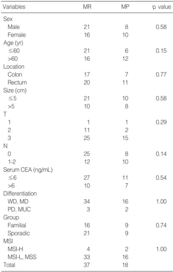
To clarify the clinical significance of the methylation sta- tus of individual genes or the extent of methylation of mul- tiple CpG islands, we compared molecular and clinicopatho- logic features of colorectal cancer patients (Table 2). We did not detect any differences in clinicopathologic features between the MP and MR groups or between groups of patients with or without a family history of colorectal cancer. In the group of sporadic colorectal cancers, however, we found that the methylation index was higher in older patients (p<0.001) and in those with right colon tumors (p=0.041).
Fig. 3. Methylation status of COX2, MGMT, hMLH1, TIMP3, p16, and MINT2 in normal mucosa and tumor tissue of patients with sporadic colorectal cancer. In tumors, the locus most frequently methylated was p16 (33.3% of cases).
No
12 10 8 6 4 2 0
COX2 hMLH1 TIMP3 MGMT p16 MINT2
6.7 20
0 6.7
0 6.7
3.4
13.3 13.3 Mucosa
33.3
0 13.3
Tumor (%)
Variables MR MP p value
Sex
Male 21 8 0.58
Female 16 10
Age (yr)
≤60 21 6 0.15
>60 16 12
Location
Colon 17 7 0.77
Rectum 20 11
Size (cm)
≤5 21 10 0.58
>5 10 8
T
1 1 1 0.29
2 11 2
3 25 15
N
0 25 8 0.14
1-2 12 10
Serum CEA (ng/mL)
≤6 27 11 0.54
>6 10 7
Differentiation
WD, MD 34 16 1.00
PD, MUC 3 2
Group
Familial 16 9 0.74
Sporadic 21 9
MSI
MSI-H 4 2 1.00
MSI-L, MSS 33 16
Total 37 18
Table 2. Clinicopathologic features of patients according to methyation status
MR, methylation-resistant; MP, methylation-prone colorectal cancer;
CEA, carcinoembryonic antigen; WD, well-differentiated; MD, moder- ately differentiated; PD, poorly differentiated; MUC, mucinous; MSI-H, microsatellite instability high frequency; MSI-L, microsatellite instability low frequency; MSS, microsatellite stable.
Fig. 2. Methylation status of COX2, MGMT, hMLH1, TIMP3, p16, and MINT2 in normal mucosa and tumor tissues of patients with a family history of colorectal cancer. In tumors, the locus most frequently methylated was MGMT (44% of cases).
No
12 10 8 6 4 2 0
COX2 hMLH1 TIMP3 MGMT p16 MINT2
28 20
0 20
0 4
20 44
16 Mucosa
8 0
16
Tumor (%)
Comparisons of methylation in patients with and without a family history of colorectal cancer
The overall frequency of methylation in tumors did not differ between patients with and without a family history of colorectal cancer (p=0.524 for methylation index; p=0.774 for MP vs. MR). When we analysed methylation of individ- ual genes, however, we found that MGMT was more fre- quently methylated in colorectal cancers of patients with a family history (p=0.016), whereas p16 was more frequently methylated in sporadic colorectal cancers (p=0.046) (Fig. 4).
We found that the normal mucosa in patients with family history showed more frequent methylation than did normal mucosa of sporadic cancer patients (p=0.016 for methyla- tion index; p=0.037 for MP vs. MR), but there was no dif- ference in methylation of any of the individual genes.
MSI
Of the 25 colorectal cancers with family history, four (16.0
%) were MSI-H and five (20.0%) were MSI-L. In contrast, of the 30 colorectal cancer without family history, two (6.7
%) were MSI-H and 28 (93.3%) were MSS (MSI-H vs. MSI- L vs. MSS, p=0.014). When we divided the microsatellite status into two groups (MSI-H vs. MSI-L plus MSS), we observed no difference between colorectal cancers with and without family history. We found that the hMLH1 gene was methylated in three of the MSI-H tumors (50.0%), three of the MSI-L tumors (60.0%), and one of the MSS tumors (2.3%). When we analysed the correlations between MSI and clinicopathologic characteristics, we found that the tumors with MSI showed right side predominance (MSI-H vs. MSI-L plus MSS; p=0.022) and were more frequently methylated at hMLH1 (p=0.022) and TIMP3 (p=0.029).
DISCUSSION
Aberrant methylation in promoter CpG islands of tumor suppressor genes is associated with transcriptional silencing of these genes and is thought to be an alternative mechanism in carcinogenesis (5, 6). Colorectal cancers with CpG island methylation are thought to have a distinct clinicopathologic phenotype and genetic profiles, including MSI and frequent mutations of the K-ras gene, but they lack p53 mutations (19, 20). There is as yet no consensus about the definition of CIMP and the panel of CpG sites that should be analysed to classify tumors as CIMP. This is not trivial, given that each study uses different methods to assay DNA methylation, assays different genes, has different definitions of CIMP, and examines a different minimal number of genes. Genes related to carcinogenesis may be tissue-dependent, and the defini- tion of CIMP for colorectal cancer might not be applicable to other cancers. CIMP has been generally defined as methyla- tion of at least two MINTs or target genes such as p16, p14, or hMLH1, when a small panel of such markers is examined (7, 21, 22).
In the present study, we have assayed six markers: MINT2, p16, TIMP3, hMLH1, MGMT, and COX2. All of these have been found to be methylated mainly in cancers and are fre- quently methylated in gastrointestinal tumors (23, 24). More- over, these genes have been reported to be closely related to colorectal carcinogenesis; specifically, cell-cycle regulation (COX2 and p16), DNA repair or protection (MGMT, and hMLH1), and metastasis and invasion (TIMP3). In our opin- ion, this selection satisfies the minimum conditions for study- ing methylation in colorectal cancer (19). We categorized colorectal tumors as MP or MR to avoid confusion with other definitions of CIMP (7, 20, 25), and calculated and compared methylation indices to reduce the bias from our selection of genes.
In this study, we have focused on a subgroup of familial colorectal cancers; i.e., tumors from patients with first-degree relatives with colorectal cancers and no germline mutations in hMLH1 and hMSH2 (16). This group of cancers may be a mixture of heterogeneous tumors with different molecular profiles. Several mechanisms of genetic predisposition to col- orectal cancer with a familial tendency have been suggested.
These include defects in MMR genes other than hMLH1 and hMSH2, low penetrance of polymorphisms, simple phe- nocopy, or technically undetectable alterations in hMLH1 and hMSH2 (16, 26, 27). Therefore, in interpreting the results of this study, our definition of familial cancers should be con- sidered. That is, we enrolled and classified patients as having familial colorectal cancer, who have been known not to have a mutation in hMLH1 and hMSH2, and have at least one first-degree relative without considering familial history of cancers in other organs and in second-degree relatives. There- fore, our results may not be representative of the methyla- tion status in hereditary cancers not fulfilling the Amster-
(%)
Fig. 4. Differences in methylation status of MGMT and p16 in tumors of patients with a family history of colorectal cancer and in those with sporadic colorectal cancer. Compared with sporadic colorec- tal cancers, MGMT was more frequently (p=0.016) and p16 was less frequently (p=0.046) methylated in colorectal cancers from patients with a family history.
No
12 10 8 6 4 2 0
COX2 hMLH1 TIMP3 MGMT p16 MINT2 CIMP+
20 20
Familial Sporadic
20
6.7 4
6.7 44
p=0.016 p=0.046
13.3 8
33.3
16 13.3 36 30
dam criteria of HNPCC. The present study was also limited by the small number of samples studied.
Several reports have described an association between the methylation status of multiple genes and a familial tenden- cy to colorectal cancer (15, 28). A recent large study, how- ever, found no evidence that patients with heavily methylat- ed colorectal cancers were more likely to develop a second malignancy or have a positive family history of cancer (29).
Aberrant methylation may result from an inherited defect in the methylation apparatus. In this study, considerable pro- portions of cancer in both groups with or without familial history of colorectal cancer presented the methylated genes that may involved in carcinogenetic pathways. It still remains to be elucidated whether promoter methylation in multiple genes is one of main mechanism to evoke cancer or simple bystander. However, our results confirm that CpG island pro- moter methylation may be a universal event in sporadic or familial colorectal cancer, and is suggested as one of the mech- anisms for ‘second hits’ by which tumor suppressor genes are inactivated. Evidence for this mechanism in familial col- orectal cancers was previously reported in studies showing aberrant methylation of individual genes, including CDH1, VHL, and hMLH1, in hereditary cancer syndromes (30-32).
Cancer is a genetic disease. Most cancer-causing mutations are somatic, occurring in the affected tissue during the course of carcinogenesis. However, most cancers also have a heredi- tary component, caused by predisposing mutations that affect the germline, are heritable, contribute to the initiation of carcinogenesis, and influence the carcinogenesis pathway.
Although hypermethylation is not a rare event, either in sporadic colorectal cancers or in colorectal cancers with fam- ily history, predisposing germline alterations can affect detail- ed aspects of methylation. Our data showed two of the genes methylated at their promoters, p16 and MGMT, differ accord- ing to the tumor type. These findings suggest that the methy- lation of p16, leading to its loss of function, may be a domi- nant and necessary event for sporadic colorectal carcinogen- esis, whereas methylation of MGMT may be dominant and necessary in colorectal carcinogenesis in individuals with a family history. It was reported that the frequency and pattern of gene methylation varied between HNPCC syndrome and sporadic adenomas, implying differences in the molecular pathogenesis of tumors (33). MGMT has been considered as a critical step of genetic instability and methylation of MGMT are proposed to show a distinctive phenotype of MSI-L or mild family history of colorectal cancer (34, 35). The pre- sent study also showed a possibility that different pathways to cancer may exit according to the molecular background and some subgroup of familial colorectal cancer are related to the loss of function of MGMT. Thus, genes selected for methylation-induced functional loss may differ according to the genetic background. While methylation seems to be a universal mechanism by which gene function is inactivated, the germline mutations in familial tumors confer a selective
advantage for their tumorigenic growth, but other genetic and epigenetic lesions are also necessary. Genetic predisposi- tion to CpG island methylation may be a modifying factor that contributes to the penetrance of HNPCC. Our findings thus expand these early observations on methylation in famil- ial colorectal cancer and highlight the selective advantage of epigenetic gene silencing.
A ‘field defect’ is an area of abnormal tissue that predispos- es to the development of cancer. The molecular basis is rela- tively simple to understand when it occurs in patients who have a genetic predisposition for cancer development or mas- sive exposure to a carcinogen. Within this defective field, a second change may confer a growth advantage on a given cell relative to other cells. In colorectal cancer patients with germline genetic defects, all cells in the colonic mucosa have the same genetic alteration, leading to the frequent develop- ment of tumors in these individuals. In some of these pati- ents, however, the selective advantage of genetic alterations is not great (36). Methylation has been proposed as a candi- date mediator of this field defect and methylation status of normal mucosa was explored to verify the role of methyla- tion as an earliest event in carcinogenetic pathway (37, 38).
A recent study reported that some colorectal cancers arise from a field defect defined by epigenetic inactivation of some genes such as MGMT (38). Detection of this abnormality may useful in predict the colorectal cancer risk. Germline defects that alter methylation machinery may increase methy- lation frequency in normal colonic mucosa of individuals with a family history of colorectal cancer, and this may be associated with the frequent and multiple incidences of col- orectal tumors in these patients. We found that methylation status of mucosa differed between in the patients of sporadic and familial colorectal cancers, although we did not detect differences in individual genes. This finding might be a small clue that field defect related to methylation has variable effects regarding selective power and involved genes for field defect is also diverse according to the specific carcinogenetic path- way. A methylation field effect in the entire whole colonic mucosa may be associated with the process of carcinogenesis in familial colorectal cancer, at least in those tumors with high methylation rates in normal mucosa.
Conclusively, alterations in the methylation machinery may also be associated with both sporadic and familial col- orectal carcinogenesis, although there are qualitative and quantitative differences in methylation between sporadic and familial tumors. Our findings, however, do not support the concept that a germline defect in the methylation machin- ery is responsible for the development of most tumors with multiple epimutation. This epigenetic mechanism can silence different genes affected by other genetic backgrounds, lead- ing to divergent pathways of development in hereditary and sporadic colorectal cancers.
REFERENCES
1. Baylin SB. Tying it all together: epigenetics, genetics, cell cycle, and cancer. Science 1997; 277: 1948-9.
2. Tomlinson I, Bodmer W. Selection, the mutation rate and cancer:
ensuring that the tail does not wag the dog. Nat Med 1999; 5: 11-2.
3. Jackson AL, Loeb LA. The mutation rate and cancer. Genetics 1998;
148: 1483-90.
4. Feinberg AP, Tycko B. The history of cancer epigenetics. Nat Rev Cancer 2004; 4: 143-53.
5. Jones PA. DNA methylation and cancer. Oncogene 2002; 21: 5358- 60.
6. Issa JP, Baylin SB. Epigenetics and human disease. Nat Med 1996;
2: 281-2.
7. Issa JP. CpG island methylator phenotype in cancer. Nat Rev Can- cer 2004; 4: 988-93.
8. Parkin DM, Bray F, Ferlay J, Pisani P. Estimating the world cancer burden: Globocan 2000. Int J Cancer 2001; 94: 153-6.
9. McGrath DR, Spigelman AD. Hereditary colorectal cancer: keep- ing it in the family--the bowel cancer story. Intern Med J 2002; 32:
325-30.
10. Salovaara R, Loukola A, Kristo P, Kaariainen H, Ahtola H, Eskeli- nen M, Harkonen N, Julkunen R, Kangas E, Ojala S, Tulikoura J, Valkamo E, Jarvinen H, Mecklin J, Aaltonen LA, de la Chapelle A.
Population-based molecular detection of hereditary nonpolyposis colorectal cancer. J Clin Oncol 2000; 18: 2193-200.
11. Olsson L, Lindblom A. Family history of colorectal cancer in a Swe- den county. Fam Cancer 2003; 2: 87-93.
12. Johns LE, Houlston RS. A systematic review and meta-analysis of familial colorectal cancer risk. Am J Gastroenterol 2001; 96: 2992- 3003.
13. Boland CR, Thibodeau SN, Hamilton SR, Sidransky D, Eshleman JR, Burt RW, Meltzer SJ, Rodriguez-Bigas MA, Fodde R, Ranzani GN, Srivastava S. A National Cancer Institute Workshop on Microsa- tellite Instability for cancer detection and familial predisposition: devel- opment of international criteria for the determination of microsatel- lite instability in colorectal cancer. Cancer Res 1998; 58: 5248-57.
14. Umar A, Risinger JI, Hawk ET, Barrett JC. Testing guidelines for hereditary non-polyposis colorectal cancer. Nat Rev Cancer 2004;
4: 153-8.
15. Frazier ML, Xi L, Zong J, Viscofsky N, Rashid A, Wu EF, Lynch PM, Amos CI, Issa JP. Association of the CpG island methylator phenotype with family history of cancer in patients with colorectal cancer. Cancer Res 2003; 63: 4805-8.
16. Kim JC, Lee KH, Ka IH, Koo KH, Roh SA, Kim HC, Yu CS, Kim TW, Chang HM, Gong GY, Kim JS. Characterization of mutator phenotype in familial colorectal cancer patients not fulfilling ams- terdam criteria. Clin Cancer Res 2004; 10: 6159-68.
17. Herman JG, Graff JR, Myohanen S, Nelkin BD, Baylin SB. Methy- lation-specific PCR: a novel PCR assay for methylation status of CpG islands. Proc Natl Acad Sci USA 1996; 93: 9821-6.
18. Kang GH, Lee S, Kim WH, Lee HW, Kim JC, Rhyu MG, Ro JY.
Epstein-barr virus-positive gastric carcinoma demonstrates frequent aberrant methylation of multiple genes and constitutes CpG island
methylator phenotype-positive gastric carcinoma. Am J Pathol 2002;
160: 787-94.
19. Toyota M, Ohe-Toyota M, Ahuja N, Issa JP. Distinct genetic pro- files in colorectal tumors with or without the CpG island methylator phenotype. Proc Natl Acad Sci USA 2000; 97: 710-5.
20. Park SJ, Rashid A, Lee JH, Kim SG, Hamilton SR, Wu TT. Frequent CpG island methylation in serrated adenomas of the colorectum. Am J Pathol 2003; 162: 815-22.
21. Jass JR, Whitehall VL, Young J, Leggett BA. Emerging concepts in colorectal neoplasia. Gastroenterology 2002; 123: 862-76.
22. Rashid A, Issa JP. CpG island methylation in gastroenterologic neo- plasia: a maturing field. Gastroenterology 2004; 127: 1578-88.
23. Laird PW. The power and the promise of DNA methylation markers.
Nat Rev Cancer 2003; 3: 253-66.
24. Jubb AM, Bell SM, Quirke P. Methylation and colorectal cancer. J Pathol 2001; 195: 111-34.
25. Rashid A, Shen L, Morris JS, Issa JP, Hamilton SR. CpG island methy- lation in colorectal adenomas. Am J Pathol 2001; 159: 1129-35.
26. Fearnhead NS, Wilding JL, Winney B, Tonks S, Bartlett S, Bicknell DC, Tomlinson IP, Mortensen NJ, Bodmer WF. Multiple rare vari- ants in different genes account for multifactorial inherited suscepti- bility to colorectal adenomas. Proc Natl Acad Sci USA 2004; 101:
15992-7.
27. de la Chapelle A. Genetic predisposition to colorectal cancer. Nat Rev Cancer 2004; 4: 769-80.
28. Esteller M, Fraga MF, Guo M, Garcia-Foncillas J, Hedenfalk I, God- win AK, Trojan J, Vaurs-Barriere C, Bignon YJ, Ramus S, Benitez J, Caldes T, Akiyama Y, Yuasa Y, Launonen V, Canal MJ, Rodriguez R, Capella G, Peinado MA, Borg A, Aaltonen LA, Ponder BA, Baylin SB, Herman JG. DNA methylation patterns in hereditary human can- cers mimic sporadic tumorigenesis. Hum Mol Genet 2001; 10: 3001-7.
29. Ward RL, Williams R, Law M, Hawkins NJ. The CpG island methy- lator phenotype is not associated with a personal or family history of cancer. Cancer Res 2004; 64: 7618-21.
30. Wheeler JM, Loukola A, Aaltonen LA, Mortensen NJ, Bodmer WF.
The role of hypermethylation of the hMLH1 promoter region in HNPCC versus MSI+ sporadic colorectal cancers. J Med Genet 2000; 37:
588-92.
31. Grady WM, Willis J, Guilford PJ, Dunbier AK, Toro TT, Lynch H, Wiesner G, Ferguson K, Eng C, Park JG, Kim SJ, Markowitz S.
Methylation of the CDH1 promoter as the second genetic hit in heredi- tary diffuse gastric cancer. Nat Genet 2000; 26: 16-7.
32. Prowse AH, Webster AR, Richards FM, Richard S, Olschwang S, Resche F, Affara NA, Maher ER. Somatic inactivation of the VHL gene in Von Hippel-Lindau disease tumors. Am J Hum Genet 1997;
60: 765-71.
33. Kaz A, Kim Y, Dzieciatkowski S, Lynch H, Watson P, Washington MK, Lin L, Grady WM. Evidence for the role of aberrant DNA methy- lation in the pathogenesis of Lynch syndrome adenomas. Int J Can- cer 2007; 120: 1922-9.
34. Jass JR. Serrated adenoma of the colorectum and the DNA-methy- latior phenotype. Nat Clin Pract Oncol 2005; 2: 398-405.
35. Jass JR, Whitehall VL, Young J, Leggett BA. Emerging concepts in colorectal neoplasia. Gastroenterol 2002; 123: 862-76.
. .
36. Bodmer W. The somatic evolution of cancer. The Harveian Oration of 1996. J R Coll Physicians Lond 1997; 31: 82-9.
37. Ye C, Shrubsole MJ, Cai Q, Ness R, Grady WM, Smalley W, Cai H, Washington K, Zheng W. Promoter methylation status of the MGMT, hMLH1, and CDKN2A/p16 genes in non-neoplastic mucosa of patients with and without colorectal adenomas. Oncol Rep 2006; 16: 429-35.
38. Shen L, Kondo Y, Rosner GL, Xiao L, Hernandez NS, Vilaythong J, Houlihan PS, Krouse RS, Prasad AR, Einspahr JG, Buckmeier J, Alberts DS, Hamilton SR, Issa JP. MGMT promoter methylation and field defect in sporadic colorectal cancer. J Natl Cancer Inst 2005;
97: 1330-8.