Effects of Omega-3 Fatty Acids on Erectile Dysfunction in a Rat Model of Atherosclerosis-induced Chronic Pelvic Ischemia
The aim of this study was to investigate whether the omega-3 fatty acids help to improve erectile function in an atherosclerosis-induced erectile dysfunction rat model. A total of 20 male Sprague-Dawley rats at age 8 weeks were divided into three groups: Control group (n = 6, untreated sham operated rats), Pathologic group (n = 7, untreated rats with chronic pelvic ischemia [CPI]), and Treatment group (n = 7, CPI rats treated with omega-3 fatty acids). For the in vivo study, electrical stimulation of the cavernosal nerve was performed and erectile function was measured in all groups. Immunohistochemical antibody staining was performed for transforming growth factor beta-1 (TGF-β1), endothelial nitric oxide synthase (eNOS), and hypoxia inducible factor 1-alpha (HIF-1α). In vivo measurement of erectile function in the Pathologic group showed significantly lower values than those in the Control group, whereas the Treatment group showed significantly improved values in comparison with those in the Pathologic group. The results of western blot analysis revealed that systemically administered omega-3 fatty acids ameliorated the cavernosal molecular environment. Our study suggests that omega-3 fatty acids improve intracavernosal pressure and have a beneficial role against pathophysiological
consequences such as fibrosis or hypoxic damage on a CPI rat model, which represents a structural erectile dysfunction model.
Keywords: Erectile Dysfunction; Fatty Acids, Omega-3; Ischemia; Rats Ji Sung Shim, Dae Hee Kim,
Jae Hyun Bae, and Du Geon Moon Department of Urology, Korea University College of Medicine, Seoul, Korea
Received: 13 September 2015 Accepted: 23 December 2015 Address for Correspondence:
Du Geon Moon, MD
Department of Urology, Korea University Guro Hospital, Korea University College of Medicine, 148 Gurodong-ro, Guro-gu, Seoul 08308, Korea
E-mail: [email protected]
http://dx.doi.org/10.3346/jkms.2016.31.4.585 • J Korean Med Sci 2016; 31: 585-589
INTRODUCTION
Erectile dysfunction (ED) is defined as the inability to achieve and maintain an erection sufficient to permit satisfactory sexu- al intercourse. It has been estimated to affect 20 to 30 million men in the United States (1). Since aging is one of the main risk factors for ED, most men will be afflicted by this condition dur- ing their lifetime (2,3). ED can be classified as psychogenic, neu- rogenic, hormonal, arterial, cavernosal, or drug-induced. How- ever, vasculogenic factors (arterial and cavernosal) are mostly related with aging.
In spite of a previously introduced arteriogenic ED model, there are few studies of atherosclerosis- or hyperlipidemia-in- duced ED models (4). Reasons for this may be that too much time is required to cause pelvic atherosclerosis by feeding cho- lesterol to rats and that it is difficult to model ED solely by ath- erosclerosis. Another reason is that rats are known to be choles- terol resistant, which is the traditional method of developing atherosclerosis (5).
Considering the points mentioned above, endothelial injury is essential to creating an atherosclerosis-induced vasculogenic ED model because it can enhance the process of atherosclero- sis (6). Therefore, in our study we examined a chronic pelvic ischemia (CPI) model, which is based on both endothelial inju-
ry and a hypercholesterol diet. The CPI model has been validat- ed in several previous studies as reported by Nomiya et al. (7,8).
Omega-3 fatty acids are limited in our diet, with the main sources being fish and plants. Long chain omega-3 fatty acids, docosahexaenoic acid and eicosapentaenoic acid, have well- described beneficial effects on cardiovascular disease, diabetes mellitus, lipid profiles, and other inflammatory diseases in hu- mans (9). Suggested potential mechanisms for their protective effects are lowering oxidative stress, reducing the secretion of pro-inflammatory cytokines, and attenuation of atherosclerotic plaques (9,10).
There have been few studies regarding the action of omega-3 fatty acids on erectile function. The aim of this study was to in- vestigate whether the omega-3 fatty acids helps to improve erec- tile function in an atherosclerosis-induced ED rat model.
MATERIALS AND METHODS
Animal groups and experimental design
Specific pathogen-free male Sprague-Dawley rats (250-280 g) were obtained from the Hanlim Inc. (Osan, Korea). Upon arriv- al, animals were randomized and housed three per cage. The animals were 8 weeks of age and were acclimated for at least 2 weeks. They were all kept under 12 hours light-dark cycles at
room temperature (22°C ± 2°C), and had free access to a stan- dard diet and water. The rats were randomly assigned to differ- ent experimental groups: 1) the Control group that underwent a sham operation and received a regular diet and water; 2) the Pathologic group that underwent endothelial injury of the iliac arteries and was provided a 2% cholesterol diet (Research Diets, New Brunswick, NJ, USA) for 4 weeks; and 3) the Treatment group, after establishment of the CPI model, that was provided a high-fat diet for 4 weeks and then treated with omega-3 fatty acids ([DHA] ethyl ester > 98% and [EPA] ethyl ester > 98%, Cayman Chemical, Ann Arbor, MI, USA) with a regular diet for 2 weeks. Omega-3 fatty acids (200 mg/kg; DHA: 100 mg/kg/day + EPA: 100 mg/kg/day) were offered every day by oral gavage.
Chronic pelvic ischemia model
Animals were anesthetized by inhalation of 2.5%-3.5% isoflu- rane. An inguinal incision was made and, through dissection into the iliac fossa, the femoral artery and superficial epigastric artery were isolated. A 2Fr Fogarty catheter (Edwards Lifescienc- es, Irvine, CA, USA) was passed into the femoral artery through a small incision and up to the common iliac artery. To induce endothelial injury, the balloon of the catheter was inflated with 0.2 mL of air and was pulled down to the femoral artery. This maneuver was repeated 10 times. The same procedure was per- formed on the contralateral side. In the control group sham op- eration, only an inguinal incision and iliac artery exposure, with subsequent skin closure, was performed.
Measurement of intracavernous pressure
Under anesthesia, polyethylene tubing (PE-50) was inserted into the left carotid artery in order to record blood pressure. A 22-gauge angiocatheter was connected to PE-50 tubing filled with hepa- rinized saline (50 U heparin/mL) and inserted into the left cor- pus cavernosum for intracavernous pressure (ICP) measure- ment. After confirming the location of the major pelvic gangli- on, which was placed on the lateral aspect of the prostate, elec- trostimulation was performed twice on each cavernous nerve.
Stimulation was performed at 5 V and 20 Hz, at more than 5-min- ute intervals, with bipolar hook electrodes using an electronic stimulator (Grass SD9, Grass Instrument, Quincy, MA, USA).
The major indices for evaluating erectile function were maxi- mum ICP and the ICP/mean arterial pressure (MAP) ratio. ICP and blood pressure were monitored using PowerLabTM (AD In- struments, Colorado Springs, CO, USA).
Quantitative western blots
Penile homogenates of frozen tissue (100 mg) were obtained using T-PER and protease inhibitors (3 µM leupeptin, 1 µM pepstatin A, 1 mM phenyl methyl sulfonyl fluoride) and centri- fuged at 100,000 × g for 10 minutes. Protein concentration was determined with the Bradford method (Bradford solution: Sig-
ma, Saint Louis, MO, USA) and 40 µg of all samples were load- ed. Equal amounts of supernatant protein (30 µg) were run on 7.5% or 10% (ACTA2) polyacrylamide gels and submitted to western blot immunodetection using the following primary an- tibodies: HIF-1α, eNOS, TGF-β1 (TBST buffer 1:200; Santa Cruz Biotechnology, Santa Cruz, CA, USA; overnight), actin (2.5%
skim milk 1:3,000; Cell Signaling, MA, USA; 1 hour, room tem- perature), and the following secondary antibodies: HIF-1α, eNOS, TGF-β1 anti-rabbit (1:30,000; Sigma). Band intensities were de- termined with densitometry and were corrected on the basis of the respective intensities of a housekeeping protein, glyceralde- hyde-3-phosphate dehydrogenase (GAPDH), upon reprobing.
Statistical analysis
Continuous variables were analyzed using one-way ANOVA and expressed as the mean ± standard deviation (SD). A Krus- kal-Wallis test was used to analyze non-parametric variables.
All analyses were performed using SPSS software (Version 20.0, SPSS Inc., Chicago, IL, USA). A P value < 0.05 was considered statistically significant.
Ethics statement
All animal care and experimental procedures were carried out with approval by the Korea University Hospital animal care and use committee according to the animal experimentation guide- lines of the Korea University Ansan Hospital Animal Laboratory (KUIACUC-2013-177).
RESULTS
No rats died during the CPI-model or sham operations. Feed- ing of omega-3 fatty acids did not cause any side effects or other unusual signs. Body and tissue weight was not significantly dif- ferent between the three groups.
Erectile response to cavernous electrostimulation
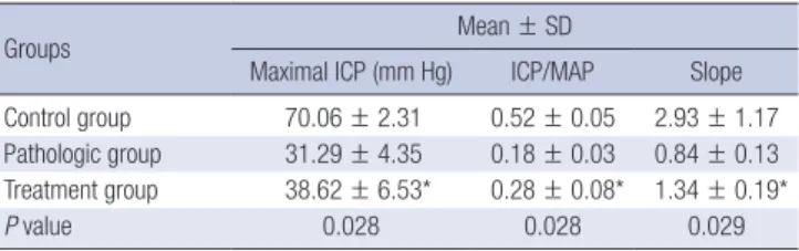
The results of cavernosal pressure measurement are shown in Table 1. In vivo erectile function (maximal ICP, ICP/MAP, and slope values) was measured. In the Control group, these values were 70.06 ± 2.31, 0.52 ± 0.05, and 2.93 ± 1.17, respectively. In
Table 1. Comparative analysis of intracavernous pressure measurements in response to cavernous nerve electrostimulation
Groups Mean ± SD
Maximal ICP (mm Hg) ICP/MAP Slope Control group 70.06 ± 2.31 0.52 ± 0.05 2.93 ± 1.17 Pathologic group 31.29 ± 4.35 0.18 ± 0.03 0.84 ± 0.13 Treatment group 38.62 ± 6.53* 0.28 ± 0.08* 1.34 ± 0.19*
P value 0.028 0.028 0.029
ICP, intracavernous pressure; MAP, mean arterial pressure.
*P < 0.05, statistically significant difference between the pathologic group and ome- ga-3 fatty acids treated group.
the Treatment group, the maximal ICP value showed significant improvement compared to in the Pathologic group (38.62 ± 6.53 vs. 31.29 ± 4.35; P = 0.028). We evaluated the ICP/MAP value in order to adjust for blood pressure. For ICP/MAP results, the Treat- ment group also showed improvement relative to the Patholog- ic group (0.28 ± 0.08 vs 0.18 ± 0.03; P = 0.028). We determined that there was significant difference between the two groups in the slope parameters (1.34 ± 0.19 vs 0.84 ± 0.13; P = 0.029).
Western blot of cavernous tissue
We performed a western blot on cavernous tissue and analyzed the results by using densitometry, which is a semi-quantitative method. The ratio divided as α-SMA are shown in Fig. 1. HIF-1α levels for the treatment group were lower than in the pathologic group (0.49 ± 0.24 vs. 0.95 ± 0.23, respectively). As expected, the lowest value was found in the control group. TGF-β1 level was highest in the pathologic group (0.48 ± 0.23). TGF-β1 was 0.42
± 0.23 in the treatment group and 0.25 ± 0.09 in the control group.
Expression of eNOS was higher in the treatment group than in the pathologic group (0.58 ± 0.24 vs 0.48 ± 0.10, respectively).
The results revealed that systemically administered omega-3 fatty acids ameliorated the cavernosal molecular environment as shown by the decreased expression of TGF-β1 and HIF-1α, and overexpression of eNOS in the treatment group relative to the pathologic group.
DISCUSSION
Our results, together with previous studies, suggest that by low- ering oxidative stress and reducing the degree of cavernosal fi- brosis, omega-3 fatty acids may protect from erectile dysfunc- tion in a rat model of CPI. First, the end point for assessing erec-
tile function is measurement of increased ICP and ICP/MAP using in vivo studies (11,12). Second, the beneficial effect of the treatment was shown by a decreased in vitro expression of TGF- β1 and HIF-1α in our study when comparing the treatment group compared to the pathologic group.
We used a CPI model, which was made by inducing endo- thelial injury and applying a hypercholesterol diet. There are two mechanisms involved in establishing our pelvic ischemia model. One is impaired arterial inflow followed by secondary cavernosal dysfunction due to hypoxia, which have been pro- posed as possible mechanisms of atherosclerotic ED. It is known that TGF-β1 has an important role in this process. Another mech- anism is the effect of endothelial injury, which represents a change in nitric oxide synthase (NOS). Nitric oxide (NO) is formed by NOS and affects erectile function owing to its role in smooth muscle relaxation. Several studies have shown that smooth mus- cle relaxation is induced by endothelial NOS and neuronal NOS, which is secreted from nonadrenergic noncholinergic nerves.
When there is a problem in the metabolism of NO, impaired smooth muscle relaxation and hypoxic effects, due to platelet aggregation, appear (13).
ED is associated with a loss of smooth muscle cells, and an increase in fibrosis has been repeatedly reported in the corpo- ral tissue of patients with ED (14,15). The percentage of smooth muscle cells steadily decreases with age. Corporal tissue dem- onstrated excessive deposits of collagen fibers, which results in corporal fibrosis. These changes can also occur in the tunica media of the penile arteries (16).
Changes in elastic fibers or collagen types can result in me- chanical alterations of the penis, which reduce its elasticity and compliance. In addition to these changes, postmortem studies have revealed that aging is associated with increasing degrees of atherosclerotic vascular alterations in the arterial bed of the penis. The exact pathophysiologic mechanism of ischemia-in- duced fibrosis of the corpus cavernosum is not clearly under- stood. However, in vitro studies have suggested that it is likely caused by hypoxia-induced overexpression of TGF-β1 (17).
TGF-β1 is a pleiotropic cytokine that has been shown to in- crease collagen synthesis in corpus cavernosum smooth mus- cle cells in vitro. Under ischemic conditions, TGF-β1 induces its own mRNA, leading to a further increase in TGF-β1 synthesis, which results in the development of severe fibrosis (17). In hu- mans, a correlation between oxygen tension in the penis and the percentage of smooth muscle fibers has been demonstrat- ed. The number of muscular fibers is therefore dependent on good oxygenation of the penis (18).
Ultimately, cavernosal fibrosis will result in corporal veno- occlusive dysfunction (CVOD), which is mainly due to failure of the corporal smooth muscle mass to achieve sufficient relax- ation. This relaxation is necessary for passive veno-occlusion of the subtunical veins to occur. Therefore, any process that de- Fig. 1. The protein expression of the experimental groups analyzed using western
blotting. TGF-β1, transforming growth factor β1; HIF-1α, hypoxia induced factor 1α;
eNOS, endothelial nitric oxide synthase. Alpha-smooth muscle actin (α-SMA) served as a control for the loading.
*P < 0.05, statistically significant difference between the pathologic group and ome- ga-3 fatty acids treated group.
Expression ratio
HIF-1α eNOS TGF-β1 1.2
1.0 0.8 0.6 0.4 0.2 0
0.29 ± 0.09 0.95 ± 0.23
0.49 ± 0.24*
0.72 ± 0.34
0.48 ± 0.10 0.58 ± 0.24*
0.25 ± 0.09 0.48 ± 0.23
0.42 ± 0.23*
Control group Pathologic group Treatment group
creases the content or function of corporal smooth muscle, or ultimately causes corporal fibrosis, will increase the likelihood of the development of CVOD.
There is significant evidence that omega-3 fatty acids supple- mentation is beneficial in diseases involving ischemic injury (19,20). Omega-3 fatty acid supplementation is reported to ele- vate superoxide dismutase and catalase activity in various or- gans such as the kidney, liver, and intestine (21). Several studies have also shown a decreased level of MDA with omega-3 fatty acids supplementation, after ischemic injury (22). These find- ings coincide with our results, which showed that TGF-β1 and HIF-1α concentrations in CPI rats given a daily administration of 200 mg/kg of omega-3 fatty acids were significantly lower than in the untreated CPI group.
Previous studies have shown that omega-3 fatty acids can decrease adhesion molecule expression in inflammatory cells and the endothelium (23). Production of inflammatory cyto- kines such as IL-6, IL-8, or TNF- α, which have been known to be up-regulated in CPI, decreased with exposure to omega-3 fatty acids in human and animal models (24). These results streng- then the evidence that omega-3 fatty acids work mainly through its anti-oxidant and anti-inflammatory properties.
It is becoming more evident that corporal fibrosis is the main underlying etiology for ED in the majority of patients (25). Re- gardless of age or etiology, 66% to 75% of cases of ED are caused by corporal fibrosis and ultimately, CVOD. Therefore, if the cause of CVOD itself can be prevented, then ED could become a pre- ventable condition. The results of our study strengthen the evi- dence that omega-3 fatty acids work mainly through its anti-ox- idant and anti-inflammatory properties and have an ameliora- tive function on fibrotic changes of cavernosum tissue.
Chronic atherosclerosis-induced pelvic ischemia induces impaired erectile function and altered protein expression. Ad- ministering omega-3 fatty acids could improve erectile function as shown by increases in ICP and the ICP/MAP ratio in the CPI treatment group. Additionally, omega-3 fatty acids have a ben- eficial role in reducing the pathophysiological consequences of fibrotic changes and the hypoxic damage to the cavernosal mo- lecular environment, in a CPI model representing a vasculogen- ic ED model.
DISCLOSURE
The authors have no potential conflicts of interest to disclose.
AUTHOR CONTRIBUTION
Conception and design of the study: Bae JH. Acquisition of data:
Kim DH. Statistical analysis: Kim DH. First draft of manuscript:
Shim JS. Revision and critical review of the manuscript: Moon DG. Manuscript approval: all authors.
ORCID
Ji Sung Shim http://orcid.org/0000-0002-6745-1776 Du Geon Moon http://orcid.org/0000-0002-9031-9845 REFERENCES
1. Benet AE, Melman A. The epidemiology of erectile dysfunction. Urol Clin North Am 1995; 22: 699-709.
2. Rosen RC, Wing R, Schneider S, Gendrano N 3rd. Epidemiology of erec- tile dysfunction: the role of medical comorbidities and lifestyle factors.
Urol Clin North Am 2005; 32: 403-17.
3. Russell S, McVary KT. Lower urinary tract symptoms and erectile dysfunc- tion: epidemiology and treatment in the aging man. Curr Urol Rep 2005;
6: 445-53.
4. Aboseif SR, Breza J, Orvis BR, Lue TF, Tanagho EA. Erectile response to acute and chronic occlusion of the internal pudendal and penile arteries.
J Urol 1989; 141: 398-402.
5. Ritskes-Hoitinga J, Beynen AC. Atherosclerosis in the rat. Artery 1988; 16:
25-50.
6. Ross R, Glomset JA. The pathogenesis of atherosclerosis (second of two parts). N Engl J Med 1976; 295: 420-5.
7. Aizawa N, Homma Y, Igawa Y. Effect of silodosin, a selective alpha1A-ad- renoceptor antagonist on primary bladder afferent activity and bladder microcontractions in rats. Neurourol Urodyn 2013; 32: 569-71.
8. Nomiya M, Yamaguchi O, Andersson KE, Sagawa K, Aikawa K, Shishido K, Yanagida T, Kushida N, Yazaki J, Takahashi N. The effect of atheroscle- rosis-induced chronic bladder ischemia on bladder function in the rat.
Neurourol Urodyn 2012; 31: 195-200.
9. Delgado-Lista J, Perez-Martinez P, Lopez-Miranda J, Perez-Jimenez F. Long chain omega-3 fatty acids and cardiovascular disease: a systematic re- view. Br J Nutr 2012; 107 Suppl 2: S201-13.
10. Calder PC. The role of marine omega-3 (n-3) fatty acids in inflammatory processes, atherosclerosis and plaque stability. Mol Nutr Food Res 2012;
56: 1073-80.
11. Magee TR, Kovanecz I, Davila HH, Ferrini MG, Cantini L, Vernet D, Zuni- ga FI, Rajfer J, Gonzalez-Cadavid NF. Antisense and short hairpin RNA (shRNA) constructs targeting PIN (Protein Inhibitor of NOS) ameliorate aging-related erectile dysfunction in the rat. J Sex Med 2007; 4: 633-43.
12. Rajasekaran M, Kasyan A, Jain A, Kim SW, Monga M. Altered growth fac- tor expression in the aging penis: the Brown-Norway rat model. J Androl 2002; 23: 393-9.
13. Williams JK, Andersson KE, Christ G. Animal models of erectile dysfunc- tion (ED): potential utility of non-human primates as a model of athero- sclerosis-induced vascular ED. Int J Impot Res 2012; 24: 91-100.
14. Iacono F, Giannella R, Somma P, Manno G, Fusco F, Mirone V. Histologi- cal alterations in cavernous tissue after radical prostatectomy. J Urol 2005;
173: 1673-6.
15. Yaman O, Yilmaz E, Bozlu M, Anafarta K. Alterations of intracorporeal structures in patients with erectile dysfunction. Urol Int 2003; 71: 87-90.
16. Ferrini MG, Davila HH, Valente EG, Gonzalez-Cadavid NF, Rajfer J. Aging- related induction of inducible nitric oxide synthase is vasculo-protective to the arterial media. Cardiovasc Res 2004; 61: 796-805.
17. Moreland RB. Is there a role of hypoxemia in penile fibrosis: a viewpoint
presented to the Society for the Study of Impotence. Int J Impot Res 1998;
10: 113-20.
18. Sattar AA, Salpigides G, Vanderhaeghen JJ, Schulman CC, Wespes E. Cav- ernous oxygen tension and smooth muscle fibers: relation and function.
J Urol 1995; 154: 1736-9.
19. Ajami M, Davoodi SH, Habibey R, Namazi N, Soleimani M, Pazoki-To- roudi H. Effect of DHA+EPA on oxidative stress and apoptosis induced by ischemia-reperfusion in rat kidneys. Fundam Clin Pharmacol 2013;
27: 593-602.
20. Castillo RL, Arias C, Farías JG. Omega 3 chronic supplementation attenu- ates myocardial ischaemia-reperfusion injury through reinforcement of antioxidant defense system in rats. Cell Biochem Funct 2014; 32: 274-81.
21. Brahmbhatt V, Oliveira M, Briand M, Perrisseau G, Bastic Schmid V, Des- taillats F, Pace-Asciak C, Benyacoub J, Bosco N. Protective effects of dietary EPA and DHA on ischemia-reperfusion-induced intestinal stress. J Nutr
Biochem 2013; 24: 104-11.
22. Avramovic N, Dragutinovic V, Krstic D, Colovic M, Trbovic A, de Luka S, Milovanovic I, Popovic T. The effects of omega 3 fatty acid supplementa- tion on brain tissue oxidative status in aged wistar rats. Hippokratia 2012;
16: 241-5.
23. Miles EA, Wallace FA, Calder PC. Dietary fish oil reduces intercellular ad- hesion molecule 1 and scavenger receptor expression on murine macro- phages. Atherosclerosis 2000; 152: 43-50.
24. De Caterina R, Cybulsky MI, Clinton SK, Gimbrone MA Jr, Libby P. The omega-3 fatty acid docosahexaenoate reduces cytokine-induced expres- sion of proatherogenic and proinflammatory proteins in human endo- thelial cells. Arterioscler Thromb 1994; 14: 1829-36.
25. Melman A, Gingell JC. The epidemiology and pathophysiology of erectile dysfunction. J Urol 1999; 161: 5-11.