INTRODUCTION
Asthma affects nearly 250 million people worldwide, of whom approximately 20%-25% have moderate-to-severe uncontrolled disease.1 Despite the benefits of standard inhaled corticoste- roid-based treatment, patients with inadequately controlled asthma remain at high risk of asthma exacerbations. Given the socioeconomic costs of asthma exacerbations, the identifica- tion of at-risk subjects is important.2
There are various precipitants of acute asthma exacerbation, such as viral infection, exercise, and exposure to allergens, oc- cupational substances, drugs, or air pollutants.1 However, respi- ratory tract viruses, such as rhinovirus, are the most common triggers and can lead to bacterial infections.3 The host response to viral or bacterial infection is likely to influence susceptibility
to asthma exacerbation. During exacerbation, airway inflam- mation is amplified by intrinsic host factors, such as bronchiec- tasis, diminished lung function, deficiency of interferon pro- duction by epithelial cells, and immunodeficiency.4
Most adult-onset primary immunodeficiency diseases (PID) are humoral immune deficiencies, such as common variable immunodeficiencies, hypogammaglobulinemia, immunoglob-
Effects of Immunoglobulin Replacement on Asthma Exacerbation in Adult Asthmatics with IgG Subclass Deficiency
Joo-Hee Kim,
1Young-Min Ye,
2Ga-Young Ban,
2Yoo-Seob Shin,
2Hyun Young Lee,
2Young-Hee Nam,
3Soo-Keol Lee,
3You Sook Cho,
4Seung-Hun Jang,
1Ki-Suck Jung,
1Hae-Sim Park
2*
1 Division of Pulmonary, Allergy, and Critical Care Medicine, Department of Medicine, Hallym University Sacred Heart Hospital, Hallym University College of Medicine, Anyang, Korea
2Department of Allergy and Clinical Immunology, Ajou University School of Medicine, Suwon, Korea
3Department of Allergy and Clinical Immunology, Dong-A University College of Medicine, Busan, Korea
4Department of Allergy and Clinical Immunology, Asan Medical Center, University of Ulsan College of Medicine, Seoul, Korea
This is an Open Access article distributed under the terms of the Creative Commons Attribution Non-Commercial License (http://creativecommons.org/licenses/by-nc/4.0/) which permits unrestricted non-commercial use, distribution, and reproduction in any medium, provided the original work is properly cited.
Purpose: Recurrent respiratory tract infection is a common manifestation of primary immunodeficiency disease, and respiratory viruses or bacteria are important triggers of asthma exacerbations. Asthma often coexists with humoral immunodeficiency in adults, and some asthmatics with immu- noglobulin (Ig) G subclass deficiency (IgGSCD) suffer from recurrent exacerbations. Although some studies suggest a benefit from Ig replacement, others have failed to support its use. This study aimed to assess the effect of Ig replacement on asthma exacerbation caused by respiratory infec- tion as well as the asthma control status of adult asthmatics with IgGSCD. Methods: This is a multi-center, open-label study of adult asthmatics with IgGSCD. All patients received monthly intravenous immunoglobulin (IVIG) for 6 months and were evaluated regarding asthma exacerbation re- lated to infection, asthma control status, quality of life, and lung function before and after IVIG infusion. Results: A total of 30 patients were en- rolled, and 24 completed the study. Most of the patients had a moderate degree of asthma severity with partly (52%) or uncontrolled (41%) status at baseline. IVIG significantly reduced the proportion of patients with asthma exacerbations, lowered the number of respiratory infections, and im- proved asthma control status, compared to the baseline values (P<0.001). The mean asthma-specific quality of life and asthma control test scores were improved significantly (P=0.009 and P=0.053, respectively); however, there were no significant changes in lung function. Conclusions: IVIG reduced the frequency of asthma exacerbations and improved asthma control status in adult asthmatics with IgGSCD, suggesting that IVIG could be an effective treatment option in this population.
Key Words: Asthma; Exacerbation; immunodeficiency; immunoglobulins; intravenous
Correspondence to: Hae-Sim Park, MD, PhD, Director, Ajou Research Institute for Innovation Medicine Department of Allergy & Clinical Immunology, Ajou University Medical Center, 164 Worldcup-ro, Yeongtong-gu, Suwon 16499, Korea.
Tel: +82-31-219-5150; Fax: +82-31-219-5154; E-mail: [email protected] Received: May 9, 2017; Revised: June 2, 2017; Accepted: June 13, 2017
•Joo-Hee Kim and Young Min Ye are equally contributed to this study.
•There are no financial or other issues that might lead to conflict of interest.
Allergy Asthma Immunol Res. 2017 November;9(6):526-533.
https://doi.org/10.4168/aair.2017.9.6.526 pISSN 2092-7355 • eISSN 2092-7363
ulin (Ig) G subclass deficiency (IgGSCD), and selective IgA defi- ciency.5 IgGSCD is common in adult asthmatic patients6 and associated with increased susceptibility to sinopulmonary in- fections.7,8 Such patients suffer from recurrent upper respirato- ry infections (rhinosinusitis and otitis media) as well as lower respiratory infections (bronchitis and pneumonia), which can cause structural changes in their airways. In fact, obstructive airway diseases, including asthma, bronchiolitis, and bronchi- ectasis, are common in patients with PID.9.10
Management of primary antibody deficiencies includes infec- tion control and Ig replacement in selected cases.11 Although few reports have suggested the efficacy of Ig replacement thera- py in patients with IgGSCD, intravenous immunoglobulin (IVIG) significantly improved quality of life, reduced the num- ber of infections, and decreased the need for antibiotics and hospitalization.7,12 Several open trials suggested that Ig replace- ment had corticosteroid-sparing effects in severe asthmat- ics13,14; however, randomized controlled studies failed to dem- onstrate the efficacy of Ig replacement in asthmatics.15,16
Therefore, we hypothesized that IVIG would reduce the fre- quency of respiratory infections/asthma exacerbations and control asthma status in asthmatics associated with IgGSCD as well as with histories of recurrent upper and lower respiratory infections.
MATERIALS AND METHODS Study design
This is an open-label, single-arm, phase III multicenter clini- cal trial conducted at 4 sites between January 2013 and Febru- ary 2016. The study was performed in accordance with the In- ternational Conference on Harmonization Good Clinical Prac- tice (ICH GCP) and applicable legal requirements, and regis- tered at ClinicalTrials.gov (NCT01992328). The study protocols and informed consent forms were reviewed and approved by the appropriate ethics committees. Written informed consent was obtained from all subjects and/or their legally authorized representatives prior to performing any study-related proce- dures.
Study population
The subjects ranged in age from 16 to 75 years and had been diagnosed with asthma more than 6 months before enrollment in the study based on clinical symptoms (such as cough, wheezing, breathlessness, chest tightness, and dyspnea), air-
way reversibility (defined as an increase of forced expiratory volume in 1 second (FEV1) >12% or 200 mL from pre-bron- chodilator use), and airway hyperresponsiveness (PC20<16 mg/mL of methacholine). They met the criteria for IgGSCD of the International Union of Immunological Societies. IgGSCD was defined as a pre-IVIG level 2 standard deviations below the mean of that subclass on at least 2 separate occasions. IgG sub- classes 1, 2, 3, and 4 were assayed by a turbidimetric enzyme immunoassay (Green Cross Corp, Yongin, South Korea). The normal ranges for IgG subclasses are: 382.4-928.6 mg/dL for IgG1, 241.8-700.3 mg/dL for IgG2, 21.8-176.1 mg/dL for IgG3, and 3.9-86.4 mg/dL for IgG4. All patients had a history of at least 2 episodes of steroid bursts or antibiotic therapies for asth- ma exacerbation due to respiratory infections in the previous year. In addition, patients were required to undergo normal complete blood count, routine chemistry, urinalysis, and elec- trocardiography at the time of screening. Exclusion criteria in- cluded treatment with IVIG 6 months before study entry, hy- persensitivity to a component of IVIG, and concomitant use of drugs, such as systemic steroids or immunomodulatory agents which affect asthma control.
Procedures
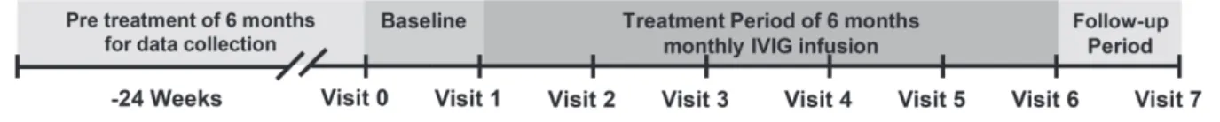
During the 6-month observation period, medical data, in- cluding infectious complications, were collected by the study physicians. After the observation period, patients received IVIG (400 mg/kg/month, Green Cross Corp) as an adjunct to their standard asthma therapy. The treatment comprised 6 infusions at monthly intervals, followed by a post-treatment observation period (Fig. 1). Pulmonary function test (PFT) results, asthma control test results (ACT), asthma-specific quality of life (AQOL), and safety variables were assessed every 4 weeks at scheduled clinic visits. Sputum and blood samples for the mea- surement of cytokines were obtained at baseline and week 21.
The trough levels of IgG subclasses were measured at baseline, and at weeks 5 and 25.
Outcomes
The primary endpoint was the proportion of subjects experi- encing asthma exacerbations related to respiratory infections during the treatment period compared to that in the prior 6 months. Asthma exacerbation was defined a condition in which: (1) a treating physician elected to administer systemic glucocorticoids (prednisolone 10 mg equivalent dose per day for at least 3 consecutive days) or (2) a patient was either hospi-
Fig. 1. Schematic of the study design.
talized or visited an emergency department/a physician’s office (unscheduled). Secondary endpoints included the change in FEV1 from baseline, use of systemic glucocorticoids, use of an- tibiotics and treatment duration, the number of acute exacer- bations, AQOL and ACT scores, and serum levels of IgG sub- classes and cytokines. Asthma severity at diagnosis and asthma control level at enrollment were assessed based on the Expert Panel Report 3 and the Global Initiative for Asthma (GINA) guidelines, respectively. Adverse events were assessed at each visit and collected its severity, duration, and causality assess- ment by the criteria of WHO-UMC (World Health Organiza- tion-The Uppsala Monitoring Centre).
Measurement of cytokine levels before and after treatment with IVIG
Serum samples were obtained at visit 1 (pre-treatment) and visit 7 (post-treatment). Cytokines—interleukin 4, 5, 6, 8, 9, and 12p70, interferon gamma (IFN)-induced protein 10 (IP-10), and IFN-γ—were quantified using a Luminex bead-based mul- tiplex assay (R&D Systems, Minneapolis, MN, USA).
Statistical analysis
A sample size of 21 had 80% power at a 5% level of signifi- cance to detect differences in the proportion of asthma exacer- bations before and after treatment, based on previous data.17 To allow for the possibility of up to 30% of participants withdraw- ing early from the study, a recruitment target of 30 participants was set. The intention-to-treat (ITT) population comprised all subjects who received at least 1 IVIG infusion; the per-protocol (PP) population comprised the subjects in the ITT population without full protocol deviations.
Analysis of the primary efficacy─the reduction in the pro- portion of subjects in the ITT and PP populations experiencing asthma exacerbations related to respiratory infection before and after treatment─was performed by the one sample t test.
The analysis of secondary outcomes was performed using Co- chran’s Q test for categorical variables and repeated ANOVA for continuous variables. Paired analysis was performed using the McNemar-Bowker test for categorical variables and the paired t test for continuous variables. Differences in serum cytokine levels before and after treatment were evaluated using Wilcox- on’s signed rank test. Safety data are reported only as descrip- tive statistics.
All analyses were two-sided and performed at a 5% signifi- cance level. The results were analyzed using SPSS (ver. 22; SPSS Inc., Chicago, IL, USA) and R software (ver. 3.2.3.; R Develop- ment Core Team, Vienna, Austria).
RESULTS Patients
In total, 30 patients were enrolled and 24 completed the study.
The demographic and clinical features of the study subjects are listed in Table 1. There was a significant female predominance (female: male, 6.5:1.0), and the mean age at diagnosis was
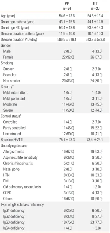
Table 1. Baseline characteristics of the study subjects PP
n=24 ITT
n=30
Age (year) 54.6±13.6 54.5±13.4
Onset age asthma (year) 43.1±15.6 44.1±14.5
Onset age PID (year) 53.4±13.6 53.5±13.3
Disease duration asthma (year) 11.5±10.8 10.4±10.3 Disease duration PID (day) 586.5±616.1 513.2±573.8 Gender
Male 2 (8.0) 4 (13.0)
Female 22 (92.0) 26 (87.0)
Smoking
Smoker 2 (8.0) 2 (7.0)
Exsmoker 2 (8.0) 4 (13.0)
Non-smoker 20 (83.0) 24 (80.0)
Severity*
Mild, intermittent 1 (5.0) 1 (4.0)
Mild, persistent 1 (5.0) 3 (11.0)
Moderate 11 (46.0) 13 (45.0)
Severe 11 (50.0) 12 (44.0)
Control status†
Controlled 1 (4.0) 2 (7.0)
Partly controlled 11 (46.0) 15 (52.0)
Uncontrolled 12 (50.0) 10 (41.0)
Baseline FEV1% 75.1±23.3 73.4 ±23.1
Underlying disease
Allergic rhinitis 16 (67.0) 19 (63.0)
Aspirin/sulfite sensitivity 9 (38.0) 9 (30.0)
Chronic rhinosinusitis 5 (21.0) 6 (20.0)
Nasal polyp 2 (8.0) 3 (10.0)
HTN 8 (33.0) 10 (33.0)
DM 3 (13.0) 3 (10.0)
Old pulmonary tuberculosis 1 (4.0) 1 (3.0)
COPD 3 (13.0) 4 (13.0)
Others 16 (67.0) 18 (60.0)
Type of IgG subclass deficiency
IgG1 deficiency 6 (25.0) 6 (20.0)
IgG2 deficiency 8 (33.0) 8 (27.0)
IgG3 deficiency 18 (75.0) 23 (77.0)
IgG4 deficiency 1 (4.0) 1 (3.0)
Data are presented as mean±SD, n (%), unless otherwise indicated.
*Severity was defined in Expert Panel Report 3; †Control status was defined ac- cording to Global Initiative for Asthma (GINA) guidelines.
ITT, intention to treat; PP, per protocol; COPD, chronic obstructive pulmonary dis- ease; DM, diabetes mellitus; FEV1, forced expiratory volume in 1 s; HTN, hyper- tension; PID, primary immunodeficiency disease.
54.5±13.4 years. The mean duration of asthma and PID was 10.4±10.3 years and 513.2±573.8 days, respectively. Most pa- tients had moderate (45.0%) to severe (44.0%) asthma and were in a partly controlled (52.0%) or uncontrolled (41.0%) state. At baseline, the mean FEV1 of the study subjects was 73.4%±
23.1%. Among the 4 IgG subtypes, deficiency in IgG3 was most common (n=23, 77%).
Primary and secondary outcomes
Ig replacement was associated with a reduction in the propor- tion of patients with asthma exacerbations caused by respirato- ry infections compared to the pre-treatment value (Table 2).
The proportion of asthma exacerbation by respiratory infec- tions was significantly reduced at each visit compared to base- line (Fig. 2). The overall asthma exacerbation rate, including
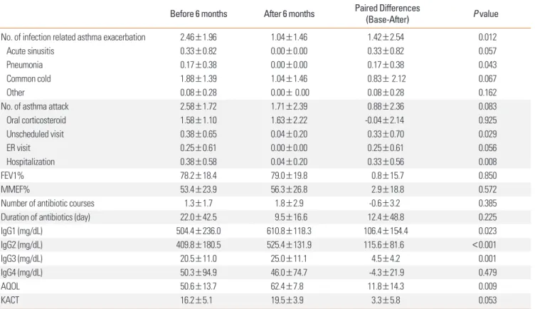
both infections and noninfectious causes, was also reduced during the treatment versus pre-treatment period. The number of infectious complications was significantly reduced, from 2.46±1.96 during the previous 6 months to 1.04±1.46 during the treatment period (Table 3). We noted significant reductions in the frequency of asthma exacerbation events requiring un- scheduled physician visits and hospital admission (P=0.029 and P=0.008, respectively).
Table 2. Reduction rate of asthma exacerbation caused by respiratory infec- tions between pre- and post-treatment of IVIG for 6 months
Mean±SD Median (min to max) P value In PP (n=24) -0.31±1.14 -0.83 (-1.00 to 3.00) 0.193 In ITT (n=30) -0.45±1.05 -1.00 (-1.00 to 3.00) 0.026 ITT, intention to treat; PP, per protocol; SD, standard deviation
Fig. 2. Proportion of asthma exacerbations during pre- and post-IVIG treatment periods as well as at each visit.
Asthma exacerbation by respiratory infections (%)
Before 6 months
V7 V6 V5 V4 V3 V2
After 6 months 100
80 60 40 20 0
Table 3. Primary and secondary efficacy outcomes
Before 6 months After 6 months Paired Differences
(Base-After) P value
No. of infection related asthma exacerbation 2.46±1.96 1.04±1.46 1.42±2.54 0.012
Acute sinusitis 0.33±0.82 0.00±0.00 0.33±0.82 0.057
Pneumonia 0.17±0.38 0.00±0.00 0.17±0.38 0.043
Common cold 1.88±1.39 1.04±1.46 0.83± 2.12 0.067
Other 0.08±0.28 0.00± 0.00 0.08±0.28 0.162
No. of asthma attack 2.58±1.72 1.71±2.39 0.88±2.36 0.083
Oral corticosteroid 1.58±1.10 1.63±2.22 -0.04±2.14 0.925
Unscheduled visit 0.38±0.65 0.04±0.20 0.33±0.70 0.029
ER visit 0.25±0.61 0.00±0.00 0.25±0.61 0.056
Hospitalization 0.38±0.58 0.04±0.20 0.33±0.56 0.008
FEV1% 78.2±18.4 79.0±19.8 0.8±15.7 0.850
MMEF% 53.4±23.9 56.3±26.8 2.9±18.8 0.572
Number of antibiotic courses 1.3±1.7 1.8±2.9 -0.6±3.2 0.385
Duration of antibiotics (day) 22.0±42.5 9.5±16.6 12.4±48.8 0.225
IgG1 (mg/dL) 504.4±236.0 610.8±118.3 106.4±154.4 0.023
IgG2 (mg/dL) 409.8±180.5 525.4±131.9 115.6±81.6 <0.001
IgG3 (mg/dL) 20.5±11.0 25.0±11.1 4.5±4.2 0.001
IgG4 (mg/dL) 50.3±94.9 46.0±74.7 -4.3±21.9 0.479
AQOL 50.6±13.7 62.4±7.8 11.8±14.3 0.009
KACT 16.2±5.1 19.5±3.9 3.3±5.8 0.053
Data are presented as mean±SD.
AQOL, asthma-related quality of life; ER, emergency room; FEV1, forced expiratory volume in 1 s; KACT, Korean asthma control test; MMEF, maximal mid-expiratory flow.
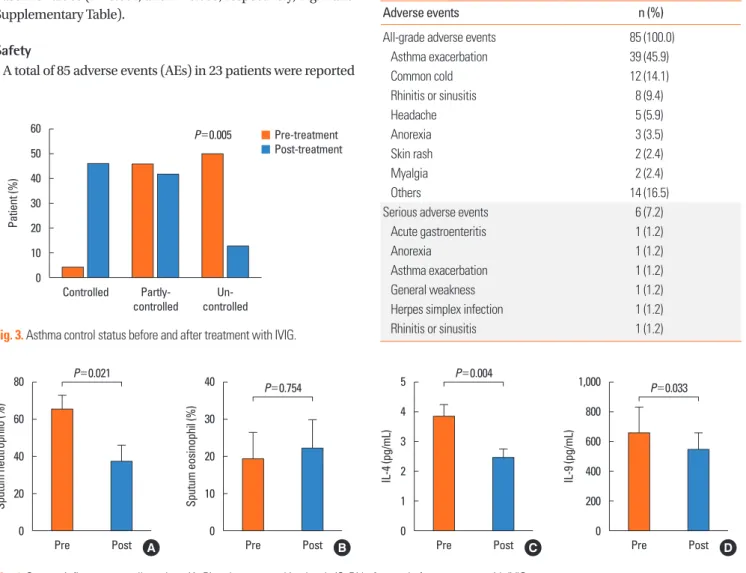
No significant changes in lung function were noted at any visit during the treatment period compared to baseline. However, there was a significant increase in the proportion of patients categorized as controlled during the study period compared to those partly or fully controlled at baseline (Fig. 3). In addition, IVIG treatment resulted in significant improvements in ACT and total AQOL scores compared to those at baseline (P=0.009 and P=0.053, respectively). Improvements in AQOL and ACT scores were observed as early as week 8 and sustained up to week 25 (P=0.009, and P=0.053, respectively) (Supplementary Figure and Table 3). Following the 6 months of IVIG, the serum level of IgG1 (P=0.023), IgG2 (P<0.001), and IgG3 (P=0.001) were significantly increased. However, IgG4 was not increased (P=0.479, Table 3). The total inflammatory cells in sputum comprised primarily neutrophils (65.8%±27.4%) rather than eosinophils (19.6%±25.7%) at baseline. IVIG was associated with a significant decrease in the number of neutrophils in spu- tum compared to baseline, but, there was no significant change in the number of eosinophils (Fig. 4). IL-4 and IL-9 levels were significantly lower at the end of the study compared to the baseline values (P=0.004, and P=0.033, respectively; Fig. 4 and Supplementary Table).
Safety
A total of 85 adverse events (AEs) in 23 patients were reported
during the study period (Table 4). Six cases of serious AEs were reported: acute gastroenteritis, anorexia, asthma exacerbation, general weakness, herpes simplex infection, and rhinitis/sinus- itis. However, none of the severe AEs were related to IVIG treat- ment according to the criteria of WHO-UMC causality assess- ment. Most of the AEs were mild (84.7%) or moderate (12.9%).
Thirty-nine cases of acute asthma exacerbation in 13 patients were reported, 25 of which were related to respiratory infec- tions; the remaining had non-infectious causes. IVIG-related adverse drug reactions were headache (n=5), skin rash (n=2), and myalgia (n=2); however, none of these patients discontin- ued treatment due to the reactions.
DISCUSSION
The results of this study showed that IVIG improved out- comes in patients with uncontrolled, moderate-to-severe asth- ma. They had a history of recurrent respiratory infections and
Fig. 3. Asthma control status before and after treatment with IVIG.
Patient (%)
Controlled Un-
controlled Partly-
controlled 60
50 40 30 20 10 0
Pre-treatment Post-treatment P=0.005
Fig. 4. Sputum inflammatory cell numbers (A, B) and serum cytokine levels (C, D) before and after treatment with IVIG.
Sputum neutrophilo (%) IL-4 (pg/mL)
Sputum eosinophil (%) IL-9 (pg/mL)
Pre Post Pre Post Pre Post Pre Post
80
60
40
20
0
5 4 3 2 1 0 40
30
20
10
0
1,000 800 600 400 200 0
P=0.021 P=0.004
P=0.754 P=0.033
A B C D
Table 4. Adverse events
Adverse events n (%)
All-grade adverse events 85 (100.0)
Asthma exacerbation 39 (45.9)
Common cold 12 (14.1)
Rhinitis or sinusitis 8 (9.4)
Headache 5 (5.9)
Anorexia 3 (3.5)
Skin rash 2 (2.4)
Myalgia 2 (2.4)
Others 14 (16.5)
Serious adverse events 6 (7.2)
Acute gastroenteritis 1 (1.2)
Anorexia 1 (1.2)
Asthma exacerbation 1 (1.2)
General weakness 1 (1.2)
Herpes simplex infection 1 (1.2)
Rhinitis or sinusitis 1 (1.2)
had been diagnosed with IgGSCD. In the primary analysis, IVIG significantly reduced the proportion of patients with asth- ma exacerbations due to respiratory infections compared to baseline. During the 24-week treatment, the number of respira- tory infections was significantly decreased, and the proportion of patients with controlled asthma status was increased. These results were achieved despite the continued use of previous asthma treatments throughout the study.
Most of the asthmatic patients have rare or intermittent exac- erbations; however, there is evidence that a subset of asthmat- ics is ‘exacerbation-prone’18 Several epidemiologic studies have reported a strong correlation between FEV1 and the risk of asthma exacerbation.19-21 Acute severe exacerbations in suscep- tible asthmatics activate pathways of inflammation and remod- eling, resulting in the deterioration of lung function. Accelerat- ed loss of lung function in turn increases the risk of exacerba- tion, resulting in a vicious cycle and the exacerbation-prone phenotype. Moreover, longitudinal studies of a PID cohort show that permanently diminished lung function due to recur- rent respiratory infections leads to the exacerbation of underly- ing asthma or an increase in asthma symptoms.22-24 In the pres- ent study, all subjects had histories of more than 2 recurrent re- spiratory infections per year, and their mean FEV1 was 73.4%.
This suggests the presence of a subset of asthmatics with an ex- acerbation-prone phenotype as well as considerable overlap between asthma and PIDs.
Several open trials using high doses (1-2 g/kg) of monthly Ig replacement showed that IVIG had a systemic steroid-sparing effect in children and adults with steroid-dependent severe asthma.13,14,25 However, randomized controlled studies failed to demonstrate the efficacy of IVIG in the same population, al- though the steroid-sparing effect was noted in a subgroup that required high daily doses of oral corticosteroids.15,16,26 In con- trast, IVIG has been shown to be effective and is used exten- sively in patients with primary humoral immunodeficiencies.27 Regarding the efficacy of IVIG in IgGSCD, several open-label studies have reported that IVIG significantly decreases the number of infections, the need for antibiotics, and hospitaliza- tion, and improves the quality of life in patients with recurrent infections.7,12 Therefore, we treated carefully selected groups of adult asthmatic patients with IgGSCD in the present study. All were ethnically Korean. We demonstrated that 400 mg/kg of monthly IVIG resulted in favorable clinical outcomes, such as improvement in infection-related asthma exacerbation, asth- ma control status, and quality of life.
Apart from its initial use as a supplementary therapy for pri- mary humoral immune deficiencies, IVIG has also benefited patients with various autoimmune or allergic diseases due to its anti-inflammatory and immunomodulatory effects at high dos- es.28 Several mechanisms by which IVIG exerts its immune modulating effects on asthma have been suggested: it can di- rectly act on T cells by enhancing the activity of CD25FoxP3+
Treg cells, leading to the production of greater amounts of transforming growth factor β and IL-10, and can inhibit IL-4 production in a murine model of allergic airway inflamma- tion.29-31 Levels of the Th2 cytokines IL-4 and IL-9 were signifi- cantly reduced after IVIG, whereas Th1 cytokine levels were not. The subjects also showed elevated numbers of neutrophils and eosinophils at baseline. Neutrophilia in airways is the con- sequence of viral or bacterial infections that can lead to acute exacerbations.32 The elevated sputum neutrophils were signifi- cantly decreased in patients whose asthma was controlled by treatment with IVIG. These data suggested that lower doses of IVIG might down-regulate IL-4 and IL-9 production, and that clinical improvements of IVIG could result from not only re- placement of Ig inhibiting respiratory pathogens, but also anti- inflammatory effects in the airways of asthmatics. However, mechanisms by which IVIG inhibits Th2 cytokine production and airway inflammation are unclear.
This study has several limitations. It used an open-label, sin- gle-arm design having inherent weaknesses. There was no con- trol group and the sample size was relatively small. To over- come these weaknesses, we evaluated the patients 6 months before and after treatment. Furthermore, to minimize the effect of potential confounders for primary and secondary endpoints, we enrolled the patients being well-compliant and receiving appropriate asthma treatments during the pre-IVIG treatment period; nonetheless, a monthly scheduled meeting with a phy- sician may detect asthma exacerbation in advance and then prevent severe asthma exacerbation during the post-IVIG treat- ment period. Furthermore, a 6-month treatment period was relatively short to assess the effect of seasonal variations in asth- ma exacerbation. However, patient-centered outcomes, such as AQOL scores, were improved by IVIG treatment and no serious adverse reactions leading to discontinuation of IVIG were re- ported. In contrast to previous trials that focused on reducing systemic steroid use as a primary end point, the efficacy of IVIG as an add-on therapy against asthma exacerbations was dem- onstrated in our study.15,26 These findings could facilitate the development of new therapeutic options for exacerbation- prone asthmatics with IgGSCD.
In conclusion, the addition of IVIG to standard therapy reduc- es asthma exacerbations and improves asthma control status as well as quality of life, especially for patients whose asthma is not controlled and who have IgGSCD, leading to recurrent re- spiratory infections. This study will help larger double-blind placebo-controlled trials define the role of IVIG in such patient populations. Further studies are needed to evaluate other out- comes, such as lung function, cost effectiveness, and the opti- mal dose of IVIG.
ACKNOWLEDGMENTS
This study was supported by Green Cross Corp. and a grant of
the Korea Health Technology R&D Project through the Korea Health Industry Development Institute, funded by the Ministry of Health & Welfare, Republic of Korea (HI14C1061 and HI16C0992). This study was supported by ARO at Clinical Trial Center of Ajou University Hospital.
REFERENCES
1. Dougherty RH, Fahy JV. Acute exacerbations of asthma: epidemi- ology, biology and the exacerbation-prone phenotype. Clin Exp Al- lergy 2009;39:193-202.
2. Fajt ML, Wenzel SE. Development of new therapies for severe asth- ma. Allergy Asthma Immunol Res 2017;9:3-14.
3. Busse WW, Lemanske RF Jr, Gern JE. Role of viral respiratory infec- tions in asthma and asthma exacerbations. Lancet 2010;376:826- 34.
4. Kurai D, Saraya T, Ishii H, Takizawa H. Virus-induced exacerba- tions in asthma and COPD. Front Microbiol 2013;4:293.
5. Azar AE, Ballas ZK. Evaluation of the adult with suspected immu- nodeficiency. Am J Med 2007;120:764-8.
6. Kim JH, Park HJ, Choi GS, Kim JE, Ye YM, Nahm DH, et al. Immu- noglobulin G subclass deficiency is the major phenotype of prima- ry immunodeficiency in a Korean adult cohort. J Korean Med Sci 2010;25:824-8.
7. Abdou NI, Greenwell CA, Mehta R, Narra M, Hester JD, Halsey JF.
Efficacy of intravenous gammaglobulin for immunoglobulin G subclass and/or antibody deficiency in adults. Int Arch Allergy Im- munol 2009;149:267-74.
8. Kim JH, Park S, Hwang YI, Jang SH, Jung KS, Sim YS, et al. Immu- noglobulin g subclass deficiencies in adult patients with chronic airway diseases. J Korean Med Sci 2016;31:1560-5.
9. Agondi RC, Barros MT, Rizzo LV, Kalil J, Giavina-Bianchi P. Allergic asthma in patients with common variable immunodeficiency. Al- lergy 2010;65:510-5.
10. Touw CM, van de Ven AA, de Jong PA, Terheggen-Lagro S, Beek E, Sanders EA, et al. Detection of pulmonary complications in com- mon variable immunodeficiency. Pediatr Allergy Immunol 2010;
21:793-805.
11. Ochs HD, Hagin D. Primary immunodeficiency disorders: general classification, new molecular insights, and practical approach to diagnosis and treatment. Ann Allergy Asthma Immunol 2014;112:
489-95.
12. Abrahamian F, Agrawal S, Gupta S. Immunological and clinical profile of adult patients with selective immunoglobulin subclass deficiency: response to intravenous immunoglobulin therapy. Clin Exp Immunol 2010;159:344-50.
13. Jakobsson T, Croner S, Kjellman NI, Pettersson A, Vassella C, Björk- stén B. Slight steroid-sparing effect of intravenous immunoglobu- lin in children and adolescents with moderately severe bronchial asthma. Allergy 1994;49:413-20.
14. Haque S, Boyce N, Thien FC, O’Hehir RE, Douglass J. Role of intra- venous immunoglobulin in severe steroid-dependent asthma. In- tern Med J 2003;33:341-4.
15. Kishiyama JL, Valacer D, Cunningham-Rundles C, Sperber K, Richmond GW, Abramson S, et al. A multicenter, randomized, double-blind, placebo-controlled trial of high-dose intravenous immunoglobulin for oral corticosteroid-dependent asthma. Clin Immunol 1999;91:126-33.
16. Salmun LM, Barlan I, Wolf HM, Eibl M, Twarog FJ, Geha RS, et al.
Effect of intravenous immunoglobulin on steroid consumption in patients with severe asthma: a double-blind, placebo-controlled, randomized trial. J Allergy Clin Immunol 1999;103:810-5.
17. Bernatowska-Matuszkiewicz E, Pac M, Skopcynska H, Pum M, Eibl MM. Clinical efficacy of intravenous immunoglobulin in patients with severe inflammatory chest disease and IgG3 subclass defi- ciency. Clin Exp Immunol 1991;85:193-7.
18. Kupczyk M, ten Brinke A, Sterk PJ, Bel EH, Papi A, Chanez P, et al.
Frequent exacerbators--a distinct phenotype of severe asthma.
Clin Exp Allergy 2014;44:212-21.
19. Koga T, Oshita Y, Kamimura T, Koga H, Aizawa H. Characterisation of patients with frequent exacerbation of asthma. Respir Med 2006;
100:273-8.
20. ten Brinke A, Sterk PJ, Masclee AA, Spinhoven P, Schmidt JT, Zwin- derman AH, et al. Risk factors of frequent exacerbations in difficult- to-treat asthma. Eur Respir J 2005;26:812-8.
21. Kim HJ, Lee J, Kim JH, Park SY, Kwon HS, Kim TB, et al. Factors af- fecting recovery time of pulmonary function in hospitalized pa- tients with acute asthma exacerbations. Allergy Asthma Immunol Res 2016;8:499-504.
22. Chen Y, Stirling RG, Paul E, Hore-Lacy F, Thompson BR, Douglass JA. Longitudinal decline in lung function in patients with primary immunoglobulin deficiencies. J Allergy Clin Immunol 2011;127:
1414-7.
23. Goldstein MF, Hilditch GJ, Dvorin DJ, Belecanech GA. Immuno- globulin replacement for selective IgM immunodeficiency, bron- chiectasis, and asthma. Ann Allergy Asthma Immunol 2016;116:
172-3.
24. Verma N, Grimbacher B, Hurst JR. Lung disease in primary anti- body deficiency. Lancet Respir Med 2015;3:651-60.
25. Landwehr LP, Jeppson JD, Katlan MG, Esterl B, McCormick D, Hamilos DL, et al. Benefits of high-dose i.v. immunoglobulin in pa- tients with severe steroid-dependent asthma. Chest 1998;114:1349- 56.
26. Niggemann B, Leupold W, Schuster A, Schuster R, v Berg A, Grübl A, et al. Prospective, double-blind, placebo-controlled, multicen- tre study on the effect of high-dose, intravenous immunoglobulin in children and adolescents with severe bronchial asthma. Clin Exp Allergy 1998;28:205-10.
27. Yong PL, Boyle J, Ballow M, Boyle M, Berger M, Bleesing J, et al. Use of intravenous immunoglobulin and adjunctive therapies in the treatment of primary immunodeficiencies: a working group report of and study by the Primary Immunodeficiency Committee of the American Academy of Allergy Asthma and Immunology. Clin Im- munol 2010;135:255-63.
28. Ballow M. The IgG molecule as a biological immune response modifier: mechanisms of action of intravenous immune serum globulin in autoimmune and inflammatory disorders. J Allergy Clin Immunol 2011;127:315-23.
29. Yamamoto M, Kobayashi K, Ishikawa Y, Nakata K, Funada Y, Kotani Y, et al. The inhibitory effects of intravenous administration of rab- bit immunoglobulin G on airway inflammation are dependent upon Fcγ receptor IIb on CD11c(+) dendritic cells in a murine mod- el. Clin Exp Immunol 2010;162:315-24.
30. Massoud AH, Guay J, Shalaby KH, Bjur E, Ablona A, Chan D, et al.
Intravenous immunoglobulin attenuates airway inflammation through induction of forkhead box protein 3-positive regulatory T cells. J Allergy Clin Immunol 2012;129:1656-1665.e3.
31. Massoud AH, Yona M, Xue D, Chouiali F, Alturaihi H, Ablona A, et al. Dendritic cell immunoreceptor: a novel receptor for intrave- nous immunoglobulin mediates induction of regulatory T cells. J
Allergy Clin Immunol 2014;133:853-863.e5.
32. Gern JE. How rhinovirus infections cause exacerbations of asthma.
Clin Exp Allergy 2015;45:32-42.