Chemotherapy
Infect Chemother 2013;45(4):387-393 pISSN 2093-2340 · eISSN 2092-6448
Received: July 2, 2013 Revised: August 23, 2013 Accepted: September 2, 2013 Corresponding Author : Tae Hyong Kim, MD.
Department of Internal Medicine, Soonchunhyang University Seoul Hospital, 59 Daesagwan-ro, Yongsan-gu, Seoul 140-743, Korea
Tel: +82-2-709-9194, Fax: +82-2-709-9083 E-mail: [email protected]
This is an Open Access article distributed under the terms of the Creative Commons Attribution Non-Commercial License (http://creativecommons.org/licenses/by-nc/3.0) which permits unrestricted non-commercial use, distribution, and repro- duction in any medium, provided the original work is properly cited.
Copyrights © 2013 by The Korean Society of Infectious Diseases | Korean Society for Chemotherapy
www.icjournal.org
Necrotizing Fasciitis in Three University Hospitals in Korea: A Change in Causative Microorganisms and Risk Factors of Mortality During the Last Decade
Shi Nae Yu
1, Tae Hyong Kim
1, Eun Jung Lee
1, Eun-Joo Choo
2, Min Hyok Jeon
3, Yung Gyu Jung
3, Tae Jin Kim
2, In Ki Mun
1, and Ji Sung Lee
41Department of Internal Medicine, Soonchunhyang University Seoul Hospital, Seoul; 2Department of Internal Medicine, Soonchunhyang University Bucheon Hospital, Bucheon; 3Department of Internal Medicine, Soonchunhyang University Cheonan Hospital, Cheonan;
4Biostatistical Consulting Unit, Soonchunhyang University Medical Center, Seoul, Korea
Background: Necrotizing fasciitis is a life-threatening infectious disease with rapidly progressive involvement of the affected site.
Because of the high mortality rate of this disease, early diagnosis, surgical exploration, and administration of appropriate antibiotics are necessary. The present study aimed to further review the changes in the clinical and microbiological characteristics of necrotizing fasciitis using patients’ medical records from consecutive databases of 3 hospitals in Korea.
Materials and Methods: In this study, we retrospectively reviewed the medical records of patients with necrotizing fasciitis who were clinically diagnosed between May 2001 and February 2012 in 3 university hospitals in Korea. In total, the data of 83 patients were analyzed, including those of 20 patients from our previous study in 2006. An organism found in a blood culture or surgical specimen was regarded as a causative organism.
Results: Of the 83 patients, 68(81.9%) had community-acquired infections. Ninety microorganism species were indentifed by culture. Streptococcus was the most commonly identified pathogen. Non-fermentative gram-negative bacteria and Candida species have recently emerged, especially in immunocompromised hosts.
Conclusions: Gram-positive organisms are still the most common pathogens of necrotizing fasciitis. However in our study, vari- ous gram-negative bacteria with different levels of susceptibility to antibiotics, as well as Candida species, were responsible for the necrotizing fasciitis. Initial empirical antimicrobial agents for necrotizing fasciitis should be considered depending on the individual patient’s condition.
Key Words: Necrotizing fasciitis, Skin and soft tissue infection
Introduction
Necrotizing fasciitis (NF) is a relatively rare but rapidly pro- gressive soft tissue infection characterized by widespread fascial necrosis. Because of the high mortality rate of NF (6–76%) [1], early diagnosis, early surgical exploration, and appropriate antibiotics are necessary. Moreover, a local epidemiological investigation of the causative organism and its susceptibility to antibiotics should be performed. In 2006, we reported the clinical characteristics and causative organism of NF cases from 3 university hospitals in Korea [2].
The present study aimed to further review the changing clin- ical and microbiological characteristics of NF using consecu- tive database records of patients from 3 hospitals. In addition, we performed an analysis to determine the contributing fac- tors to NF-related mortality.
Materials and Methods
We retrospectively reviewed the medical records of patients with NF who were clinically diagnosed between May 2001 and February 2012 in 3 university hospitals, which located in Seoul, Bucheon, and Cheonan. Patients who were readmitted for secondary treatment such as skin grafting were excluded.
In our previous study, we enrolled 22 patients with NF diag- nosed between May 2001 and May 2005. In addition, we re- viewed the medical records of 30 and 38 patients with NF di- agnosed between June 2005 and April 2009, and between May 2009 and February 2012, respectively. Of the 90 patients, 7 were not subjected to analysis because they either discharged against medical recommendation or transferred to another hospital.
Therefore, the records of a total of 83 patients were analyzed.
Age, sex, comorbidities, affected sites, clinical signs including bulla, microbiological findings in sterile specimens, antibiot- ics susceptibility, antibiotics used, type of surgery, clinical outcome, and cause of death were reviewed for each patient.
Hospital-acquired infection was defined according to the definition of the Centers for Disease Control and Prevention (CDC) [3], as the onset of signs or symptoms 48 h after hospi- tal admission. The organism isolated from blood cultures or surgical specimens was regarded as the causative organism. If blood culture result was not available, aspiration culture re- sults of a skin bulla specimen were used. Open pus culture re- sults were not considered for microbiological analysis.
The time of performing initial curative surgical procedures
such as incision and drainage, fasciotomy, or amputation was regarded as the starting time of operation.
Data are presented as mean [SD] for normally distributed variables and as median (interquartile range) or number (%) of subjects for categorical variables. Comparisons of the base- line and disease characteristics between the patients who died and those who survived were performed using the Pear- son Chi-square test, Fisher exact test, or Student t-test accord- ing to the type of variable. To determine the risk factors for outcome, we performed a multiple logistic regression analysis.
All statistical analyses were performed using SAS version 9.3 (SAS Institute Inc., Cary, NC). A 2-sided P-value of < 0.05 was considered statistically significant.
Results
Of the 83 patients included in this study, 59 (71%) were men, with a mean age of 58 years. There were 68 (81.9%) patients with community-acquired infection. The most common co- morbidity among the patients was diabetes mellitus (n = 38;
45.8%), followed by chronic liver disease (n = 22; 26.5%), and chronic kidney disease (n = 22; 26.5%). The most frequently affected site was the lower extremities (n = 42; 50.6%). Thirty- one patients presented with bulla at the time of diagnosis. In the records of 5 patients, there was no information regarding the occurrence of bulla. Septic shock, severe sepsis, and sepsis occurred in 23 (27.7%), 12 (14.5%), and 12 (14.5%) patients, respectively (Table 1).
Radiological imaging (magnetic resonance imaging, com- puted tomography, or ultrasonography) was performed in 66.2% (55/83) of the patients. Of these patients, 52.7% (29/55) had NF-positive results, of which 34.9% (29/83) were con- firmed by positive imaging study results.
Sixty-eight patients (81.9%) had a microbiologically defined infection. Monomicrobial infection was confirmed in 50 pa- tients (73.5%), and polymicrobial infection was confirmed in 18 patients (26.5%). Twenty-three organisms were isolated from blood cultures, and 59 from surgical specimens. Gram- positive bacteria (GPB) were found in 45 patients (54.2%), and gram-negative bacteria (GNB) in 40 patients (48.2%). Esche- richia coli was the most frequently identified pathogen (n = 13, 14.4%; extended-spectrum beta-lactamase [ESBL]-produc- ing microbes: 30.8%), followed by Staphylococcus aureus (n = 12, 13.3%; methicillin-resistant Staphylococcus aureus [MRSA]: 41.6%), and Streptococcus pyogenes (n = 12, 12%;
penicillin susceptibility: 100%; Table 1). None of the Strepto-
coccus pyogenes infection cases was hospital acquired. How- ever, 2 patients with Streptococcus pyogenes infection had a history of intramuscular injection or acupuncture, which fits the definition of health-care-associated infection. The GPB-to-
GNB ratio in the present study was similar to that in our previ- ous study (Table 2).
Of the 4 cases of ESBL-producing Escherichia coli, 2 were hospital acquired, 1 was health-care associated, and 1 was Table 1. Comparison of the baseline characteristics of patients with necrotizing fasciitis who survived and those who died
Variables N (%)
P-valuea Alive (N = 55) Died (N = 28) Total (N = 83)
Sex (male) 39 (70.9) 20 (71.4) 59 (71.1) 0.96
Age (yr) 53.7 (17.7) 62.1 ± 14.4 0.031
Associated diseases
Diabetes mellitus 24 (43.6) 14 (50.0) 38 (45.8) 0.582
Malignancies 6 (10.9) 6 (21.4) 12 (14.5) 0.198
Immunosuppressive agent 0 (0) 4 (14.3) 4 (4.8) 0.011
Chronic liver disease 11 (20) 11 (39.3) 22 (26.5) 0.060
Chronic kidney disease 10 (18) 12 (42.9) 22 (26.5) 0.016
Acquisition of infection 0.571
Community acquired 46 (83.6) 22 (78.6) 68 (81.9)
Hospital acquired 9 (16.4) 6 (21.4) 15 (18.1)
Site of infection
Trunk 10 (18.2) 7 (25) 17 (20.5) 0.467
Groin or buttock 11 (20) 0 (0) 11 (13.3) 0.013
Head and neck 6 (10.9) 1 (3.6) 7 (8.4) 0.414
Upper extremities 9 (16.4) 3 (10.7) 12 (14.5) 0.743
Lower extremities 24 (43.6) 18 (64.3) 42 (50.6) 0.075
Initial manifestations
Fever (body temperature > 38oC) 13 (23.6) 8 (28.6) 21 (25.3) 0.625
Bullae 17 (30.9) 14 (50) 31 (37.3) 0.166
Septic shock 8 (14.5) 15 (53.6) 23 (27.7) < 0.001
Laboratory examination
Serum albumin (g/dL) 3.0 ± 0.95 2.4 ± 0.65 0.001
Serum sodium (mmol/L) 137.6 ± 6.92 133.4 ± 6.63 0.011
Serum creatinine (mg/dL) 1.10 (0.7-1.63) 2.15 (1.33-3.35) <0.001
C-reactive protein (mg/dL) 13.2 (4.05-24.18) 20.9 ± 9.7 0.02
Thrombocytopenia 0.005
≥ 100,000/mm3 46 (83.6) 15 (53.6) 61 (73.5)
50,000-100,000/mm3 5 (9.1) 4 (14.3) 9 (10.8)
< 50,000/mm3 4 (7.3) 9 (32.1) 13 (15.7)
Cause
Aerobic gram-positive monomicrobial infection 17 (30.9) 9 (32.1) 26 (31.3) 0.909
Aerobic gram-negative monomicroibal infection 12 (21.8) 11 (39.3) 23 (27.7) 0.093
Polymicrobial infection 12 (21.8) 6 (21.4) 18 (21.7) 0.968
Treatment 0.007
Antibiotics only 6 (10.8) 10 (35.7) 16 (19.3)
Surgery plus antibiotics 49 (89.0) 18 (64.3) 67 (80.7)
The data are expressed as number of patients (%) or mean [SD], unless otherwise indicated.
aThe P-values were calculated using the Pearson Chi-square test, Fisher exact test, or Student t-test, as appropriate.
community acquired. However, the patient with community- acquired infection had a history of intramuscular injection. Of the 5 cases of MRSA infection, 2 were hospital acquired, 2 were health-care associated, and 1 was of an unknown cause.
Of the 2 cases of infection with Candida species, 1 was health-care associated and occurred in a patient with a pres- sure sore due to paraplegia; the patient visited the hospital be- cause the sore worsened despite self-medication. MRSA was confirmed by surgical specimen analysis, and the presence of Candida tropicalis and Streptococcus sanguis was confirmed by blood culture. The patient was treated with antibiotics, in- cluding amphotericin B for candidemia. The other 2 patients
with confirmed Candida species infection had a history of liv- er transplantation and immunosuppressive agent use. Unfor- tunately, acute transplant rejection occurred, leading to gen- eral edema that caused the NF on the patient’s trunk and leg.
Candida tropicalis was isolated from the surgical specimen.
The patient was treated with empirical antibiotics, including amphotericin B but eventually died.
A total of 67 patients underwent surgery, and amputation was performed in 5. Two amputation procedures were per- formed initially, and the other 3 were performed after simple fasciotomy or debridement. Sixteen patients did not undergo surgery because of rapid deterioration leading to death in 10 Table 2. Comparison of the causative organisms of necrotizing fasciitis
Identified organisms N (%) Total
N (%) 2001–2005 2005–2009 2009–2012
Polymicrobial infection 3 (15) 8 (38.1) 7 (25.3) 18 (26.5)
Aerobic gram positive 11 (50) 15 (50) 19 (50) 45 (50)
Streptococcus species a 3 (13.6) 6 (20) 10 (26.3) 19 (21.1)
Staphylococcus aureus 3 (13.6) 4 (13.3) 5 (13.2) 12 (13.3)
MSSA 2 (9.1) 3 (10) 2 (5.3) 7 (7.8)
MRSA 1 (4.5) 1 (3.3) 3 (7.9) 5 (5.6)
Coagulase-negative staphylococci 3 (13.6) 1 (3.3) 3 (7.9) 7 (7.8)
Enterococcus species 2 (9.1) 4 (13.3) 1 (2.6) 7 (7.8)
Aerobic gram negative 10 (45.5) 15 (50) 15 (39.5) 40 (44.4)
Escherichia coli 3 (13.6) 6 (20) 4 (10.5) 13 (14.4)
Klebsiella pneumoniae 1 (4.5) 3 (10) 2 (5.3) 6 (6.7)
Pseudomonas aeruginosa 2 (9.1) 0 2 (5.3) 4 (4.4)
Serratia marcescens 1 (4.5) 1 (3.3) 2 (5.3) 4 (4.4)
Vibrio vulnificus 1 (4.5) 0 2 (5.3) 3 (3.3)
Acinetobacter baumannii 0 0 3 (7.9) 3 (3.3)
Yersinia enterocolitica 1 (4.5) 0 0 1 (1.1)
Enterobacter agglomerans 0 1 (3.3) 0 1 (1.1)
Aeromonas hydrophila 1 (4.5) 0 0 1 (1.1)
Proteus vulgaris 0 1 (3.3) 0 1 (1.1)
Vibrio parahaemolyticus 0 1 (3.3) 0 1 (1.1)
Citrobacter freundii 0 1 (3.3) 0 1 (1.1)
Prevotella loescheii 0 1 (3.3) 0 1 (1.1)
Anaerobesb 1 (4.5) 0 1 (2.6) 2 (2.2)
Fungus
Candida tropicalis 0 0 2 (5.3) 2 (2.2)
Trichosporon beigelii 0 0 1 (2.6) 1 (1.1)
Total 22 30 38 90
MSSA, methicillin-sensitive Staphylococcus aureus; MRSA, methicillin-resistant Staphylococcus aureus.
aStreptococcus pyogenes, Streptococcus agalactiae, viridans group streptococci, Streptococcus milleri group, and group F streptococci.
bClostridium subterminale and anaerobic gram-positive bacilli: not included in the gram-positive group.
of the patients and a relatively mild disease progression in the other 6 patients. Seven of the 10 patients who died without surgery developed septic shock, and the other 3 had severe sepsis. The patients who did not undergo surgery had a signifi- cantly higher mortality rate than those who underwent sur- gery (62.5% vs. 26.8%; P = 0.007).
Of the 83 patients, 55 survived and 28 died (in-hospital mor-
tality: 33.7%) due to NF (n = 23; 27.7%), pneumonia during hospitalization (n = 3; 3.6%), and progression of malignancy (n
= 2; 2.4%).
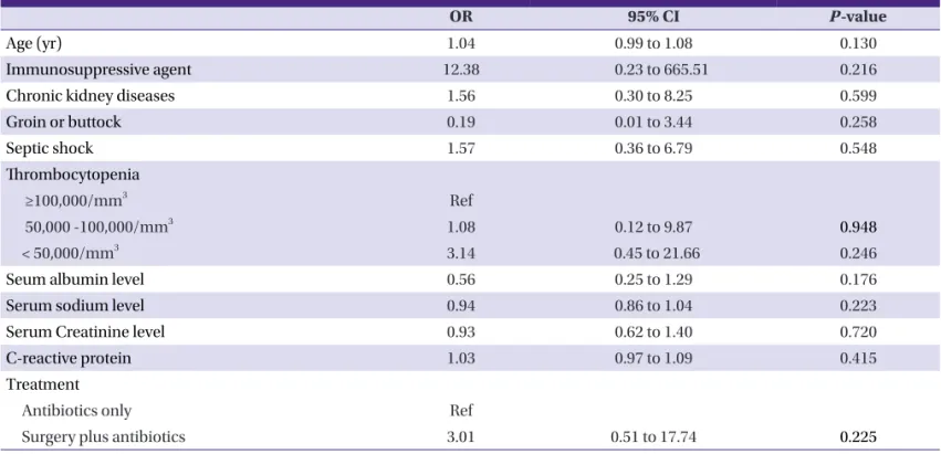
Age, chronic kidney disease, septic shock, hyponatremia, thrombocytopenia (< 50,000/mm3), hypoalbuminemia, high C-reactive protein level, and surgery were significantly associ- ated with mortality (Table 3).
Table 3. Univariate analysis of risk factors of death in necrotizing fasciitis
OR 95% CI P-value
Age (yr) 1.03 1.00 to 1.06 0.036
Immunosuppressive agent 20.38 0.75 to 553.16 0.074
Chronic kidney diseases 3.38 1.22 to 9.31 0.019
Groin or buttock 0.07 0.003 to 1.357 0.078
Septic shock 6.78 2.36 to 19.47 <0.001
Thrombocytopenia
≥100,000/mm3 Ref
50,000~100,000/mm3 2.45 0.58 to 10.34 0.221
< 50,000/mm3 6.90 1.85 to 25.68 0.004
Seum albumin level 0.42 0.22 to 0.79 0.007
Serum sodium level 0.91 0.84 to 0.98 0.016
Serum Creatinine level 1.31 0.99 to 1.74 0.063
C-reactive protein 1.06 1.01 to 1.10 0.021
Treatment
Antibiotics only Ref
surgery plus antibiotics 4.54 1.44 to 14.29 0.010
OR, odds ratio; 95% CI, 95% confidence interval.
Table 4. Multivariate analysis of risk factors of death in necrotizing fasciitis
OR 95% CI P-value
Age (yr) 1.04 0.99 to 1.08 0.130
Immunosuppressive agent 12.38 0.23 to 665.51 0.216
Chronic kidney diseases 1.56 0.30 to 8.25 0.599
Groin or buttock 0.19 0.01 to 3.44 0.258
Septic shock 1.57 0.36 to 6.79 0.548
Thrombocytopenia
≥100,000/mm3 Ref
50,000 -100,000/mm3 1.08 0.12 to 9.87 0.948
< 50,000/mm3 3.14 0.45 to 21.66 0.246
Seum albumin level 0.56 0.25 to 1.29 0.176
Serum sodium level 0.94 0.86 to 1.04 0.223
Serum Creatinine level 0.93 0.62 to 1.40 0.720
C-reactive protein 1.03 0.97 to 1.09 0.415
Treatment
Antibiotics only Ref
Surgery plus antibiotics 3.01 0.51 to 17.74 0.225
OR, odds ratio; 95% CI, 95% confidence interval.
Discussion
According to previous studies, the cumulative mortality rate of NF is 24% (6–76%) [4]. In medical practice, the clinician’s judgment is an important element in diagnosis [5]. However, NF is not easy to diagnose because of its ambiguous clinical manifestation.
In 1977, Gialinano et al. [6] classified NF into 2 subtypes:
type I, a polymicrobial infection (usually caused by a combi- nation of gram-positive cocci, gram-negative rods, and anaer- obes), and type II, a monomicrobial infection (caused by Streptococcus and/or Staphylococcus aureus). A polymicrobi- al infection (type I) is often diagnosed in immunocompro- mised patients and usually occurs in the perineum and trunk area. In contrast, a monomicrobial infection (type II) usually affects otherwise healthy, young, immunocompetent patients and is typically limited to the extremities. A recent clinical clas- sification distinguished 4 NF types: types I (70–80%, polymi- crobial/synergistic), II (20% of cases; usually monomicrobial), III (gram-negative monomicrobial, including marine-related organisms), and IV (fungal) [7]. In our study, monomicrobial infections were more common. Of the 18 patients with con- firmed polymicrobial infection, 4 acquired the infection dur- ing hospitalization and 8 had diabetes. Furthermore, 2 pa- tients had a malignancy, and 6 had a chronic liver disease. In a recent multi-center study of community-acquired NF in Ko- rea, monomicrobial infections were more common than poly- microbial infections [8].
Streptococcus species, especially Streptococcus pyogenes, are still common pathogens of NF. Moreover, E.coli is the most common pathogen among GNB. This was also reported in the above-mentioned multicenter study [8]. In our study, despite that GPB-to-GNB ratio was almost similar during whole study period, the causative organisms became more diverse and in- cluded fungus and non-fermentative GNB like Acinetobacter baumannii, as well as highly resistant organisms such as ES- BL-producing E.coli and MRSA recently. Infections caused by community-associated MRSA were recently reported to be a health issue in North America [9]. However, such outbreaks have not been encountered in Korea yet.
The recommendation of the Infectious Diseases Society of America (IDSA) for early surgical treatment was based on the fact that the limited role of antibiotic treatment alone by the thrombogenic nature of microvessels [10-12]. Scheduled de- bridements at intervals of 6–48 hours should be performed until no further necrosis or infected tissue is seen. Most ex- perts suggest continuation of antibiotic therapy until no addi-
tional surgical debridement is needed and the patient is no longer manifesting signs of systemic inflammation [11].
The 2011 IDSA guidelines recommend penicillin and clinda- mycin for NF caused by group A streptococci. In addition, treatment of polymicrobial infections should include antimi- crobial agents effective against aerobes and anaerobes [5].
In our previous report of the clinical characteristics of NF, we indicated an increase in the number of cases of NF caused by third-generation cephalosporin resistant GNB; therefore, the use of antibiotics against Pseudomonas aeruginosa is rec- ommended [2]. In the present study, the incidence rates of third-generation cephalosporin resistant GNB and ciprofloxa- cin-resistant GNB were 17.5% (7 cases) and 12.5% (5 cases).
Therefore, on the basis of these results, the issue of GNB resis- tance to third-generation cephalosporin should be considered when dealing with NF caused by GNB. As noted above, the causative organisms of NF are becoming more diverse; there- fore, we should consider each patient’s individual condition before administering antibiotics.
In summary, Streptococcus species are still the most com- mon causative pathogen of NF. However, diverse organisms, including GNB and Candida species, have been reported to cause NF as well. Owing to the complexity of the causative or- ganisms, Candida species or highly resistant bacteria such as Acinetobacter baumannii have recently emerged as causative agents especially in immunocompromised hosts. Judicious consideration of the most appropriate antimicrobial therapy is necessary in such cases.
References
1. McHenry CR, Piotrowski JJ, Petrinic D, Malangoni MA.
Determinants of mortality for necrotizing soft-tissue infec- tions. Ann Surg 1995;221:558-63; discussion 563-5.
2. Lee MW, Kim TH, Choo EJ, Kang JH, Kim DW, Kim DK, Park SW, An JH, Yoon HG, Eo SJ, Lee GW, Lee YH, Lee JY, Cheon KI. Characteristics of necrotizing fasciitis in three university hospitals in Korea. Korean J Med 2006;70:681-7.
3. Garner JS, Jarvis WR, Emori TG, Horan TC, Hughes JM.
CDC definitions for nosocomial infections, 1988. Am J In- fect Control 1988;16:128-40.
4. Miller SA, Forrest JL. Translating evidence-based decision making into practice: appraising and applying the evi- dence. J Evid Based Dent Pract 2009;9:164-82.
5. Stevens DL, Bisno AL, Chambers HF, Everett ED, Dellinger P, Goldstein EJ, Gorbach SL, Hirschmann JV, Kaplan EL,
Montoya JG, Wade JC; Infectious Diseases Society of America. Practice guidelines for the diagnosis and man- agement of skin and soft-tissue infections. Clin Infect Dis 2005;41:1373-406.
6. Guturu P, Shah V, Urrutia R. Interplay of tumor microenvi- ronment cell types with parenchymal cells in pancreatic cancer development and therapeutic implications. J Gas- trointest Cancer 2009;40:1-9.
7. Lenarz T, Lim H, Joseph G, Reuter G, Lenarz M. Central auditory prosthesis. HNO 2009;57:551-62.
8. Choi SH, Choi SH, Kwak YG, Chung JW, Choo EJ, Kim KH, Yun NR, Lee S, Kwon KT, Cho JH, Kim NJ. Clinical charac- teristics and causative organisms of community-acquired
necrotizing fasciitis. Infect Chemother 2012;44:180-4.
9. Miller LG, Perdreau-Remington F, Rieg G, Mehdi S, Perl- roth J, Bayer AS, Tang AW, Phung TO, Spellberg B. Necro- tizing fasciitis caused by community-associated methicil- lin-resistant Staphylococcus aureus in Los Angeles. N Engl J Med 2005;352:1445-53.
10. Macedo E, Mehta RL. Prerenal failure: from old concepts to new paradigms. Curr Opin Crit Care 2009;15:467-73.
11. Franklin D. Forensic age estimation in human skeletal re- mains: current concepts and future directions. Leg Med (Tokyo) 2010;12:1-7.
12. Dong Y, Tian B, Kempa TJ, Lieber CM. Coaxial group III- nitride nanowire photovoltaics. Nano Lett 2009;9:2183-7.