1
통신저자:최 영 준
218-711, 강원도 강릉시 사천면 방동리 415 강릉아산병원 정형외과
TEL: 033-610-3249, FAX: 033-641-8050 E-mail: yjchoi@gnah.co.kr
3차원 전산화 단층 촬영을 통한 슬개-대퇴 관절 정렬의 분석
이기원ㆍ최영준ㆍ안형선ㆍ김정환ㆍ황재광ㆍ하정기ㆍ한희돈ㆍ최지호 울산대학교 의과대학 강릉아산병원 정형외과학교실
Patellofemoral Evaluation in Korean Patients using Three-dimensional Computed Tomography
Ki-Won Lee, M.D., Young-Joon Choi, M.D., Hyung-Sun Ahn, M.D., Chung-Hwan Kim, M.D., Jae-Kwang Hwang, M.D., Jung-Ki Ha, M.D., Hee-Don Han, M.D. and Ji-Ho Choi, M.D.
Department of Orthopedic Surgery, Gangneung Asan Hospital, Ulsan University College of Medicine, Gangneung, Korea Purpose: The purpose of this study was to analyze sex- and laterality-specific patellofemoral alignment using three-dimensional computed tomography (3D-CT) in normal Korean patients.
Materials and Methods: The study included 90 patients (45 men, 45 women; 180 knees) with no history of anterior knee pain or malalignment by physical examination. The mean patient age was 42.2 years (Range:
24∼66 years). 3D-CT scanning was performed with each patient in the supine position with 15o of knee flexion.
Patellofemoral joint alignment was evaluated by measuring the sulcus angle, congruence angle, lateral patello- femoral angle, condyle-patellar angle, and condyle-lateral angle.
Results: Comparing men and women, respectively, the sulcus angles were 145.9o±8.9 and 149.4o±9.7, the congruence angles were 12.6o±22.7 and 12.0o±19.6, the lateral patellofemoral angles were 9.9o±6.0 and 8.5o±4.3, the condyle-patellar angles (lateral facets) were 14.2o±7.1 and 11.8o±4.8, the condyle-patellar angles (patellar axes) were −8.5o±7.7 and −10.6o±6.1, and the condyle-lateral angles were 15.5o±7.6 and 16.4o±4.0.
There was no significant difference in these measurements between left and right knees, but there was a significant difference in the sulcus angle and condyle-patellar angle between men and women (p<0.05).
Conclusion: These data will hopefully serve as a basis for evaluating normal patellofemoral alignment and for diagnosing patellofemoral malalignment in Korean patients.
Key Words: Patellofemoral joint alignment, Three-dimensional computed tomography
서 론
슬개-대퇴 관절에서 부정정렬은 슬개골 연화증, 슬개골 탈구증, 재발성 슬개골 아탈구증 등을 일으켜 전방 슬관 절 동통의 원인이 된다5). 슬개-대퇴 관절의 부정정렬의 진단은 중증인 경우에는 어렵지 않으나 경증인 경우에는 어려움이 따른다4). 슬개-대퇴 관절의 이상을 찾기 위하여
병력, 이학적 검사, 단순 방사선 검사, 전산화 단층 촬영 등으로 슬개골의 기울기 및 전위 이상 여부를 측정하는 방법들이 사용되어 왔으며 주로 사용되는 슬개골의 기울 기 및 전위 측정 방법으로는 구각(sulcus angle)9), 일치각 (congruence angle)8), 슬개골 경사각(lateral patellofe- moral angle)6) 등이 있다. 하지만 이제까지 슬개-대퇴 관 절 정렬의 측정값의 보고가 각 저자마다 많은 차이를 보 이고 있으며 이는 일관되지 않은 검사 방법 및 결과의 분 석 때문이다. 또한 검사 시 슬관절 굴곡 및 대퇴사두근의 수축 정도도 결과에 영향을 미치는 주요한 원인이라 지적 되고 있다3). 슬개-대퇴 관절 정렬의 측정은 단순 방사선 검사보다 전산화 단층 촬영을 이용하여 하는 것이 더 정
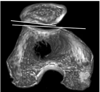
Fig. 1. Sulcus angle of both femoral facet.
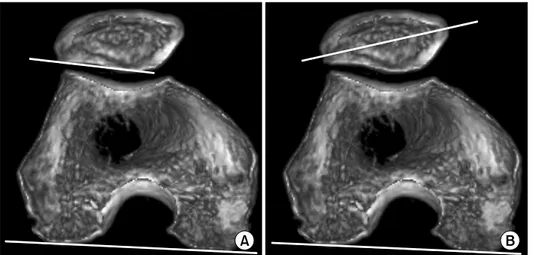
Fig. 2. Congruence angle between halving sulcus angle and line between sulcus and inferior dome of patella (dotted line).
Fig. 3. Lateral patellofemoral angle between lateral patellar facet and line over highest part of both condyle.
한 기준점을 잡을 수 있는 영상으로 3차원적으로 재구성 하여 원위 대퇴부의 양측 상과(medial & lateral epi- condyle) 및 내측구(intercondylar sulcus)가 명확히 보이 는 단면을 얻어 슬개-대퇴 관절 정렬을 측정하고 일관된 검사 방법 및 결과 분석을 통해 정상 한국 성인에서의 슬 개-대퇴 관절 정렬값을 알아보고자 하였다.
대상 및 방법
2005년 3월부터 2007년 10월까지 병력 상 슬관절에 증 상이 없으며 이학적 검사 상 정상 소견을 보인 한국 성인 90명, 180예의 슬관절을 대상으로 하였다. 남자가 45명 90예였고 여자가 45명 90예였으며 평균 연령은 42.2세 (범위: 24∼66)였다. 3차원 전산화 단층 촬영(Light- speed Ultra 16 Computed Tomography, GE medical system, USA)은 앙와위에서 슬관절을 15도 굴곡하여 대 퇴사두근이 이완된 상태에서 시행하였다. 대퇴사두근의 이완은 검사자가 대퇴사두근을 촉지하여 이완된 상태를 확인 후 시행하였다. 3차원 전산화 단층 촬영은 0.625 mm의 간격으로 시행하였으며 촬영된 단면 자료를 이용
이용하였다. 슬개-대퇴 관절 정렬의 평가로 3차원 전산화 단층 촬영에서 구각(sulcus angle) (Fig. 1), 일치각(con- gruence angle) (Fig. 2), 슬개골 경사각(lateral patellofe-
Fig. 5. Condyle-lateral angle between lateral femoral facet and line over posterior condyles.
Fig. 4. Condyle patellar angle. (A) Lateral facet between lateral patellar fa- cet and line over posterior condyles. (B) Patellar axis between longest line in pa- tellar major axis and line over posterior condyles.
moral angle) (Fig. 3), 후과-슬개골 간 경사각(condylar facet angle 및 condylar patellar axis angle) (Fig. 4A, B)1), 후과-대퇴외과 간 경사각(condyle lateral angle) (Fig. 5)1)의 값을 얻어 측정하였으며 모든 각은 Picture Archiving and Communication System (PACS)을 이용 하여 측정하였다. 측정은 2인에 의해 이루어졌으며 각 측 정자는 독립적으로 동일 영상에 대해 1일 간격으로 각각 2회를 시행하여 이의 평균치를 평가하였다. 이상과 같은 방법으로 측정하여 정상 성인 한국인에서의 평균치 및 표 준 편차를 구하였고 측정한 각각의 각을 좌우 및 성별에 따라 비교 분석하였다. 통계적 유의성은 SPSS 10.0 (SPSS Inc.)을 통해 paired t-test를 이용하여 검증하였으며 등급 내 상관 계수(intraclass correlation coefficient)를 이용한 검사자 간 계측 신뢰도를 평가하였다.
결 과
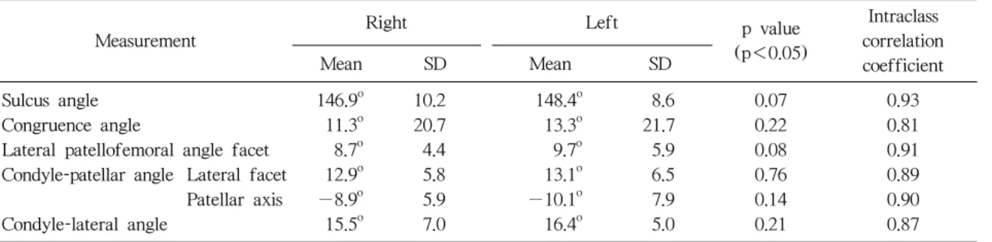
3차원 전산화 단층 촬영을 이용하여 측정한 결과 구각 의 평균치는 남자 145.9o±8.9, 여자 149.4o±9.7이었으며 좌우 차이는 없었으나 남녀 간에는 여자가 각이 큰 것으 로 유의한 차이를 보였다(p<0.05). 일치각의 평균치는 남자 12.6o±22.7, 여자 12.0o±19.6이었으며 좌우 및 남 녀간의 차이는 발견되지 않았다. 슬개골 경사각의 평균치 는 남자 9.9o±6.0, 여자 8.5o±4.3이었으며 좌우 및 남녀 간의 차이는 발견되지 않았다. 후과-슬개골 간 경사각 중 condylar-patellar facet angle은 남자 14.2o±7.1, 여자 11.8o±4.8이었으며 condylar-patellar axis angle은 남자
−8.5o±7.7, 여자 −10.6o±6.1이었으며 좌우 차이는 없
었으나 남녀 간에는 여자가 각이 큰 것으로 유의한 차이 를 보였다(p<0.05). 후과-대퇴외과 간 경사각의 평균치 는 남자 15.5o±7.6, 여자 16.4o±4.0이었으며 좌우 및 남 녀 간의 차이는 발견되지 않았다. 등급 내 상관 계수를 이용한 검사자 간 계측 신뢰도 평가에서는 통계학적으로 유의한 차이가 없었다(Table 1, 2).
고 찰
슬개-대퇴 관절의 이상을 찾기 위한 검사 방법은 단순 방사선 검사, 전산화 단층 촬영 등으로 슬개골의 기울기 및 전위의 이상 여부를 측정하는 방법들이 사용되어 왔
Sulcus angle 146.9 10.2 148.4 8.6 0.07 0.93
Congruence angle 11.3o 20.7 13.3o 21.7 0.22 0.81
Lateral patellofemoral angle facet 8.7o 4.4 9.7o 5.9 0.08 0.91 Condyle-patellar angle Lateral facet 12.9o 5.8 13.1o 6.5 0.76 0.89
Patellar axis −8.9o 5.9 −10.1o 7.9 0.14 0.90
Condyle-lateral angle 15.5o 7.0 16.4o 5.0 0.21 0.87
Measurement Men Women p value
(p<0.05)
Intraclass correlation coefficient
Mean SD Mean SD
Sulcus angle 145.9o 8.9 149.4o 9.7 0.01 0.92
Congruence angle 12.6o 22.7 12.0o 19.6 0.84 0.80
Lateral patellofemoral angle facet 9.9o 6.0 8.5o 4.3 0.27 0.87 Condyle-patellar angle Lateral facet 14.2o 7.1 11.8o 4.8 0.09 0.91
Patellar axis −8.5o 7.7 −10.6o 6.1 0.04 0.86
Condyle-lateral angle 15.5o 7.6 16.4o 4.0 0.29 0.88
Table 2. Summary of Data (between Men and Women)
다. Merchant 등8)은 슬관절을 45도 굴곡하여 슬개-대퇴 관절을 축성 촬영하는 법을 소개하였고, Laurin 등6)은 슬 관절을 20도 굴곡하여 촬영하는 것이 슬개-대퇴 관절의 실제 구조에 근접한다고 하였다. 그러나 단순 방사선 촬 영은 적당한 기준면 결정이 어렵고 영상이 중첩되며, 슬 관절 굴곡 25도 이하에서는 관찰의 어려움이 있어 평가 의 신뢰도가 떨어지기 때문에 전산화 단층 촬영을 이용한 슬개-대퇴 관절의 평가가 더 민감하고 특이하다고 인정하 게 되었다2). Martinez 등7)에 의하면 슬개-대퇴 관절은 슬관절이 완전 신전 상태에 가까울수록 불안정성이 증가 하므로 슬관절 굴곡 30도 이내의 검사가 중요하다고 하 였다. 따라서 전산화 단층 촬영은 단순 방사선 검사에 비 해 슬관절 굴곡 30도 이하에서도 측정 및 평가가 가능하 며 대퇴과를 기준면으로 사용하기 때문에 슬개골 축성 영 상의 변화가 적어 정확한 검사 결과를 얻을 수 있다.
Song 등10)은 전산화 단층 촬영을 이용한 정상 한국인 20 명의 슬개-대퇴 관절 정렬의 평가에서 슬관절 20도 굴곡 시 구각은 남자 148o±5, 여자 148o±7, 일치각은 남자 2o±10도, 여자 −6o±14, 슬개골 경사각은 남자 8o±5, 여자 8o±6이라고 하였다. 본 연구에서는 Song 등의 연구 와 구각 및 슬개골 경사각의 평균치는 비슷하게 나타났으
나 구각이 여자에서 큰 것으로 유의한 차이를 보였으며 일치각의 평균치가 더 큰 것으로 측정되었다. 최근 Alemparte 등1)은 정상 남녀 각각 15명의 좌우 슬관절 총 60개를 전산화 단층 촬영하여 슬개골 정렬을 평가하여 평균치 및 표준오차를 구하고 남녀 간 차이를 비교하였 다. 이들은 슬관절 굴곡 15도 상태에서 촬영을 시행하여 구각, 일치각 및 슬개골 경사각을 측정하였고 추가로 후 과-슬개골 간 경사각 및 후과-대퇴외과 간 경사각을 측정 하였다. Alemparte 등의 결과는 구각은 남자 137o±10.1, 여자 142o±9.66, 일치각은 남자 3.03o±17.4, 여자 7.27o
±15.1, 슬개골 경사각은 남자 −7o±4.55, 여자 −9.3o
±5.05, 후과 슬개골 간 경사각 중 condylar patellar fac- et angle은 남자 16.1o±7.33, 여자 12.2o±6.18, condylar patellar axis angle은 남자 −9.3o±4.63, 여자 −13o± 5.42, 후과-대퇴외과 간 경사각은 남자 22.8o±5.01, 여자 21.3o±3.87였으며, 이중 구각과 후과-슬개골 간 경사각 이 남녀 간에 유의한 차이를 보였다. 본 연구에서는 Alemparte 등의 연구에 비해 일치각이 좀 더 크게 측정 되었으며 구각과 condylar patellar axis angle이 남녀 차 이를 보인 점이 비슷하였다. 이제까지 슬개-대퇴 관절 정 렬을 연구한 각 저자마다 이전 연구 보고들의 한계점 및
각 연구결과마다 측정값이 많은 차이를 보임을 지적하며 계측 방법 및 분석 방법의 오차를 줄이기 위해 많은 노력 을 시행하였다. 현재까지는 슬개-대퇴 관절의 정렬 분석 시 전산화 단층 촬영을 시행하며 슬관절 20도 이내의 굴 곡과 이완된 대퇴사두근 상태의 표준화된 지침을 가지고 검사와 분석을 시행하는 것이 오차를 줄이는 방법이라 보 고되었다3). 저자들은 전산화 단층촬영 시 검사대상의 누 운 자세에 따라 축성 영상의 각이 조금씩 변하고 또한 축 성 영상을 얻는 부위가 검사할 때 다를 수 있다는 점에 착안하여 조금 더 일관된 방법으로 영상을 얻고자 3차원 전산화 단층촬영을 이용하였고 이는 촬영된 슬관절을 컴 퓨터 상에서 3차원적 영상으로 재구성한 후 원하는 방향 으로 회전을 시행하여 원하는 부위에서 일정한 축성영상 을 얻을 수 있어 좀 더 정확한 결과를 얻을 수 있다고 생 각한다.
하지만 본 연구는 이전 검사와 비교하여 좀 더 정확한 해부학적 측정은 할 수 있었으나 결과에 있어서 아주 큰 차이는 보이지 않았다. 또한, 연령 분포가 넓고 그 대상수 가 많지 않으며 연구 대상의 선택에 있어서 정확한 표본 크기 추정(sample size estimation)을 시행하지 않고 대상 을 임의로 선택하였다는 점에서 대표성에 한계점이 있어 앞으로 적절한 크기의 좀 더 많은 대상을 포함한 각 연령 별의 평균치를 측정하는 연구가 필요할 것으로 생각한다.
결 론
3차원 전산화 단층 촬영을 이용하여 일정한 기준점을 잡을 수 있는 영상으로 재구성하여 슬개-대퇴 관절 정렬 을 측정하는 방법은 과거 conventional CT의 축성 단면 을 통해 얻은 영상보다 해부학적 실제에 근접한 측정을 할 수 있을 것으로 생각되었으나 결과에서는 이제까지의 연구들과 큰 차이는 보이지 았았다. 본 연구 결과를 바탕 으로 슬개골 연화증, 슬개골 탈구증, 재발성 슬개골 아탈 구증 등 슬개-대퇴 관절의 부정정렬로 인한 질환을 진단 하는 데 많은 도움이 될 것으로 생각한다.
REFERENCES
1. Alemparte, J, Ekdahl M, Burnier L, et al: Patellofemoral evauation with radiographs and computed tomography scans in 60 knees of asymptomatic subjects. Arthroscopy, 23: 170-177, 2007.
2. Delgado-Martins H: A study of the position of the patella using computerised tomography. J Bone Joint Surg Br, 61:
443-444, 1979.
3. Delgado-Martinez AD, Estrada C, Rodriquez-Merchánt EC, Atienza M, Ordóñez JM: CT scanning of the patellofe- moral joint. The quadriceps relaxed or contracted? Int Orthop, 20: 159-162, 1996.
4. Inoue M, Shino K, Hirose H, Horibe S, Ono K:
Subluxation of the patella. Computed tomography analysis of patellofemoral congruence. J Bone Joint Surg Am, 70: 1331- 1337, 1988.
5. Jung YB, Park YJ: Patellofemoral malalignment syndrome:
distal realignment (modified maquet op). J Korean Knee Society, 4: 21-25, 1992.
6. Laurin CA, Dussault R, Levesque HP: The tangential x-ray investigation of the patellofemoral joint: X-ray technique, diag- nostic criteria and their interpretation. Clin Orthop Relat Res, 144: 16-26, 1979.
7. Martinez S, Korobkin M, Fondren FB, Hedlund LW, Goldner JL: Computed tomography of the normal patellofe- moral joint. Invest Radiol, 18: 249-253, 1983.
8. Merchant AC, Mercer RL, Jacobson RH, Cool CR:
Roentgenographic analysis of patellofemoral congruence. J Bone Joint Surg Am, 56: 1391-1396, 1974.
9. Schutzer SF, Ramsby GR, Fulkerson JP: The evaluation of patellofemoral pain using computerized tomography. A prelimi- nary study. Clin Orthop Relat Res, 204: 286-293, 1986.
10. Song EK, Kim HS, Kim MJ, Ryang DH: Analysis of kine- matic patellar motions in normal Korean by computerized tomography. J Korean Orthop Assoc, 27: 1147-1155, 1992.
목적: 3차원 전산화 단층 촬영을 이용하여 정상 한국인에서 슬개-대퇴 관절 정렬의 평균치를 알아보고 좌우 및 남녀 차이를 알아보고자 하였다.
대상 및 방법: 슬관절 증상의 병력이 없으며 이학적 검사에서 정상 소견을 보인 한국 성인 남자 45명, 여자 45명, 180예의 슬관절을 대상으로 하였다. 평균 연령은 42.2세(범위: 24∼66세)였으며 3차원 전산화 단층 촬영은 앙와위에서 슬관절을 15도 굴곡하여 시행하였다. 슬개-대퇴 정렬은 구각, 일치각, 슬개골 경사 각, 후과-슬개골 간 경사각, 후과-대퇴외과 간 경사각을 이용하여 측정하였다.
결과: 구각 평균치는 남자 145.9o±8.9, 여자 149.4o±9.7, 일치각은 12.6o±22.7과 12.0o±19.6, 슬개 골 경사각은 9.9o±6.0과 8.5o±4.3, 후과-슬개골 간 경사각 중 facet angle은 14.2o±7.1과 11.8o±4.8, 후과-슬개골 간 경사각 중 axis angle은 -8.5o±7.7과 -10.6o±6.1, 후과-대퇴외과 간 경사각은 15.5o± 7.6과 16.4o±4.0으로 측정되었다. 좌우 측정값 간의 통계학적 유의한 차이는 없었으며, 남녀 각각의 측정 평균치는 구각과 후과-슬개골 간 경사각 중 axis angle이 남녀 간에 통계학적으로 유의한 차이를 보였다(p
<0.05).
결론: 3차원 전산화 단층 촬영을 이용하여 정상 한국인의 슬개-대퇴 관절 정렬을 측정하여 한국 성인 평균치를 알 수 있었으며 이는 슬개-대퇴 관절 부정 정렬의 진단에 도움이 될 것으로 생각한다.
색인 단어: 슬개-대퇴 관절 정렬, 3차원 전산화 단층 촬영