210
책임저자 : 박정수, 서울시 서대문구 성산로 250
120-752, 연세대학교 의과대학 외과학교실 Tel: 02-2228-2100, Fax: 02-313-8289
E-mail: [email protected] 게재승인일:2008년 9월 13일
원발성 유두 갑상선암의 수술적 치료 후 측경부 전이 림프절에서 발생한 미분화 갑상선암 3예
연세대학교 의과대학 외과학교실, 1진단병리과학교실
성태연ㆍ홍순원1ㆍ강상욱ㆍ이승철ㆍ정종주ㆍ이용상ㆍ남기현ㆍ장항석ㆍ정웅윤ㆍ박정수
Anaplastic Transformation of Metastatic Papillary Thyroid Carcinomas in the Cervical Lymph Nodes: Report of 3 Cases
Tae-Yon Sung, M.D., Soon-Won Hong
1, M.D., Sang- Wook Kang, M.D., Seung Chul Lee, M.D., Jong Ju Jeong, M.D., Yong Sang Lee, M.D., Kee-Hyun Nam,
M.D., Hang-Seok Chang, M.D., Woong Youn Chung, M.D. and Cheong Soo Park, M.D.Anaplastic thyroid carcinoma (ATC) is a rare disease that shows very aggressive behavior. Most ATCs arise from pre-existing thyroid carcinomas. However, anaplastic trans- formation occurring in metastatic cervical nodes is ex- tremely rare. We report herein on 3 cases of anaplastic transformation of metastatic lateral cervical lymph nodes from primary papillary thyroid carcinoma (PTC), which hap- pened long after the initial surgical treatment. All the pa- tients died of disease within 4 months in spite of aggressive treatment for the lesions. Our experience supports that ap- propriate lymph node dissection is mandatory at the time of initial surgery even for differentiated thyroid carcinomas.
(Korean J Endocrine Surg 2008;8:210-214)
Key Words: Anaplastic transformation, Papillary thyroid
carcinoma, Anaplastic thyroid carcinoma 중심 단어:미분화 전환, 유두 갑상선암, 미분화 갑상선암
Departments of Surgery, 1Pathology, Yonsei University College of Medicine, Seoul, Korea
서 론
미분화 갑상선암은 가장 공격적인 성향을 보이는 질환들 중 하나로 매우 불량한 예후를 보인다. 전체 갑상선암 중 1% 내외를 차지하고 있으나, 다양한 치료법의 적용에도 불 구하고 치료 성적은 현재까지도 실망스러운 상태이다.(1,2) 현재까지 미분화 갑상선암이 발생하는 기전에 대해서는 확 실하지는 않으나 기존에 있던 분화 갑상선암이 전환되어 발생한다는 설이 가장 유력하다.(1-7)
일반적으로 미분화 갑상선암은 갑상선 자체의 유두암이 전환되어 발생한 것이 대부분이다.(8-11) 그러나, 유두 갑상 선암 수술 후 전이된 측경부 림프절에서 발생된 미분화 갑 상선암(anaplastic transformation)은 극히 드물다.(12-14) 저자들은 원발성 유두 갑상선암의 수술적 치료 후, 장기 추적 관찰 중에 측경부 전이 림프절에서 발견된 미분화 갑 상선암 3예를 경험하여 보고하는 바이다.
증 례 증례 1
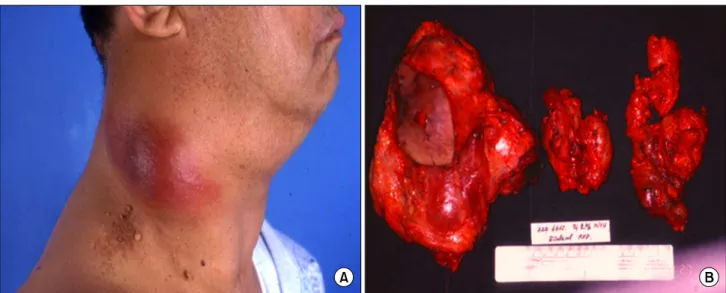
64세 남자 환자로 우측 경부에 빠른 속도로 커지는 종괴 를 주소로 내원하였다(Fig. 1A). 환자는 39년 전 유두 갑상 선암으로 갑상선 우엽과 협부 절제술을 받은 과거력이 있 었다. 내원시 시행한 경부 컴퓨터 단층 촬영 상 남아있는 갑상선의 전반적인 석회화 소견, 양측 경부 다발성 림프절 전이 및 우측 경정맥의 폐쇄 소견을 보였다. 또한, 흉부 컴 퓨터 단층 촬영에서 다발성 폐전이 소견이 보였다. 수술은 갑상선 완결 절제술 및 중앙 경부 림프절 절제술과 우측 측경부 림프절 청소술 및 좌측 측경부 변형적 림프절 청소 술을 시행하였다(Fig. 1B). 병리 조직 소견은 유두 갑상선암 이 측경부 림프절로 다발성으로 전이된 것이었으나 일부가 미분화 갑상선암으로 전환되어 있었다. 수술 후 외부 방사 선 치료와 200 mCi의 방사선동위원소 치료를 받을 예정 이 였으나, 갑작스럽게 경부 종괴가 새로이 성장하기 시작하 여 세침흡입세포검사를 한 결과, 재발성 미분화 갑상선암
Fig. 1. Specimen of case 1. (A) Rapid growing mass in right lateral cervical area and (B) gross specimen after second operation for recurrent thyroid carcinoma showing anaplastic transformation.
Fig. 2. Histopathology and cytology of case 2. (A) Seven years ago, the tumor showed a typical and conventional papillary micro-carcinoma (H&E, ×40). (B) Histology of recurrent tumor shows reminiscent of papillary carcinoma with epithelioid and pleomorphic cytology (H&E, ×400).
소견을 보였다. 환자는 급격히 진행되는 호흡 부전으로 수 술 4개월 후 사망하였다.
증례 2
63세 남자로 우측 경부 종괴와 우측 견갑 부위 통증을 주 소로 내원하였다. 환자는 7년 전 0.8 cm 크기의 미세유두 갑상선암으로 갑상선 좌엽 전절제 및 우엽 부분 절제술과 중앙 경부 림프절 절제술을 시행 받은 과거력이 있었다. 그 당시 종양은 전형적인 유두암 소견을 보였다(Fig. 2). 내원 시 시행한 경부 초음파 및 양전자 방사-컴퓨터 단층 촬영 상 우측 쇄골 상부에 3.6 cm 크기의 종괴와 다발성 측경부
림프절 전이 소견을 보였다(Fig. 3). 우측 쇄골 상부의 결절 에서 세침흡입세포검사를 시행한 결과, 유두 갑상선암을 포함한 미분화 갑상선암 소견을 보였다. 이는 조직 검사로 도 확인되었다(Fig. 2). 환자는 총 4,724 cGy 용량의 방사선 치료가 진행되는 동안 경부와 흉부에 심한 통증을 호소하 였으며 진단 3개월 후 폐전이로 인한 호흡기능부전으로 사 망하였다.
증례 3
27세 남자로 통증을 동반한 경부 종괴를 주소로 내원하 였다. 환자는 8년 전 4 cm 크기의 유두 갑상선암으로 갑상
Fig. 3. Positron emission techne- tium-computer tomography (PET-CT) images of case 2. Images showing intense uptake of irregular tumor mass from right supra- clavicular fossa to superi- or mediastinum.
Fig. 4. Histopathology of case 3. (A) Eight years ago, the tumor showed a typical and conventional papillary carcinoma (H&E, ×40).
(B) High power view of anaplastic area shows solid growth of tumor and epithelioid and pleomorphic cytology (H&E, ×400).
Fig. 5. CT and ultrasonography of case 3. Bulging mass in right upper lesion suggest- ing recurrent thyroid carci- noma or metastatic nodes.
선 전절제술 및 중앙 경부 림프절 절제술과 우측 내경정맥 림프절 절제술을 받은 과거력이 있었다. 그 당시 종양은 전 형적인 유두암 소견을 보였다(Fig. 4). 외래 추적 관찰 5년째 경부 초음파 및 컴퓨터 단층 촬영에서 양측 경부 다발성 림프절 비대가 발견되어 세침흡입세포검사로 유두 갑상선
암이 전이된 것으로 확인되어 좌측 측경부 림프절 청소술 과 우측 측경부 변형 선택적 림프절 절제술을 동시에 시행 하였다. 수술 6주 후 200 mCi 방사성동위원소 치료를 하였 다. 환자는 이 후 외래 추적 소실되었다가 2차 수술 3년째, 상기 주소로 내원하였으며 경부 컴퓨터 단층 촬영 및 초음
파에서 우측 상경부에 약 4.0 cm 크기의 종양이 발견되어 (Fig. 5) 이 종괴를 광범위하게 절제하였다. 병리 조직 검사 결과, 종괴의 대부분은 미분화 갑상선암 조직으로 되어 있 었으며, 일부분에서 유두 갑상선암 소견도 보였다(Fig. 4).
환자는 수술 후 외부 방사선 치료 중 수술 부위 근처에서 다시 새로운 종괴가 성장하기 시작하여, 환자의 거절로 외 부 방사선치료를 중단하고 고주파(radiofrequency ablation) 치료를 1회 시행하였다. 고주파 치료 후 종괴의 크기가 약 간 작아지는 듯 하다가 다시 악화되어 수술 후 4개월 만에 폐전이로 추정되는 호흡기능 부전으로 사망하였다.
고 찰
갑상선암의 약 90% 이상은 분화 갑상선암이며, 이는 대 부분 양호한 예후를 보인다.(1) 그러나 미분화 갑상선암은 1% 내외의 낮은 발병율로 최근 감소 추세를 보이고 있으나, 매우 공격적인 임상 양상과 불량한 예후를 보인다. 미분화 갑상선암은 진단 시 이미 절반 이상이 갑상선외 침범 (extrathyroidal extension), 림프절 전이 및 원격 전이를 보인 다.(15) 미분화 갑상선암은 수술 및 방사선치료 등 여러 복 합 요법에도 불구하고 극소수의 환자만이 1년 이상의 생존 율을 보이며 일반적으로 진단 후 6개월 이내에 사망하는 것으로 알려져 있다.(1,2,6)
효과적인 치료 방안을 모색하기 위한 임상 양상, 병리 조 직 소견 및 분자학적 기전에 대한 연구는 여전히 계속되고 있으며,(17,18) 최근에는 미분화 갑상선암과 연관된 특이 분 자학적 종양표지자에 대한 연구가 진행 중이다.(1,2,7) 대부분의 미분화 갑상선암은 분화 갑상선암에서 전환되 어 발생하는 것으로 보고되고 있으나, 몇몇 보고에서는 미 분화 갑상선암에서 분화 갑상선암의 병리 소견이 발견되지 않는 경우도 있다고 하였다.(17,19) 소수에서는 외부 방사선 치료 및 방사성동위원소 치료가 미분화 갑상선암으로의 변 형 위험 인자로 보고하고 있다.(12-14,20)
저자들이 경험한 환자들 중 증례 1은 39년 전 분화도가 좋은 유두 갑상선암으로 갑상선 우엽과 협부 절제술만 받 았던 환자로 오랜 기간이 지난 후에 잔존 갑상선과 경부 림프절에 재발 및 원격 전이를 보였다. 이는 분화 갑상선암 도 오랜 기간이 지나면 저분화 또는 미분화 갑상선암으로 전환될 수 있음을 시사한다. 또한, 일차 진단 시 종양의 병 리 조직 소견 및 전이 여부만으로는 추후 암종의 변이 및 예후를 예측 할 수 없음을 보여준다. 증례 2는 첫 번째 환자 와 마찬가지로 갑작스런 크기 변화를 보이는 새로이 발견 된 종괴를 주소로 내원한 환자이며 7년 전 유두 갑상선암으 로 갑상선 아전절제술을 받았었다. 이 환자의 병리 조직학 적 소견 또한 분화 갑상선암이 미분화 갑상선암으로 전환 된다는 것을 뒷받침한다.(1-6,16) 세 번째 환자는 젊은 남자 로 첫 내원 당시 이미 갑작스런 팽창을 동반한 종괴를 호소
하였으며 유두 갑상선암에 대한 적절한 수술에도 불구하고 여러 차례 재발 및 경부 림프절의 다발성 전이가 반복되었 다. 재발이 반복되는 과정에서 조직분화도의 변이를 보였 으며 마지막 병리 조직 결과는 미분화 갑상선암 소견을 보 였다. 저자들이 경험한 증례 3 외에도 지금까지 보고된 연 구들에 의하면 유두 갑상선암의 발견 당시 경부 림프절에 다발성 전이가 있거나 수술 후 여러 차례 재발이 반복 될수 록 조직분화도가 나빠지는 양상을 보이는 것으로 되어 있 다.(21-23) 특히, 유두 갑상선암은 측경부 림프절 전이가 가 장 많은 암으로 재발도 경부 림프절에서 가장 많이 발견된 다. 이런 반복적인 재발 위험 요소를 줄이기 위해 첫 수술시 적절한 범위의 경부 림프절 절제술이 중요하며 이는 미분 화 갑상선암으로의 전환을 감소시키는 방법이 될 수 있을 것이다.(23,24) 저자들이 경험한 예를 보면, 이들 모두 새로 이 발견된 종괴가 통증을 야기할 정도로 매우 빠른 속도로 자라는 것을 볼 때, 원발암이 비록 분화가 잘된 유두 갑상선 암이라 할지라도 재발 시 종괴의 성장 속도가 빠르면 미분 화암으로의 전환을 생각해야 할 것이다. Ito 등(24)은 림프 절 전이 부위에서 발생한 미분화암은 완전히 절제만 되면 장기 생존을 기대할 수 있다고 하였으나, 저자들의 경험으 로는 절제를 했더라도 곧 재발하는 양상을 보아 일반 미분 화 갑상선암과 다를 바가 없다고 생각되었다. 따라서, 이와 같은 미분화 갑상선암으로의 전환을 예방하기 위하여 유두 갑상선암이라 하더라도 초기 단계에서 적절한 중앙 경부 림프절 절제술과 임상적으로 의심되는 측경부 림프절의 포 괄적 절제술은 매우 중요하다고 사료되었다.
REFERENCES
1) Wang HM, Huang YW, Huang JS, Wang CH, Kok VC, Hung CM, et al. Anaplastic carcinoma of the thyroid arising more often from follicular carcinoma than papillary carcinoma. Ann Surg Oncol 2007;14:3011-8.
2) Wiseman SM, Griffith OL, Deen S, Rajput A, Masoudi H, Gilks B, et al. Identification of molecular markers altered during transformation of differentiated into anaplastic thyroid carcinoma. Arch Surg 2007;142:717-27.
3) Aldinger KA, Samaan NA, Ibanez M, Hill CS Jr. Anaplastic carcinoma of the thyroid: a review of 84 cases of spindle and giant cell carcinoma of the thyroid. Cancer 1978;41:2267-75.
4) Nishiyama RH, Dunn EL, Thompson NW. Anaplastic spindle- cell and giant-cell tumors of the thyroid gland. Cancer 1972;30:113-27.
5) Spires JR, Schwartz MR, Miller RH. Anaplastic thyroid carcinoma. Association with differentiated thyroid cancer.
Arch Otolaryngol Head Neck Surg 1988;114:40-4.
6) Wiseman SM, Loree TR, Hicks WL Jr, Rigual NR, Winston JS, Tan D, et al. Anaplastic thyroid cancer evolved from papillary carcinoma: demonstration of anaplastic transforma-
tion by means of the inter-simple sequence repeat polymerase chain reaction. Arch Otolaryngol Head Neck Surg 2003;
129:96-100.
7) Takano T, Ito Y, Hirokawa M, Yoshida H, Miyauchi A.
BRAF V600E mutation in anaplastic thyroid carcinomas and their accompanying differentiated carcinomas. Br J Cancer 2007;96:1549-53.
8) Vinette DS, MacDonald LL, Yazdi HM. Papillary carcinoma of the thyroid with anaplastic transformation: diagnostic pitfalls in fine-needle aspiration biopsy. Diagn Cytopathol 1991;7:75-8.
9) Ohsumi S, Urakami J, Matsumori H, Sasaki S, Murakami M, Nose S. A case of two primary carcinomas: thyroid papillary carcinoma with anaplastic transformation of metastatic cervical lymph node and breast cancer. Gan No Rinsho 1990;36:
2439-44.
10) Fortson JK, Durden FL Jr, Patel V, Darkeh A. The coexistence of anaplastic and papillary carcinomas of the thyroid: a case presentation and literature review. Am Surg 2004;70:1116-9.
11) Sato K, Waseda R, Tatsuzawa Y, Soma R, Ueda Y, Katsuda S. Papillary thyroid carcinoma with anaplastic transformation showing a rhabdoid phenotype solely in the cervical lymph node metastasis. Pathol Res Pract 2006;202:55-9.
12) Gál I, Hatházi L, Nemes Z, Solymosi T. Anaplastic trans- formation of papillary carcinoma of the thyroid. Orv Hetil 1994;135:581-4.
13) Igase M, Yamamoto Y, Kohara K, Miki T. An autopsy case of anaplastic thyroid carcinoma that transformed from pa- pillary carcinoma within 5 years from initial diagnosis. Nippon Ronen Igakkai Zasshi 2000;37:819-22.
14) Sotome K, Onishi T, Hirano A, Nakamaru M, Furukawa A, Miyazaki H, et al. A rare case of anaplastic transformation within the metastatic site of the retroperitoneal region in a patient 17 years after total thyroidectomy for papillary carcinoma of the thyroid beginning with multiple bone metastases. Thyroid 2007;17:1309-11.
15) Kebebew E, Greenspan FS, Clark OH, Woeber KA, McMillan
A. Anaplstic thyroid carcinoma: treatment outcome and prognostic factors. Cancer 2005;103:1330-5.
16) Rosai J, Carcangiu ML, DeLellis RA. Tumors of the thyroid gland. In: Rosai J, Sobin LH, editors. Atlas of Tumor Patho- logy. Washington DC: AFIP; 1992.
17) Wiseman SM, Loree TR, Rigual NR, Hicks WL Jr, Douglas WG, Anderson GR, et al. Anaplastic transformation of thyroid cancer: review of clinical, pathologic, and molecular evidence provides new insights into disease biology and future therapy.
Head Neck 2003;25:662-70.
18) Are C, Shaha AR. Anaplastic thyroid carcinoma: biology, pathogenesis, prognostic factors, and treatment approaches.
Ann Surg Oncol 2006;13:453-64.
19) McIver B, Hay ID, Giuffrida DF, Dvorak CE, Grant CS, Thompson GB, et al. Anaplastic thyroid carcinoma: a 50-year experience at a single institution. Surgery 2001;130:1028-34.
20) Shingu K, Kobayashi S, Yokoyama S, Fujimori M, Asanuma K, Ito KI, et al. The likely transformation of papillary thyroid carcinoma into anaplastic carcinoma during postoperative radioactive iodine-131 therapy: report of a case. Surg Today 2000;30:910-3.
21) Ozaki O, Ito K, Mimura T, Sugino K, Ito K. Anaplastic transformation of papillary thyroid carcinoma in recurrent disease in regional lymph nodes: a histologic and immuno- histochemical study. J Surg Oncol 1999;70:45-8.
22) Kawahara E, Ooi A, Oda Y, Katsuda S, Terahata S, Michigishi T. Papillary carcinoma of the thyroid gland with anaplastic transformation in the metastatic foci. An immuno- histochemical study. Acta Pathol Jpn 1986;36:921-7.
23) Ito Y, Miyauchi A. Lateral lymph node dissection guided by preoperative and intraoperative findings in differentiated thyroid carcinoma. World J Surg 2008;32:729-39.
24) Ito Y, Higashiyama T, Hirokawa M, Fukushima M, Inoue H, Yabuta T, et al. Prognosis of patients with papillary carci- noma showing anaplastic transformation in regional lymph nodes that were curatively resected. Endocr J 2008 Jul 9.
[Epub ahead of print]