10.3988/jcn.2011.7.2.53 J Clin Neurol 2011;7:53-59
Introduction
Systemic cancer and ischemic stroke are both common condi- tions, and are two of the most common causes of death among the elderly. The number of people living with cancer is increas- ing, and the steadily increasing proportion of elderly people in the world is predicted to result in an increase of approximate- ly 50% in new cancer cases over the next 20 years, with the number of new cases each year rising from 10.9 million in 2002
to 16 million in 2020 (World Health Organization, Global ac- tion against cancer, 2005). In addition, improvements in treat- ment practice (cancer medicine) have the potential of improv- ing survival by up to 15%. Therefore, the number of stroke patients with cancer is expected to rise with increases in the number of people living with cancer. Moreover, associations between can- cer and stroke have been reported. Cerebrovascular disease oc- curs commonly in cancer patients, with 15% of cancer patients experiencing a thromboembolic event during their clinical
Ischemic Stroke and Cancer: Stroke Severely Impacts Cancer Patients, While Cancer Increases the Number of Strokes
Oh Young Bang,a Jin Myoung Seok,a Seon Gyeong Kim,a Ji Man Hong,b Hahn Young Kim,c Jun Lee,d Pil-Wook Chung,e Kwang-Yeol Park,f Gyeong-Moon Kim,a Chin-Sang Chung,a Kwang Ho Leea
aDepartment of Neurology, Samsung Medical Center, Sungkyunkwan University School of Medicine, Seoul, Korea
bDepartment of Neurology, Ajou University School of Medicine, Suwon, Korea
cDepartment of Neurology, Konkuk University School of Medicine, Seoul, Korea
dDepartment of Neurology, Yeungnam University School of Medicine, Daegu, Korea
eDepartment of Neurology, Kangbuk Samsung Hospital, Sungkyunkwan University School of Medicine, Seoul, Korea
fDepartment of Neurology, Chung-Ang University School of Medicine, Seoul, Korea
Received October 27, 2010 Revised November 22, 2010 Accepted November 22, 2010 Correspondence Oh Young Bang, MD, PhD Department of Neurology, Samsung Medical Center, Sungkyunkwan University School of Medicine, 50 Irwon-dong, Gangnam-gu, Seoul 135-710, Korea Tel +82-2-3410-3599 Fax +82-2-3410-0052 E-mail [email protected]
BackgroundzzCancer and ischemic stroke are two of the most common causes of death among the elderly, and associations between them have been reported. However, the main pathomecha- nisms of stroke in cancer patients are not well known, and can only be established based on accu- rate knowledge of the characteristics of cancer-related strokes. We review herein recent studies concerning the clinical, laboratory, and radiological features of patients with cancer-related stroke.
Main ContentszzThis review covers the epidemiology, underlying mechanisms, and acute and preventive treatments for cancer-related stroke. First, the characteristics of stroke (clinical and ra- diological features) and systemic cancer (type and extent) in patients with cancer-specific stroke are discussed. Second, the role of laboratory tests in the early identification of patients with can- cer-specific stroke is discussed. Specifically, serum D-dimer levels (as a marker of a hypercoagu- lable state) and embolic signals on transcranial Doppler (suggestive of embolic origin) may pro- vide clues regarding changes in the levels of coagulopathy related to cancer and anticoagulation.
Finally, strategies for stroke treatment in cancer patients are discussed, emphasizing the importance of preventive strategies (i.e., the use of anticoagulants) over acute revascularization therapy in can- cer-related stroke.
ConclusionzzRecent studies have revealed that the characteristics of cancer-related stroke are distinct from those of conventional stroke. Our understanding of the characteristics of cancer-relat- ed stroke is essential to the correct management of these patients. The studies presented in this re- view highlight the importance of a personalized approach in treating stroke patients with cancer.
J Clin Neurol 2011;7:53-59 Key Wordszz cancer, stroke, ischemic, hypercoagulopathy, embolism, anticoagulants.
course.1 As a consequence, the proportion of patients who have cancer is expected to increase among stroke patients.
The treatment of stroke in cancer patients can be challenging, requiring the development of specific treatment strategies. Al- though patients with systemic cancer usually have poor outcomes, their survival rate is increasing with the development of more effective cancer medicines. Early identification of stroke mech- anisms may be important in cancer patients, because stroke mech- anisms in cancer patients may differ from those in stroke patients without cancer. However, the mechanisms underlying stroke in patients with cancer are largely unknown.2-4 The prevention and appropriate treatment of stroke in cancer patients require an ac- curate understand of its clinicoradiologic characteristics and pa- thogenic mechanisms.
Herein we review recent studies in which modern methods of stroke evaluation have been applied to identify the character- istics of stroke in cancer patients, such as biomarkers, multimod- al magnetic resonance imaging (MRI), and embolism monitor- ing using transcranial Doppler ultrasound (TCD). This review covers the epidemiology, mechanisms, and acute and preven- tive treatments for cancer-related stroke.
Subtypes of Stroke and Cancer Differ between Patients with and
without Conventional Stroke Mechanisms
The controversies regarding the characteristics of stroke in pa- tients with cancer may be due to the involvement of both can- cer-unrelated and cancer-related mechanisms in the develop-
ment of stroke in cancer patients (Table 1). In cancer patients with- out conventional stroke mechanisms (CSM; e.g., atherosclerosis, cardioembolism, or lacunar), a cancer-specific mechanism can be considered as the main cause of stroke. We recently prospec- tively studied 161 patients registered from 6 centers in South Korea with active cancer who experienced acute ischemic stroke.5 Stroke outside CSM occurred in a large proportion of cancer patients: CSM were absent in -40% of stroke patients with cancer, and occurred with a higher frequency of crypto- genic mechanisms than in stroke patients without cancer (18%).
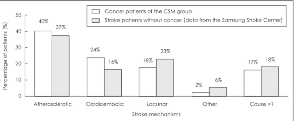
Interestingly, tumor-specific mechanisms were unlikely to play a role in the development of stroke among patients exhibiting CSM, given that the distribution of stroke subtype among can- cer patients with CSM was similar to that in stroke patients with- out cancer (Fig. 1).
The characteristics of cancer, including the type (primary can- cer and pathologic type) and extent of cancer and the time in- terval from diagnosis of cancer and stroke, may be important in the development of stroke in patients with cancer. Patients with stroke had different primary cancers; lung cancer being the most common, followed by gastric and colorectal cancer (Table 2).
When we compared this to the data from the Samsung Cancer Center, the proportion of primary cancer types did not differ be- tween patients with and without stroke. The one exception was lung cancer, which was significantly more prevalent among stroke patients than among those without stroke. Among the pa- thologic type of lung cancer, adenocarcinoma was significantly more prevalent in patients without CSM than in those with CSM or those without stroke; about 70% of patients without CSM had adenocarcinoma, whereas about 70% of patients with cancer-un- Table 1. Mechanisms underlying stroke in patients with cancer
Cancer-unrelated mechanisms Conventional stroke mechanisms
Atherosclerosis, cardioembolic, lacunar, etc.
Cancer-related mechanisms Coagulopathy by
Tumor cell (especially adenocarcinoma)-derived cytokines or microparticles Tissue factor and cancer procoagulants
Cytokines such as tumor necrosis factor-α, interleukins
Intravascular coagulation or nonbacterial thrombotic endocarditis Tumor occlusion
Tumor embolism (lung or cardiac), intravascular lymphoma Direct tumor-related (metastasis or central nervous system tumor)
Vessel compression or infiltration Treatment-related mechanisms
Chemotherapy causing coagulopathy, such as cisplatin, methotrexate, l-asparaginase, bevacizumab Radiation or surgery causing vascular stenosis
Medical comorbidities, such as fungal infection or infective endocarditis
related stroke or those without stroke had nonadenocarcinoma types. Moreover, metastasis at the time of stroke was more prev- alent among patients without CSM than among those with CSM.5 These findings suggest that although occult tumor may cause coagulopathy and thromboembolism, patients with certain types of advanced-stage cancer are prone to cancer-related stroke.
Characteristics of Cancer-Related Stroke
The clinical and radiological features and laboratory findings may help to identify patients with cancer-related stroke mech- anisms. There have been conflicting reports concerning wheth- er or not risk factors in cancer patients (vs. noncancer patients) differ.6-9 However, in previous studies, CSM were pooled with cancer-related mechanisms. In our study, patients with CSM were older and were more likely to have vascular risk factors than were patients without CSM.5
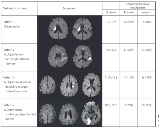
It has been reported that the pattern of the lesion identified on diffusion-weighted imaging (DWI) is correlated with the pa- thogenic mechanism underlying the stroke as well as the out- come after stroke.10,11 The infarct pattern in cancer patients with
stroke is seldom reported. DWI patterns of multiple lesions in- volving multiple arterial territories were more frequently ob- served in patients without CSM, whereas single/multiple lesions involving one arterial territory were observed more frequently in patients with CSM (Fig. 2).5 Recent studies have demonstrat- ed that concealed cancer should be considered in patients who exhibit multiple infarcts on DWI.12
Laboratory findings suggesting coagulopathy may also pre- dict possible cancer-specific stroke mechanisms. The level of D- dimer, a plasmin-derived degradation product of cross-linked fi- brin, is a direct measure of activated coagulation, and has been used in many previous studies as a measure of hypercoagulabil- ity.4,13 Most patients without CSM had elevated D-dimer levels, and the levels were higher in patients with multiple embolic strokes than in patients with a single infarct or multiple infarcts within one vascular territory.5 Our results and those of others sug- gest that there is a strong correlation between D-dimer level and the tumor burden and stage.5,14-17
Thus, the aforementioned parameters may predict possible cancer-related stroke mechanisms. Most patients with both DWI patterns of multiple lesions involving multiple arterial territo- ries (pattern 3 or 4) and D-dimer levels of >1.11 μg/mL had can-
Atherosclerotic Cardioembolic Lacunar Other Cause >1
50 40%
40 30 20 10 0
37%
24%
16% 18%
23%
2% 6%
17% 18%
Percentage of patients
(%)
Stroke mechanisms Cancer patients of the CSM group
Stroke patients without cancer (data from the Samsung Stroke Center)
Fig. 1. Stroke subtypes in patients with vs. without cancer (data from the Sam- sung Stroke Center). Figure modified from Kim et al.5 CSM: conventional stroke mech- anisms.
Table 2. Cancer types in patients with vs. without stroke
Cancer type Stroke patients with cancer, n (%)
Cancer patients without stroke, n (%)*
Cryptogenic group CSM group Total
Gastric 12 (18.8%) 13 (13.4%) 25 (15.5%) 1,764 (15.9%)
Colorectal 5 (7.8%) 18 (18.6%) 23 (14.3%) 1,229 (11%)0.
Breast 1 (1.6%) 1 (1.0%) 2 (1.2%) 999 (9%)0
Hepatic 4 (6.3%) 9 (9.3%) 13 (8.1%)0 1,274 (11.5%)
Lung 21 (32.8%) 26 (26.8%) 47 (29.2%) 1,276 (11.5%)†
Adenocarcinoma 15 (71.4%)† 6 (23.1%) 21 (44.7%) 421 (33.0%)
Cervical 0 (0%)0. 2 (2.1%) 2 (1.2%) 332 (3%)0
Other 21 (32.8%) 28 (28.9%) 49 (30.4%) 4,228 (38.1%)
Total 64 97 161 11,097
*Data from the Samsung Cancer Center, †p<0.001, stroke patients with cancer vs. cancer patients without stroke.
CSM: conventional stroke mechanisms.
cer-specific stroke (31 of 36 patients, 86.1%), whereas CSM were found in most patients with none of these findings (36 of 40 pa- tients, 90%). After adjusting for other factors, the DWI lesion pattern of multiple vascular territories and D-dimer levels of >1.11 μg/dL were independently associated with the possibility of cancer-related stroke mechanisms.5 The area under the receiver operating characteristic curve for this stroke mechanism accord- ing to the presence of DWI pattern of multiple vascular territo- ries or D-dimer levels of >1.11 μg/mL was 0.781 (95% confi- dence interval, 0.715-0.838).
Embolism Caused by Coagulopathy as the Main Mechanism of Stroke
A recent study that monitored embolic signals using TCD show- ed that embolisms caused by coagulopathy could be the main pathomechanism underlying cancer-related stroke.14 A routine TCD study to detect embolism cannot generally be recommend- ed for routine diagnostics in stroke patients due to its low sen- sitivity; in one study, embolic signals were detected in only 5.7%
of unselected stroke patients.18 However, the frequency of em- bolic signals on TCD suggestive of embolic origin is very high in cancer patients with acute ischemic stroke.14 An embolic sig- nal was observed in almost 50% of cancer patients with acute ischemic stroke, but more frequently in patients without CSM (58%) than in those with CSM (33%)(Fig. 3A). Moreover, the number of embolic signals was correlated with the D-dimer lev- els in patients without CSM but not in those with CSM (Fig.
3C and D), and the use of anticoagulation dramatically de- creased the D-dimer levels (supplementary Fig. 1).14 The detec- tion of an embolic signal by TCD may provide clues regarding the cancer-specific mechanism related to hypercoagulopathy, and may be used to monitor the effect of treatment in the acute stroke period.
The source of the embolism causing multiple embolic strokes in cancer patients is unknown. Nonbacterial thrombotic endo- carditis (NBTE) involves the deposition of small sterile vege- tations on the heart valve leaflets and is most commonly found in patients with cancer. DWI has revealed NBTE patterns, with all patients with NBTE exhibiting multiple widely distributed large and small strokes (pattern 4).19 In our data, more than 40%
of patients without CSM showed disseminated small lesions (pattern 3) on DWI, and transesophageal echocardiography did not usually reveal vegetations. These findings suggest that in- travascular clot formation is one of the main sources of such embolisms.
Thrombosis as a complication of cancer was first proposed by Trousseau in 1865, but the precise mechanisms underlying coagulopathy in cancer patients remain to be established. It has been suggested that substances in tumor cells, such as cysteine proteases, tissue factor, and sialic acid moieties of mucin, ex- hibit procoagulant activity, resulting in the activation of factors X and VII.2,20 In addition, aggressive antitumor therapy may also increase the risk of thrombosis.21
Membrane-derived microvesicles are reported to be function- al in that they support the tumor environment, such as neovas-
Conventional stroke mechanism
DWI lesion pattern Examples
D-dimer Present Absent
Pattern 1 Single lesion
1.2±1.9 26 (27%) 5 (8%)
Pattern 2 Multiple lesions in a single arterial territory
2.8±5.6 51 (53%) 14 (22%)
Pattern 3
Multiple small lesions involving multiple arterial territories
11.7±14.3 11 (11%) 26 (41%)
Pattern 4 Multiple small and large disseminated lesions
10.3±20.0 9 (9%) 19 (30%)
Fig. 2. Diffusion-weighted imaging (DWI) lesion patterns in patients with and with- out conventional stroke mechanisms.
cularization in cancer patients.22,23 Tissue-factor-bearing micro- vesicles were associated with the activation of coagulation in patients with colorectal cancer.24-26 The levels of tissue-factor- positive microvesicles were elevated in cancer patients and were correlated with D-dimer levels, suggesting that tissue-factor-bear- ing microvesicles are involved in the activation of coagulation in cancer patients.25 Tissue factor is not only the primary cellu- lar initiator of blood coagulation, it is also a modulator of angio- genesis and metastasis in cancer.24 Further studies are needed on preventive strategies targeting tissue factors to prevent coagu- lopathy and to control the tumor environment.
Acute and Preventive Treatment Strategies for Cancer-Related Stroke
Stroke patients with CSM should be treated according to the
stroke subtype (e.g., atherosclerotic or lacunar), because the mech- anisms underlying the stroke in these patients are unlikely to dif- fer from those of stroke patients without cancer.5 However, the optimal acute treatment and preventive strategies for cancer-re- lated stroke remain to be established.
In the setting of acute ischemic stroke, recanalization thera- py remains the principal therapeutic approach. The use of throm- bolytics within the therapeutic time window is not contraindi- cated in cancer patients under the current guidelines for acute stroke therapy. However, the response to thrombolysis may dif- fer between stroke patients with and without cancer. Multimodal MRI, including DWI and perfusion-weighted imaging, may help in the selection of patients for recanalization therapy.27 Patients who exhibited a target mismatch pattern (substantial penumbra and small core) had a favorable clinical response to recanaliza- tion therapy.28 However, the target mismatch profile is seldom A
C D
B
50
50 50
100
40 80
30
10 10
60
20 40
10
1 1
20
0
0 0
0 Cryptogenic
0.0 20.0 40.0 60.0 0.0 10.0 20.0
D-dimer level (μg/mL) D-dimer level (μg/mL)
80.0 30.0
Spearman’s correlation analysis
r=0.732 p<0.001 Spearman’s correlation analysis
r=0.152 p=0.375
100.0 40.0
Cryptogenic
Other Other
Large-artery
atherosclerosis Large-artery
atherosclerosis Cardio-
embolism Cardio-
embolism Small-artery
occlusion Small-artery
occlusion
Number of ESNumber of ES Number of ESD-dimer levels
(μg/mL
)
Fig. 3. Numbers of embolic signals (ES) on transcranial Doppler ultrasound (A) and D-dimer levels (B) for each stroke subtype. The scatterplot shows the correlation between the number of ES and D-dimer levels by subtype of ischemic stroke. (C) Patients without conventional stroke mechanisms (CSM) and (D) those with CSM. Figure modified from Seok et al.14
observed in cancer-related stroke. Patients with cancer-related stroke often exhibit normal perfusion-weighted imaging and an- giographic findings, even in the presence of multiple infarcts and severe neurological deficits. Patients with higher D-dimer lev- els are less likely to exhibit the target mismatch pattern (unpub- lished data). Moreover, patients with cancer-related stroke of- ten present with progressive neurological deficits over hours to days (or even weeks) rather than sudden catastrophic events with initial maximum deficits at onset (a representative case is shown in supplementary Fig. 2). In many patients, multifocal thromboembolism culminates in widespread infarcts of vari- ous sizes, producing confusion, lethargy, or dementia.19 Thus, it is conceivable that patients with cancer-related stroke will often be ineligible for thrombolysis (outside the therapeutic time win- dow) or unlikely to have a favorable response to thrombolysis (absence of penumbrae) at the time of presentation of ischemic symptoms.
In contrast, preventing recurrent embolism is important in cancer-related stroke. Considering the characteristics of the pre- senting symptoms (i.e., encephalopathy), the ischemic zone as- sessed by MRI (i.e., relative lack of ischemic penumbrae), and a higher rate of recurrent embolism in cancer-related stroke pa- tients, strategies for stroke treatment in cancer patients should focus on correction of the coagulopathy using appropriate an- ticoagulants, rather than the resolution of a target mismatch profile.
Standard strategies for anticoagulants to prevent recurrent em- bolism are not yet established.29 Intravenous (or subcutaneous) unfractionated heparin is the preferred treatment, but life-long maintenance on unfractionated heparin is not practical and may result in serious problems, especially hemorrhagic complica- tions. Oral vitamin K antagonists (such as warfarin) and low- molecular-weight heparin could be appropriate alternatives.
Several studies have successfully substituted low-molecular- weight heparin for unfractionated heparin in managing Trous- seau’s syndrome. Thromboprophylaxis using heparin or low- molecular-weight heparin is currently recommended in cancer patients as a prophylactic to prevent venous thromboembolism (deep venous thrombosis or pulmonary embolism).30,31 There have been two large clinical trials of the use of low-molecular- weight heparin to prevent thrombosis in patients with cancer.32,33 In patients with venous thromboembolism, low-molecular- weight heparin was more effective than an oral anticoagulant in reducing the risk of recurrent thromboembolism without the risk of bleeding.32 In patients with metastatic or locally advanced can- cer who were receiving chemotherapy, the prophylactic use of low-molecular-weight heparin reduced the incidence of throm- boembolic events.33 In addition, a risk model predictive of che- motherapy-associated venous thromboembolism has been vali- dated based on laboratory findings and the characteristics of
cancer.30 However, nether direct evidence nor guidelines are available in stroke patients with cancer. Further studies are therefore needed in the field of stroke.
Conclusion
The studies presented in this review highlight the importance of a personalized approach in treating stroke patients with can- cer. The current knowledge can be summarized as follows:
1) Cancer is a prothrombotic condition that often manifests as a stroke.
2) Stroke with a cancer-specific mechanism occurs in a large proportion of cancer patients. With the increase in the number of people living with cancer, this type of stroke could become one of the prevalent stroke subtypes in the future.
3) The characteristics of cancer-related stroke are very dis- tinct from those of conventional stroke. Embolism caused by cancer-related coagulopathy is the main mechanism underlying cancer-related stroke.
4) Improving our understanding of the characteristics of stroke in cancer patients using modern diagnostic evaluations is essen- tial to the correct management of these patients.
Conflicts of Interest
The authors have no financial conflicts of interest.
Acknowledgements
This study was supported by the Korean Healthcare Technology R & D Project, Ministry of Health & Welfare (A110280) and CRS110-13-1.
REFERENCES
1. Graus F, Rogers LR, Posner JB. Cerebrovascular complications in pa- tients with cancer. Medicine (Baltimore) 1985;64:16-35.
2. Bick RL. Cancer-associated thrombosis. N Engl J Med 2003;349:109- 111.
3. Rogers LR. Cerebrovascular complications in patients with cancer.
Semin Neurol 2004;24:453-460.
4. Grisold W, Oberndorfer S, Struhal W. Stroke and cancer: a review. Acta Neurol Scand 2009;119:1-16.
5. Kim SG, Hong JM, Kim HY, Lee J, Chung PW, Park KY, et al. Isch- emic stroke in cancer patients with and without conventional mecha- nisms: a multicenter study in Korea. Stroke 2010;41:798-801.
6. Chaturvedi S, Ansell J, Recht L. Should cerebral ischemic events in cancer patients be considered a manifestation of hypercoagulability?
Stroke 1994;25:1215-1218.
7. Zhang YY, Chan DK, Cordato D, Shen Q, Sheng AZ. Stroke risk factor, pattern and outcome in patients with cancer. Acta Neurol Scand 2006;
114:378-383.
8. Zhang YY, Cordato D, Shen Q, Sheng AZ, Hung WT, Chan DK. Risk factor, pattern, etiology and outcome in ischemic stroke patients with cancer: a nested case-control study. Cerebrovasc Dis 2007;23:181-187.
9. Cestari DM, Weine DM, Panageas KS, Segal AZ, DeAngelis LM. Stroke in patients with cancer: incidence and etiology. Neurology 2004;62:
2025-2030.
10. Baird AE, Lövblad KO, Schlaug G, Edelman RR, Warach S. Multiple acute stroke syndrome: marker of embolic disease? Neurology 2000;
54:674-678.
11. Bang OY, Lee PH, Heo KG, Joo US, Yoon SR, Kim SY. Specific DWI lesion patterns predict prognosis after acute ischaemic stroke within the MCA territory. J Neurol Neurosurg Psychiatry 2005;76:1222-1228.
12. Kwon HM, Kang BS, Yoon BW. Stroke as the first manifestation of concealed cancer. J Neurol Sci 2007;258:80-83.
13. ten Wolde M, Kraaijenhagen RA, Prins MH, Büller HR. The clinical usefulness of D-dimer testing in cancer patients with suspected deep venous thrombosis. Arch Intern Med 2002;162:1880-1884.
14. Seok JM, Kim SG, Kim JW, Chung CS, Kim GM, Lee KH, et al. Co- agulopathy and embolic signal in cancer patients with ischemic stroke.
Ann Neurol 2010;68:213-219.
15. Dirix LY, Salgado R, Weytjens R, Colpaert C, Benoy I, Huget P, et al.
Plasma fibrin D-dimer levels correlate with tumour volume, progression rate and survival in patients with metastatic breast cancer. Br J Cancer 2002;86:389-395.
16. Buccheri G, Torchio P, Ferrigno D. Plasma levels of D-dimer in lung carcinoma: clinical and prognostic significance. Cancer 2003;97:3044- 3052.
17. Blackwell K, Hurwitz H, Liebérman G, Novotny W, Snyder S, Dewhirst M, et al. Circulating D-dimer levels are better predictors of overall sur- vival and disease progression than carcinoembryonic antigen levels in patients with metastatic colorectal carcinoma. Cancer 2004;101:77-82.
18. Poppert H, Sadikovic S, Sander K, Wolf O, Sander D. Embolic signals in unselected stroke patients: prevalence and diagnostic benefit. Stroke 2006;37:2039-2043.
19. Singhal AB, Topcuoglu MA, Buonanno FS. Acute ischemic stroke pat- terns in infective and nonbacterial thrombotic endocarditis: a diffusion- weighted magnetic resonance imaging study. Stroke 2002;33:1267-1273.
20. Rickles FR, Patierno S, Fernandez PM. Tissue factor, thrombin, and can- cer. Chest 2003;124:58S-68S.
21. Li SH, Chen WH, Tang Y, Rau KM, Chen YY, Huang TL, et al. Inci- dence of ischemic stroke post-chemotherapy: a retrospective review of 10,963 patients. Clin Neurol Neurosurg 2006;108:150-156.
22. Ratajczak J, Wysoczynski M, Hayek F, Janowska-Wieczorek A, Ratajc- zak MZ. Membrane-derived microvesicles: important and underappre- ciated mediators of cell-to-cell communication. Leukemia 2006;20:
1487-1495.
23. Mostefai HA, Andriantsitohaina R, Martínez MC. Plasma membrane
microparticles in angiogenesis: role in ischemic diseases and in cancer.
Physiol Res 2008;57:311-320.
24. Yu JL, May L, Lhotak V, Shahrzad S, Shirasawa S, Weitz JI, et al. On- cogenic events regulate tissue factor expression in colorectal cancer cells: implications for tumor progression and angiogenesis. Blood 2005;
105:1734-1741.
25. Hron G, Kollars M, Weber H, Sagaster V, Quehenberger P, Eichinger S, et al. Tissue factor-positive microparticles: cellular origin and associa- tion with coagulation activation in patients with colorectal cancer. Thro- mb Haemost 2007;97:119-123.
26. Yu JL, Rak JW. Shedding of tissue factor (TF)-containing microparti- cles rather than alternatively spliced TF is the main source of TF activ- ity released from human cancer cells. J Thromb Haemost 2004;2:2065- 2067.
27. Bang OY. Multimodal MRI for ischemic stroke: from acute therapy to preventive strategies. J Clin Neurol 2009;5:107-119.
28. Albers GW, Thijs VN, Wechsler L, Kemp S, Schlaug G, Skalabrin E, et al. Magnetic resonance imaging profiles predict clinical response to early reperfusion: the diffusion and perfusion imaging evaluation for understanding stroke evolution (DEFUSE) study. Ann Neurol 2006;60:
508-517.
29. Varki A. Trousseau’s syndrome: multiple definitions and multiple mech- anisms. Blood 2007;110:1723-1729.
30. Sousou T, Khorana AA. New insights into cancer-associated thrombo- sis. Arterioscler Thromb Vasc Biol 2009;29:316-320.
31. Carrier M, Lee AY. Prophylactic and therapeutic anticoagulation for thrombosis: major issues in oncology. Nat Clin Pract Oncol 2009;6:74- 32. Lee AY, Levine MN, Baker RI, Bowden C, Kakkar AK, Prins M, et al. 84.
Low-molecular-weight heparin versus a coumarin for the prevention of recurrent venous thromboembolism in patients with cancer. N Engl J Med 2003;349:146-153.
33. Agnelli G, Gussoni G, Bianchini C, Verso M, Mandalá M, Cavanna L, et al. Nadroparin for the prevention of thromboembolic events in ambu- latory patients with metastatic or locally advanced solid cancer receiv- ing chemotherapy: a randomised, placebo-controlled, double-blind study.
Lancet Oncol 2009;10:943-949.