© Copyright
Keimyung University School of Medicine 2016
Inflammatory pseudotumor (IPT) is a relatively rare and benign disease characterized by chronic inflammatory cell infiltration with fibrosis. It is difficult to diagnose IPT because of the absence of specific symptoms or unique radiological findings. IPT can be mistaken for a malignant tumor, such as cholangiocarcinoma or hepatocellular carcinoma, due to their similar clinical and radiological findings.
Unfortunately, hepatic resection is often performed due to misdiagnosis.
Presented here is a case of a woman aged over 60 years who presented with general weakness lasting more than one month and unexplained body weight loss.
Key Words: Cancer, Inflammatory pseudotumor, Liver
Introduction
Inflammatory pseudotumor (IPT) is an unusual lesion and rarely occurs in the liver [1,2]. IPT is a benign disease with microscopically chronic inflammatory cell infiltration and fibrous stroma [3-8]. IPT is often accompanied by fever, malaise, and abdominal pain, but has no specific symptoms or signs as well as having no characteristic hematologic findings [3,9]. No definite radiological findings have been found that are characteristic of IPT [8]. Hence, the diagnosis of hepatic IPT by radiology is difficult. IPT can be mistaken for a malignant tumor, such as cholangiocarcinoma or hepatocellular carcinoma, due to their similar radiologic appearance. However, it is important to discriminate it from malignant hepatic tumor to avoid unnecessary surgery. We report a rare case of IPT involving the liver mimicking infiltrative malignancy or cholangiocarcinoma, which was difficult to diagnose by hematological and radiological study, but was confirmed following a percutaneous liver
Received: April 20, 2016 Revised: May 23, 2016 Accepted: June 02, 2016
Corresponding Author: Won Hyeong Park, M.D., Department of Internal Medicine,
Veterans Health Service Medical Center, 6-2 Dunchon-dong, Gangdong-gu, Seoul 05368, Korea
Tel: +82-2-2225-1394
E-mail: [email protected]
• The authors report no conflict of interest in this work.
Department of Internal Medicine, Veterans Health Service Medical Center, Seoul, Korea
Ji Hwan Chung, M.D., Won Hyeong Park, M.D., Ji Won Lee, M.D., Jae Yun Yang, M.D., Seong Yeong Ahn, M.D., Sang Hee Lee, M.D., Hui Seo Kim, M.D., Seung Moon Han, M.D.
Inflammatory Pseudotumor of the Liver Mimicking an Infiltrative
Malignancy
biopsy.
Case Report
A 62-year-old woman was referred to our hospital emergency room for further investigation of a 7.5 cm liver cystic mass and presenting with fatigue and general weakness that had lasted for 1 month. She had also been suffering from unexplained body weight loss (about 5 kg over two months). She had a 10 year history of diabetes and had never smoked or consumed alcohol. At the time of the referral, the initial physical examination was unremarkable, and her vital signs were normal. She also had no fever.
The blood chemical test showed the following findings: leukocyte count, 8,140/μL with 65%
neutrophil; hemoglobin, 11.2 g/dL; platelet count, 322,000/μL; C-reactive protein, 84.19 mg/L;
erythrocyte sedimentation rate, 92 mm/hr; protein, 6.5 g/dL; albumin, 4.0 g/dL; total bilirubin, 0.5 mg/dL; aspartate aminotransferase, 25 IU/L; alanine aminotransferase, 32 IU/L; alkaline phosphatase, 151
U/L; γ-glutamyl transpeptidase, 96 U/L; prothrombin time, 11.5 seconds. Tumor markers revealed 15 ng/mL of a-fetoprotein (AFP), 24 U/mL of CA-19-9, and 2 ng/mL of CEA. The patient’s results were negative for hepatitis B surface antigen (HBsAg) and hepatitis C virus antibody (anti-HCV). All auto- antibodies were negative, including antinuclear antibody (ANA), antimitochondrial antibody (AMA), liver and kidney microsomal antibody (LKMA), and liver cytosolic antibody type 1 (LCA1).
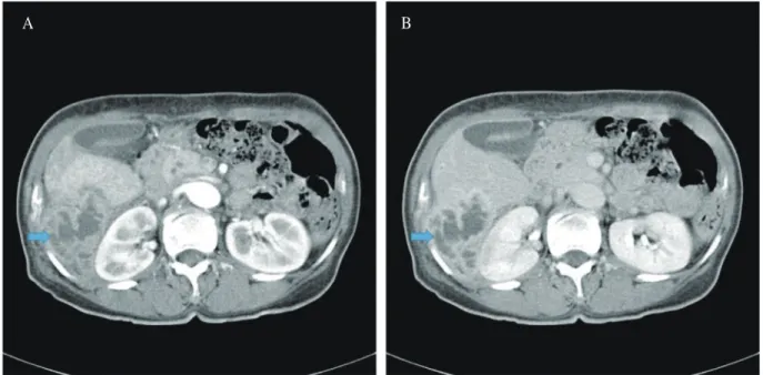
Initial contrast computed tomography (CT) of the abdomen by a private practitioner at a local clinic revealed a 7.5 cm necrotic mass located in Morrison's pouch with a peripheral contrast enhancing wall and washout with rim enhancement in the delayed phase (Fig. 1). The spleen size was slightly increased.
However, there was no definite evidence of liver cirrhosis, such as lobulation of the lateral edge of the liver or loss of hepatic volume. Ultrasonography (US) revealed the lesion was a heterogeneous hypo- echogenic mass with central necrosis in the right liver
Fig. 1. (A) Contrast-enhanced abdominal computed tomographic findings. On the arterial phase, the heterogeneously defined 7.5 cm sized necrotic mass with peripheral enhancing wall (arrow) is noted. (B) On the delayed phase, it is washed out with rim enhancement (arrow) is shown.
A B
(Fig. 2).
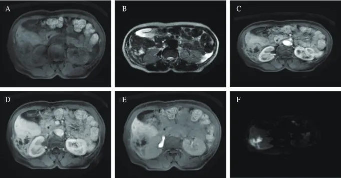
For further evaluation, abdominal magnetic resonance imaging (MRI) of the liver was performed 5 days later with unenhanced T1-weighted (T1W) and
T2-weighted (T2W) images with and without fat suppression and contrast-enhanced sequences using Gd-EOB-DTPA (Primovist; Bayer Healthcare, Berlin, Germany) as a liver-specific contrast agent. Early dynamic images were acquired first, followed by delayed hepatobiliary imaging after 20 minutes (Fig.
3). In the T1W images, the lesion with irregular borders appeared hypointense. The T2W images showed a heterogonous, slightly hyperintense lesion with an ill-defined, moderately hyperintense rim. In the arterial phase following contrast injection, the lesion was rim-enhanced inhomogeneously, as demonstrated by MRI, and in the delayed phase, the lesion had washout with rim enhancement. In the hepatobiliary phase, the lesion did not show contrast material accumulation. Diffusion-weighted imaging showed that the peripheral lesion was bright, while the center showed dark signal intensity.
Fig. 2. Abdominal sonographic finding. The lesion is heterogeneous hypoechogenic mass in the right liver.
Fig. 3. Liver MRI findings. (A) On T1W images the lesion with irregular borders appears hypointense. (B) T2W images shows a heterogenous slightly hyperintense lesion with an ill-defined moderately hyperintense rim. (C) On the arterial phase following contrast injection, the lesion is rim enhanced inhomogeneously as demonstrated on CT. (D) And on the delayed phase the lesion is washout with rim enhancement. (E) In the hepatobiliary phase, the lesion do not show contrast material accumulation. (F) Diffusion weighted image shows that the peripheral lesion was bright but the center showed dark signal intensity.
D A
E B
F C
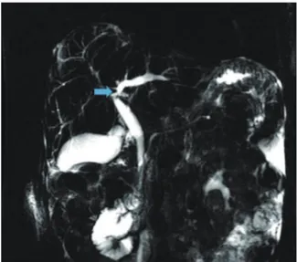
At the same time as the MRI, magnetic retrograde cholangiopancreatography (MRCP) showed luminal narrowing of intrahepatic duct (IHD) at the hilar portion with unproportional dilatation of IHD to the periphery. However, there was no evidence of an obstructing mass or stone on the duct (Fig. 4).
Collectively, these radiological findings indicated the presence of liver necrosis, with the possibility of a differential diagnosis of primary malignancy of the liver, metastatic cancer of the liver, or IPT.
To achieve a more definite diagnosis, a US-guided percutaneous liver biopsy was performed (Fig. 5).
Microscopically, the liver specimen showed marked fibrosis and inflammatory cell infiltration, mainly consisting of plasma cells and eosinophils in the background of stroma composed of myofibroblast,
Fig. 4. Magnetic retrograde cholangiopancreato- graphy finding. The lesion shows luminal narrowing of intra hepatic duct at hilar portion with unproportional dilatation of IHD to the periphery.
Fig. 5. Histologic findings. A marked fibrosis and inflammatory cell infiltration, mainly consisting of plasma cells, and eosinophils are seen in background of stroma composed of a myofibroblast, fibroblast, and collagen bundle. (A) H&E, × 400. (B) SMA, × 40. (C) IgG, × 400. (D) IgG4, × 400.
A
C
B
D
fibroblast, and collagen bundles. However, there was no evidence of malignant cells and no definite evidence of cirrhosis in the surrounding liver parenchyma. Immunohistochemical examination of the liver specimen showed that the lesion was positive for α-smooth muscle actin (SMA), CD34, and cytokeratin (CK) and negative staining of CD 117, IgG, and IgG4. Finally, based on the results of our microscopic examination of the liver specimen, we diagnosed the pathological status of this patient as IPT.
The patient was treated with intravenous antibiotics, including cefotaxime and metronidazole, for 3 weeks.
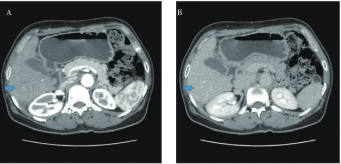
The C-reactive protein levels decreased from 84.1 mg/L to 0.14 mg/L. Follow-up CT imaging after 3 weeks of intravenous antibiotics showed that the necrotic mass size decreased from 5 × 7 cm to 3 × 3 cm, except for mild splenomegaly. On hospital day 45, the patient was in good health and free of intravenous antibiotic treatment. She was discharged with oral antibiotics, and a follow-up examination was scheduled for a month later. Two months after
the initial intravenous antibiotic therapy, CT imaging revealed nearly complete resolution of the hepatic lesion, except unproportional dilatation of the IHD to the periphery (Fig. 6). Therefore, she had been successfully cured with antibiotics and conservative management.
Discussion
IPT was first described in the lung by Brunn in 1939 and has been subsequently reported in various organs [1]. IPT of the liver is a rare disease [1,2] and was first reported by Pack and Baker [10] in 1953.
Several studies from Western countries have reported that it is common in males in their thirties and forties [3,9]. However, in Eastern countries, it tends to show an older age of onset [11,12]. Although the incidence of hepatic IPT has been reported to be 0.7% [11], the etiology of IPT remains uncertain. Several hypotheses have been introduced [3,4,9,13]. First, IPT can occur with exaggerated or inadequate inflammation by
Fig. 6. Follow-up dynamic abdominal CT findings. 6 months after the initial CT (Fig. 1). The lesion (arrow) was shrunken and healed.
A B
infection by an existing micro-organism in the portal vein. However, any association between IPT and organisms has not been identified. Additionally, biliary drainage disturbances, such as biliary stones, cholangitis, liver abscess, primary sclerosing cholangitis, history of liver resection, and hepatobiliary malignancy, have been related to IPT of the liver [14,15]. Furthermore, several studies have demon-strated that viral infection, autoimmune disease, and vascular disease are often associated with hepatic IPT.
Unfortunately, the diagnosis of hepatic IPT is very difficult because there are no specific symptoms, typical radiological features, or laboratory findings associated with it. Usually, patients with IPT can present with fever, abdominal pain, myalgia, or general weakness, but these symptoms are not specific to IPT [3,4,9,12]. Our patient also felt only chronic fatigue and general weakness. The radiological findings of IPT are non-specific.
Although IPT is often seen as a hypoechoic mass with well-defined borders on US, this is not a typical finding. On a dynamic CT scan, IPT shows low attenuation in the noncontrast phase and peripheral enhancement in the delayed stage. Previous studies have reported that the proportion or the distribution of inflammatory cells and fibrosis according to the cause and the period of inflammation can affect the images of IPT in contrast-enhanced CT [2,16,17]. In MRI, IPT is frequently hypointense in T1-weighted images, hyperintense in T2-weighted images, and shows heterogenic uptake after gadolinium injection [11,12]. However, not only are these findings non- specific, but they are also easily confused with malignant tumors [1,8,9,18-21]. Moreover, laboratory findings, including tumor markers, such as CA19-9 and AFP, do not help to distinguish IPT from malignant tumors [15]. Therefore, a histopathological needle biopsy is mandatory to differentiate between IPT and malignant tumors.
In our case, the mass was hypoechoic by US, but also had peripheral enhancement following contrast administration in the delayed stage on the dynamic CT scan. In MRI, the T1W and T2W images showed hypointensity and hyperintensity, respectively, and these MRI findings could not exclude metastatic malignancy, such as cholangiocarcinoma.
Additionally, the MRCP findings showed unproportional dilatation of the left intrahepatic bile ducts caused by narrowing of the hilar bile duct as well as sequelae of chronic cholangitis likely due to Clonorchis sinensis in the right intrahepatic bile ducts.
All these radiologic findings do not correspond to typical IPT, as previously recognized [11,12];
therefore, it was difficult to make a definitive diagnosis. Additionally, it is known that chronic cholangitis is a risk factor for cholangiocarcinoma, which is associated with IPT. As in this case, the image findings showed the infiltrative hepatic mass and sequelae of chronic cholangitis; while the laboratory findings were normal, the histopathological needle biopsy was necessary. Thus, we performed a percutaneous needle biopsy to exclude malignancy, such as cholangiocarcinoma or infiltrative hepatic malignancy. Microscopically, the histologic finding was marked fibrosis and inflammatory cell infiltration, mainly consisting of plasma cells and eosinophils in the background of stroma composed of a myofibroblast, fibroblast, and collagen bundle.
However, there was no evidence of malignant cells.
Additionally, the immunohistochemical examination showed negative staining of IgG4; therefore, fibrohistiocytic-type IPT was favored in our case [3-8,15]. This biopsy finding prevented unnecessary resection.
It has been known that the IPT is favorable prognosis and can be successfully treated using antibiotics or non-steroidal anti-inflammatory drugs in most of patients [15]. As shown in our case, the patient had been successfully responded to con-
servative management using antibiotics without surgery or steroids. And on follow-up CT imaging revealed nearly complete resolution of the hepatic lesion, except unproportional dilatation of the IHD to the periphery. Collectively, these findings supported that IPT was favored rather than malignant tumor.
In conclusion, hepatic IPT is a benign lesion, but demonstrates various radiological findings and lacks specific symptoms. When differential diagnosis from other malignant tumors is clinically difficult and malignancy is mimicked, a biopsy is frequently needed to make the diagnosis and to avoid unnecessary surgery. Especially a biopsy is essential in patients who have chronic cholangitis with not increased tumor marker.
References
1. Zamir D, Jarchowsky J, Singer C, Abumoch S, Groisman G, Ammar M, et al. Inflammatory pseudotumor of the liver--a rare entity and a diagnostic challenge. Am J Gastroenterol 1998;93:1538-40.
2. Fukuya T, Honda H, Matsumata T, Kawanami T, Shimoda Y, Muranaka T, et al. Diagnosis of inflammatory pseudotumor of the liver: value of CT. Am J Roentgenol 1994;163:1087-91.
3. Torzilli G, Inoue K, Midorikawa Y, Hui AM, Takayama T, Makuuchi M. Inflammatory pseudotumors of the liver:
prevalence and clinical impact in surgical patients.
Hepatogastroenterology 2001;48:1118-23.
4. Horiuchi R, Uchida T, Kojima T, Shikata T. Inflammatory pseudotumor of the liver. Clinicopathologic study and review of the literature. Cancer 1990;65:1583-90.
5. Yamamoto H, Yamaguchi H, Aishima S, Oda Y, Kohashi K, Oshiro Y, et al. Inflammatory myofibroblastic tumor versus IgG4-related sclerosing disease and inflammatory pseudotumor: a comparative clinicopathologic study. Am J Surg Pathol 2009;33:1330-40.
6. Zen Y, Fujii T, Sato Y, Masuda S, Nakanuma Y. Pathological
classification of hepatic inflammatory pseudotumor with respect to IgG4-related disease. Mod Pathol 2007;20:884- 94.
7. White JE, Chase CW, Kelley JE, Brock WB, Clark MO.
Inflammatory pseudotumor of the liver associated with extrahepatic infection. South Med J 1997;90:23-9.
8. Milias K, Madhavan KK, Bellamy C, Garden OJ, Parks RW. Inflammatory pseudotumors of the liver: experience of a specialist surgical unit. J Gastroenterol Hepatol 2009;24:1562-6.
9. Akatsu T, Wakabayashi G, Tanimoto A, Kameyama K, Kitajima M. Inflammatory pseudotumor of the liver masquerading as hepatocellular carcinoma after a hepatitis B virus infection: report of a case. Surg Today 2006;36:1028-31.
10. Pack GT, Baker HW. Total right hepatic lobectomy;
report of a case. Ann Surg 1953;138:253-8.
11. Tang L, Lai EC, Cong WM, Li AJ, Fu SY, Pan ZY, et al.
Inflammatory myofibroblastic tumor of the liver: a cohort study. World J Surg 2010;34:309-13.
12. Park JY, Choi MS, Lim YS, Park JW, Kim SU, Min YW, et al. Clinical features, image findings, and prognosis of inflammatory pseudotumor of the liver: a multicenter experience of 45 cases. Gut liver 2014;8:58- 63.
13. Schmid A, Janig D, Bohuszlavizki A, Henne-Bruns D.
Inflammatory pseudotumor of the liver presenting as incidentaloma: report of a case and review of the literature. Hepatogastroenterology 1996;43:1009-14.
14. Koea JB, Broadhurst GW, Rodgers MS, McCall JL.
Inflammatory pseudotumor of the liver: demographics, diagnosis, and the case for nonoperative management. J Am Coll Surg 2003;196:226-35.
15. Ahn KS, Kang KJ, Kim YH, Lim TJ, Jung HR, Kang YN, et al. Inflammtory pseodutumors mimicking intra hepatic cholangiocarcinoma of the liver; IgG4-positivity and its clinical significance. J Hepatobiliary Pancreat Sci 2012;19:405-12.
16. Yoon KH, Ha HK, Lee JS, Suh JH, Kim MH, Kim PN, et al. Inflammatory pseudotumor of the liver in patients
with recurrent pyogenic cholangitis: CT-histopathologic correlation. Radiology 1999;211:373-9.
17. Nam KJ, Kang HK, Lim JH. Author’s correction.
Inflammatory pseudotumor of the liver: CT and sonographic findings. Am J Roentgenol 1996;167:1598.
18. Yoshida T, Nishimori I, Kumon M, Kohsaki T, Taniuchi K, Ohtsuki Y, et al. Inflammatory pseudotumor of the liver: report of a case diagnosed by needle biopsy.
Hepatol Res 2003;27:83-6.
19. Kai K, Matsuyama S, Ohtsuka T, Kitahara K, Mori D, Miyazaki K. Multiple inflammatory pseudotumor of the liver, mimicking cholangiocarcinoma with tumor
embolus in the hepatic vein: report of a case. Surg Today 2007;37:530-3.
20. Kim SR, Hayashi Y, Kudo M, Matsuoka T, Imoto S, Sasaki K, et al. Inflammatory pseudotumor of the liver in a patient with chronic hepatitis C: difficulty in differentiating it from hepatocellular carcinoma. Pathol Int 1999;49:726-30.
21. Jeong JY, Sohn JH, Kim TY, Jeong WK, Kim J, Pyo JY, et al. Hepatic inflammatory pseudotumor misinterpreted as hepatocellular carcinoma. Clin Mol Hepatol.
2012;18:239-44.