328 Copyright © 2017 The Korean Society of Cardiology Korean Circulation Journal
Introduction
Percutaneous coronary intervention (PCI) has emerged as an important modality to treat coronary artery disease (CAD). As research improves pharmacology and clinical approaches for patient treatment, more complex CADs are being successfully treated with PCI and clinical outcomes are consistently improving. PCI can now be performed in most patients that require revascularization and it is also offered to subjects that have a high operative risk.1)
Guideline-based strategies and monitoring of PCI procedures are strongly recommended to improve in-hospital and long-term clinical outcomes of patients that undergo PCI.2-5) Specifically, performance
Print ISSN 1738-5520 • On-line ISSN 1738-5555
The Current Status of Percutaneous Coronary Intervention in Korea –Based on Year 2014 Cohort of Korean Percutaneous Coronary
Intervention (K-PCI) Registry–
Jae-Sik Jang, MD
1, Kyoo-Rok Han, MD
2, Keon-Woong Moon, MD
3, Dong Woon Jeon, MD
4, Dong-Ho Shin, MD
5, Jung-Sun Kim, MD
5, Duk-Woo Park, MD
6, Hyun-Jae Kang, MD
7, Juhan Kim, MD
8, Jang-Whan Bae, MD
9,
Seung-Ho Hur, MD
10, Byung Ok Kim, MD
11, Donghoon Choi, MD
5, Hyeon-Cheol Gwon, MD
12, and Hyo-Soo Kim, MD
71Division of Cardiology, Busan Paik Hospital, University of Inje College of Medicine, Busan, 2Department of Internal Medicine, Kangdong Sacred Heart Hospital, Hallym University Medical Center, Seoul, 3Division of Cardiology, St. Vincent’s Hospital, The Catholic University of Korea, Suwon, 4Division of Cardiology, National Health Insurance Service Ilsan Hospital, Goyang, 5Division of Cardiology, Severance Cardiovascular Hospital, Yonsei University College of Medicine, Seoul, 6Division of Cardiology, Asan Medical Center, University of Ulsan College of Medicine, Seoul, 7Department of Internal Medicine, Seoul National University Hospital, Seoul, 8Division of Cardiology, Heart Center of Chonnam National University Hospital, Gwangju, 9Department of Internal Medicine, Chungbuk National University College of Medicine, Cheongju, 10Division of Cardiology, Keimyung University Dongsan Medical Center, Daegu, 11Division of Cardiology, Sanggye-Paik Hospital, University of Inje College of Medicine, Seoul, 12Heart Vascular and Stroke Institute, Samsung Medical Center, Sungkyunkwan University School of Medicine, Seoul, Korea
Background and Objectives: Although several multicenter registries have evaluated percutaneous coronary intervention (PCI) procedures in Korea, those databases have been limited by non-standardized data collection and lack of uniform reporting methods. We aimed to collect and report data from a standardized database to analyze PCI procedures throughout the country.
Materials and Methods: Both clinical and procedural data, as well as clinical outcomes data during hospital stay, were collected based on case report forms that used a standard set of 54 data elements. This report is based on 2014 Korean PCI registry cohort data.
Results: A total of 92 hospitals offered data on 44967 PCI procedures. The median age was 66.0 interquartile range 57.0-74.0 years, and 70.3% were men. Thirty-eight percent of patients presented with acute myocardial infarction and one-third of all PCI procedures were performed in an urgent or emergency setting. Non-invasive stress tests were performed in 13.9% of cases, while coronary computed tomography angiography was used in 13.7% of cases prior to PCI. Radial artery access was used in 56.1% of all PCI procedures. Devices that used PCI included drug-eluting stent, plain old balloon angioplasty, drug-eluting balloon, and bare-metal stent (around 91%, 19%, 6%, and 1% of all procedures, respectively). The incidences of in-hospital death, non-fatal myocardial infarction, and stroke were 2.3%, 1.6%, and 0.2%, respectively.
Conclusion: These data may provide an overview of the current PCI practices and in-hospital outcomes in Korea and could be used as a foundation for developing treatment guidelines and nationwide clinical research. (Korean Circ J 2017;47(3):328-340)
KEY WORDS: Percutaneous coronary intervention; Registry.
Received: March 31, 2017 Revision Received: April 17, 2017 Accepted: April 18, 2017
Correspondence: Hyo-Soo Kim, MD, PhD, Department of Internal Medicine, Seoul National University Hospital, 101 Daehak-ro, Jongno-gu, Seoul 03080, Korea Tel: 82-2-2072-2226, Fax: 82-2-766-8904
E-mail: [email protected]
• The authors have no financial conflicts of interest.
This is an Open Access article distributed under the terms of the Creative Commons Attribution Non-Commercial License (http://creativecommons.org/
licenses/by-nc/3.0) which permits unrestricted non-commercial use, distribution, and reproduction in any medium, provided the original work is properly cited.
measures for PCI and ST-elevation myocardial infarction (STEMI)/
non-ST-elevation myocardial infarction (NSTEMI) were established to guide physicians with approaches for these interventions.6-8)
Although several multicenter registries have evaluated PCI procedures in Korea,9-13) no nationwide contemporary PCI registries have been collected with standardized collection forms and unified reporting methods. The Korean Society of Cardiology (KSC) and Korean Society of Interventional Cardiology (KSIC) established the Korean percutaneous coronary intervention (K-PCI) registry to collect and report results in a standardized database for PCI case analysis throughout the country. The purpose of this study was to construct baseline data standards to set- up treatment guidelines that reflect relevant clinical situations and to facilitate clinical research for cardiologists that treat CAD in Korea.
Materials and Methods
Participation and data definitions
The K-PCI registry final protocol was created in the task force team meeting and the K-PCI registry governing committee conference, it was designed for enrolling participants with respect to clinical diagnosis information, procedural data, and by considering major complications after PCI procedures performed to treat ischemic heart disease. The hospitals that could perform PCI were asked to participate and 92 hospitals voluntarily took part in this registry. Demographic characteristics, clinical history, procedural information and PCI complications that occurred before hospital discharge were collected retrospectively using case report forms (CRFs) comprised of a standard set of 54 data elements. When multiple interventions were performed
in a subject, individual procedures were considered as single cases and were entered in the database. All variables corresponded with written definitions in the CRF dictionary (K-PCI registry 2014 CRF v1.4., Supplementary Table 1 in the online-only Data Supplement) to standardize the data, and were provided by using common specifications with question and answer cases for participants.
This study was approved by the local institutional review board (IRB) at each participating site. The written consent document was waived in participating centers, because of the retrospective nature of the study, with no clinical follow-up. Data were collected using a web-based reporting system.
Data collection and management
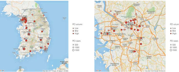
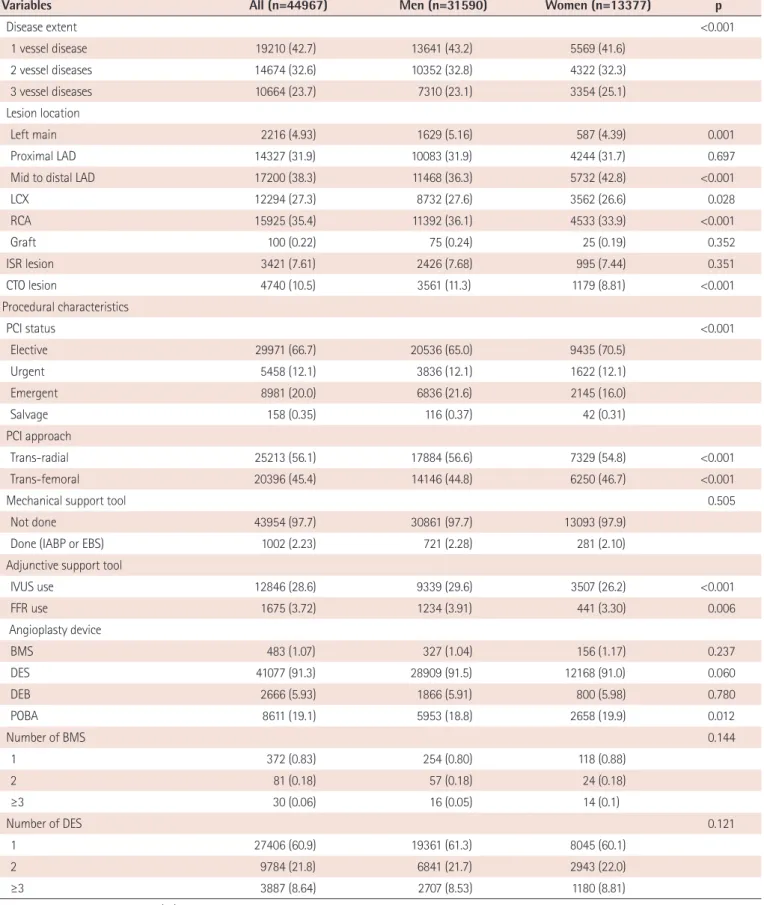
This report represents PCI data of the participating hospitals from January 1, 2014, through December 31, 2014. The K-PCI registry governing committee centrally managed the web-based registered study data. After the K-PCI registry was approved by the IRBs, demographic, procedural, and in-hospital outcomes data for 44967 PCI cases from 92 hospitals were collected. About half (45.7%) of the PCI centers were located in the capital area and high-volume facilities that performed more than 781 PCI procedures annually were concentrated in metropolitan areas, including Seoul (Fig. 1).
The K-PCI data management and inspection program determined whether collected data were accurate and complete and the CRF variables were refined based on the program results. The K-PCI registry governing committee periodically monitored the enrollment status and shared the results with participating hospitals via newsletters, the committee also performed data management quality verification through the query process.
PCI volume Low Mid High
PCI cases 500 1000 1500
PCI volume Low Mid High
PCI cases 500 1000 1500
Fig. 1. Geographic distribution of PCI facilities participating in the K-PCI registry. Ninety-two hospitals submitted data for 44967 PCI cases. High- volume facilities were concentrated in Seoul and metropolitan areas. PCI: percutaneous coronary intervention, K-PCI: Korean percutaneous coronary intervention.
Statistical analysis
Discrete data for demographic, clinical, procedural and outcome variables were presented as numbers and percentages and compared using Chi-square tests. Continuous variables are presented as medians
with interquartile ranges (IQRs) unless otherwise specified, and were compared using the Student’s t-test, analysis of variance (ANOVA) or the Wilcoxon rank-sum test, where applicable. All p values were 2-tailed, and a p value<0.05 was considered statistically significant.
All analyses were conducted using R version 3.3.2 (R Foundation for Statistical Computing, Vienna, Austria).
Results
Study populations
A total of 92 hospitals submitted data on 44967 PCI procedures performed from January through December 2014. The data presented in this report provide a contemporary assessment of PCI, as performed in Korea during the collection period, and represent important aspects of coronary interventions, including use of diagnostic support tools and procedural devices, as well as clinical outcomes before hospital discharge. Among the hospitals included in the K-PCI registry, 62%
performed 500 or fewer PCI procedures and 11% performed more than 1000 PCIs during the collection period. Twelve hospitals (13%) performed 200 or fewer PCIs for one year and these hospitals accounted for 3.2% of all PCI procedures (Fig. 2).
Clinical characteristics and presentation
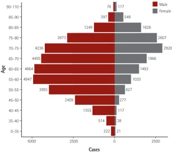
Baseline demographic and clinical characteristics of included patients are presented in Table 1. Median age was 66.0 [IQR 57.0-74.0]
1700-1800 1400-1500 1200-1300 1100-1200 1000-1100 800-900 700-800 600-700 500-600 400-500 300-400 200-300 100-200
<100
Seoul Province
5 0 5 10
Cases per year
Number of centers
90-110 85-90 80-85 75-80 70-75 65-70 60-65 55-60 50-55 45-50 40-45 35-40 0-35
Age
Cases
5000 2500
222 514 1355 2409 3993
4947 4864
4455 4236
2873
1249 397
76
21 38 117
277 627
1020 1493
1966 2920 2607 1626 548 117
0 2500
Male Female
STEMI
NSTEMI
Unstable angina
Stable angina
Silent ischemia
Sex Female (29.7%)
Female
Male (70.3%)
Male 13.5%
20.5%
19.6%
34.0%
22.4%
3.5%
19.8%
40.4%
23.0%
3.3%
Fig. 2. Distribution of facilities with different PCI volume. Sixty-two percent of the included hospitals performed 500 or fewer PCI procedures and 11% performed more than 1000 PCIs during the collection period.
PCI: percutaneous coronary intervention.
Fig. 3. Age and gender distribution. Median age was 66 years and the proportion of male patients was 70.3%.
Fig. 4. Gender-specific clinical indications for PCI. The width of the bars in the histogram indicates the number of patients. NSTEMI: non-ST- elevation myocardial infarction, STEMI: ST-elevation myocardial infarction, PCI: percutaneous coronary intervention.
years and the proportion of male patients was 70.3% (Fig. 3). Among patients undergoing PCI, 35.9% had diabetes mellitus, 9.2% had
history of prior myocardial infarction, 24% had history of PCI, 1.3%
had prior coronary artery bypass surgery, 6.4% had renal failure, 8.8%
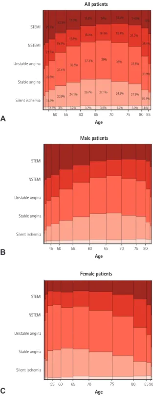
had prior history of cerebrovascular disease, and 2.7% had peripheral vascular disease. Unstable angina was the most common initial presentation (35.9%) that was a clinical indication for PCI, followed by stable angina (22.6%), NSTEMI (19.7%), STEMI (18.4%), and silent ischemia (3.5%) (Fig. 4). Patients that were between 60-80 years, were more likely to present with stable angina compared with other age groups, whereas patients in younger groups and elderly groups were more likely to present with acute coronary syndrome (ACS) (Fig 5A).
However, when this age-diagnosis interaction was sorted by gender, there was a trend toward a higher incidence of ACS in young male patients and elderly female patients (Fig. 5B, C). Cardiogenic shock was identified in 3.1% and cardiac arrest was present in 2.3% of patients in the 24 hours prior to admission. Beta blocking agents were the most frequently prescribed antianginal medication (53.5%) within 2 weeks of the procedure, followed by calcium channel blockers (36.5%), nicorandil (24.1%), long-acting nitrates (17.6%), and trimetazidine (15.2%).
Non-invasive test and imaging studies
From the total number of patients undergoing PCI, only 13.9%
(9.8% in ACS patients, 25.8% in non-ACS patients) underwent certain types of non-invasive tests before the PCI (Table 1). The treadmill test (TMT) was the dominant stress test (6.7% in ACS patients, 19.5% in non-ACS patients). Of the non-ACS patients who underwent different types of non-invasive stress tests, 13.3% demonstrated positive TMT and 5.1% demonstrated positive thallium single-photon emission computed tomography. Stress echocardiography and stress magnetic resonance imaging studies were done in less than 1% of patients in this registry. Coronary computed tomography (CT) angiography was performed in 13.7% (10.0% in ACS, 24.1% in non-ACS) of patients and 42.7% of those patients had one vessel involved. Pre-PCI evaluation of left ventricular function was performed in 67.6% of patients and the mean left ventricular ejection fraction was 60.0%.
Procedural characteristics
Angiographic and procedural characteristics for each PCI procedure are presented in Table 2. More than half of the patients had multi- vessel CAD, whereas 42.7% of patients had single vessel involvement.
The left anterior descending artery was the most frequently involved vessel (70.1%), followed by the right coronary artery (35.4%), left circumflex artery (27.3%), and left main coronary artery (4.9%).
Among patients that underwent PCI, a chronic total occlusion lesion was present in 10.5% and an in-stent restenosis lesion was present in 7.6%. Graft vessel PCI was done in only 0.2 % of all procedures. Two- thirds of the PCI procedures were performed in an elective setting. In this registry the trans-radial approach was more frequently (56.1%)
50 55 60 65 70 75 80 85
Age STEMI
NSTEMI
Unstable angina
Stable angina
Silent ischemia 16.9%
29.5%
33.4%
21.7%
29.2% 23.3% 19.5% 15.8% 14% 13.5% 14.9% 18%
19.4%
16.8% 16.4% 16.3% 19.4% 21.7%
28.4%
36.5% 37.3% 39% 39% 37.6%
33.9%
20.9% 24.1% 26.7% 24.5% 21.9%
15.8%
27.1%
3.8%
3.9%
3.7%
3.7% 3.6%
3.2%
3%
2.7%
All patients
45 50 55 60 65 70 75 80
Age STEMI
NSTEMI
Unstable angina
Stable angina
Silent ischemia
Male patients
55 60 65 70 75 80 85 90
Age STEMI
NSTEMI
Unstable angina
Stable angina
Silent ischemia
Female patients
Fig. 5. Age-specific clinical indications for PCI. The width of the bars in the histogram indicates the number of patients. NSTEMI: non-ST-elevation myocardial infarction, STEMI: ST-elevation myocardial infarction, PCI:
percutaneous coronary intervention.
A
B
C
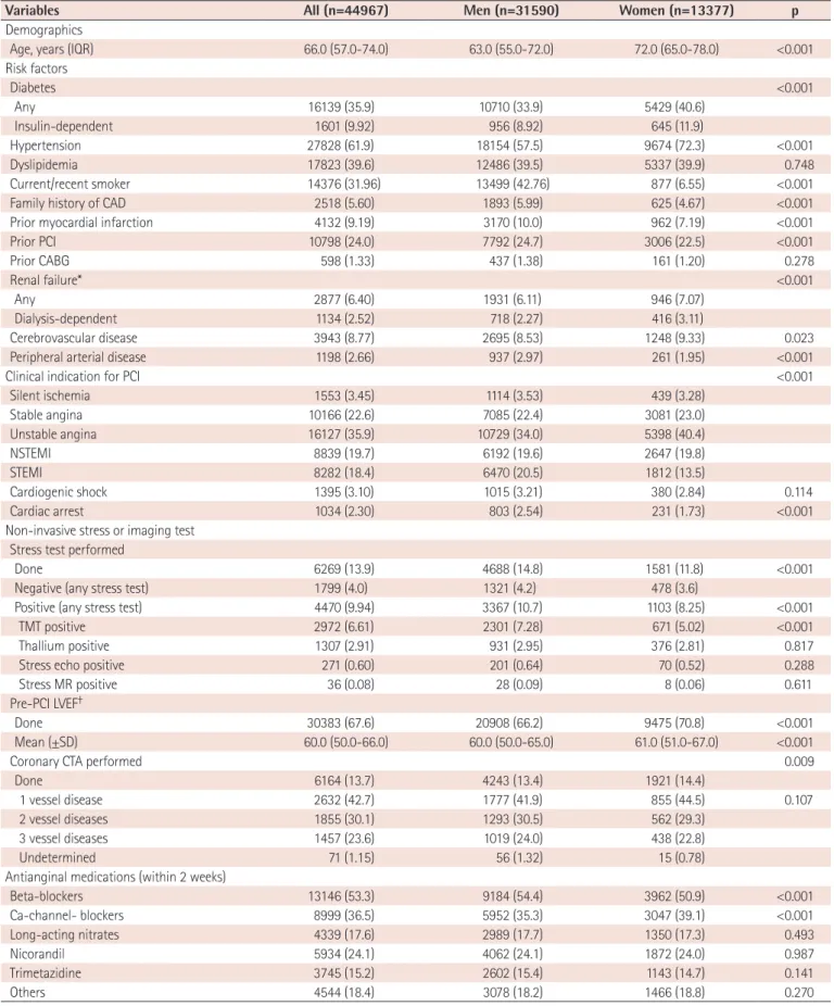
Table 1. Baseline patient and clinical characteristics
Variables All (n=44967) Men (n=31590) Women (n=13377) p
Demographics
Age, years (IQR) 66.0 (57.0-74.0) 63.0 (55.0-72.0) 72.0 (65.0-78.0) <0.001
Risk factors
Diabetes <0.001
Any 16139 (35.9) 10710 (33.9) 5429 (40.6)
Insulin-dependent 1601 (9.92) 956 (8.92) 645 (11.9)
Hypertension 27828 (61.9) 18154 (57.5) 9674 (72.3) <0.001
Dyslipidemia 17823 (39.6) 12486 (39.5) 5337 (39.9) 0.748
Current/recent smoker 14376 (31.96) 13499 (42.76) 877 (6.55) <0.001
Family history of CAD 2518 (5.60) 1893 (5.99) 625 (4.67) <0.001
Prior myocardial infarction 4132 (9.19) 3170 (10.0) 962 (7.19) <0.001
Prior PCI 10798 (24.0) 7792 (24.7) 3006 (22.5) <0.001
Prior CABG 598 (1.33) 437 (1.38) 161 (1.20) 0.278
Renal failure* <0.001
Any 2877 (6.40) 1931 (6.11) 946 (7.07)
Dialysis-dependent 1134 (2.52) 718 (2.27) 416 (3.11)
Cerebrovascular disease 3943 (8.77) 2695 (8.53) 1248 (9.33) 0.023
Peripheral arterial disease 1198 (2.66) 937 (2.97) 261 (1.95) <0.001
Clinical indication for PCI <0.001
Silent ischemia 1553 (3.45) 1114 (3.53) 439 (3.28)
Stable angina 10166 (22.6) 7085 (22.4) 3081 (23.0)
Unstable angina 16127 (35.9) 10729 (34.0) 5398 (40.4)
NSTEMI 8839 (19.7) 6192 (19.6) 2647 (19.8)
STEMI 8282 (18.4) 6470 (20.5) 1812 (13.5)
Cardiogenic shock 1395 (3.10) 1015 (3.21) 380 (2.84) 0.114
Cardiac arrest 1034 (2.30) 803 (2.54) 231 (1.73) <0.001
Non-invasive stress or imaging test Stress test performed
Done 6269 (13.9) 4688 (14.8) 1581 (11.8) <0.001
Negative (any stress test) 1799 (4.0) 1321 (4.2) 478 (3.6)
Positive (any stress test) 4470 (9.94) 3367 (10.7) 1103 (8.25) <0.001
TMT positive 2972 (6.61) 2301 (7.28) 671 (5.02) <0.001
Thallium positive 1307 (2.91) 931 (2.95) 376 (2.81) 0.817
Stress echo positive 271 (0.60) 201 (0.64) 70 (0.52) 0.288
Stress MR positive 36 (0.08) 28 (0.09) 8 (0.06) 0.611
Pre-PCI LVEF†
Done 30383 (67.6) 20908 (66.2) 9475 (70.8) <0.001
Mean (±SD) 60.0 (50.0-66.0) 60.0 (50.0-65.0) 61.0 (51.0-67.0) <0.001
Coronary CTA performed 0.009
Done 6164 (13.7) 4243 (13.4) 1921 (14.4)
1 vessel disease 2632 (42.7) 1777 (41.9) 855 (44.5) 0.107
2 vessel diseases 1855 (30.1) 1293 (30.5) 562 (29.3)
3 vessel diseases 1457 (23.6) 1019 (24.0) 438 (22.8)
Undetermined 71 (1.15) 56 (1.32) 15 (0.78)
Antianginal medications (within 2 weeks)
Beta-blockers 13146 (53.3) 9184 (54.4) 3962 (50.9) <0.001
Ca-channel- blockers 8999 (36.5) 5952 (35.3) 3047 (39.1) <0.001
Long-acting nitrates 4339 (17.6) 2989 (17.7) 1350 (17.3) 0.493
Nicorandil 5934 (24.1) 4062 (24.1) 1872 (24.0) 0.987
Trimetazidine 3745 (15.2) 2602 (15.4) 1143 (14.7) 0.141
Others 4544 (18.4) 3078 (18.2) 1466 (18.8) 0.270
Values are presented as number (%) if not otherwise specified. *GFR ≤60 mL/min/1.73 m², †can be assessed via invasive (i.e. left ventriculography) or non-invasive (i.e. echocardiography, magnetic resonance, computed tomography, or nuclear) testing. IQR: interquartile range, CAD: coronary artery disease, PCI: percutaneous coronary intervention, CABG: coronary artery bypass graft, NSTEMI: non-ST-elevation myocardial infarction, STEMI: ST-elevation myocardial infarction, TMT:
treadmill test, MR: magnetic resonance, LVEF: left ventricular ejection fraction, SD: standard deviation, CTA: computed tomography angiography
Table 2. Baseline angiographic and procedural characteristics
Variables All (n=44967) Men (n=31590) Women (n=13377) p
Disease extent <0.001
1 vessel disease 19210 (42.7) 13641 (43.2) 5569 (41.6)
2 vessel diseases 14674 (32.6) 10352 (32.8) 4322 (32.3)
3 vessel diseases 10664 (23.7) 7310 (23.1) 3354 (25.1)
Lesion location
Left main 2216 (4.93) 1629 (5.16) 587 (4.39) 0.001
Proximal LAD 14327 (31.9) 10083 (31.9) 4244 (31.7) 0.697
Mid to distal LAD 17200 (38.3) 11468 (36.3) 5732 (42.8) <0.001
LCX 12294 (27.3) 8732 (27.6) 3562 (26.6) 0.028
RCA 15925 (35.4) 11392 (36.1) 4533 (33.9) <0.001
Graft 100 (0.22) 75 (0.24) 25 (0.19) 0.352
ISR lesion 3421 (7.61) 2426 (7.68) 995 (7.44) 0.351
CTO lesion 4740 (10.5) 3561 (11.3) 1179 (8.81) <0.001
Procedural characteristics
PCI status <0.001
Elective 29971 (66.7) 20536 (65.0) 9435 (70.5)
Urgent 5458 (12.1) 3836 (12.1) 1622 (12.1)
Emergent 8981 (20.0) 6836 (21.6) 2145 (16.0)
Salvage 158 (0.35) 116 (0.37) 42 (0.31)
PCI approach
Trans-radial 25213 (56.1) 17884 (56.6) 7329 (54.8) <0.001
Trans-femoral 20396 (45.4) 14146 (44.8) 6250 (46.7) <0.001
Mechanical support tool 0.505
Not done 43954 (97.7) 30861 (97.7) 13093 (97.9)
Done (IABP or EBS) 1002 (2.23) 721 (2.28) 281 (2.10)
Adjunctive support tool
IVUS use 12846 (28.6) 9339 (29.6) 3507 (26.2) <0.001
FFR use 1675 (3.72) 1234 (3.91) 441 (3.30) 0.006
Angioplasty device
BMS 483 (1.07) 327 (1.04) 156 (1.17) 0.237
DES 41077 (91.3) 28909 (91.5) 12168 (91.0) 0.060
DEB 2666 (5.93) 1866 (5.91) 800 (5.98) 0.780
POBA 8611 (19.1) 5953 (18.8) 2658 (19.9) 0.012
Number of BMS 0.144
1 372 (0.83) 254 (0.80) 118 (0.88)
2 81 (0.18) 57 (0.18) 24 (0.18)
≥3 30 (0.06) 16 (0.05) 14 (0.1)
Number of DES 0.121
1 27406 (60.9) 19361 (61.3) 8045 (60.1)
2 9784 (21.8) 6841 (21.7) 2943 (22.0)
≥3 3887 (8.64) 2707 (8.53) 1180 (8.81)
Values are presented as number (%) and percent if not otherwise specified. LAD: left anterior descending, LCX: left circumflex, RCA: right coronary artery, ISR: in-stent restenosis, CTO: chronic total occlusion, PCI: percutaneous coronary intervention, IABP: intra-aortic balloon pump, EBS: emergency bypass system, IVUS: intravascular ultrasound, FFR: fractional flow reserve, BMS: bare-metal stents, DES: drug-eluting stents, DEB: drug-eluting balloon, POBA: plain old balloon angioplasty
used for PCI procedures, compared to the trans-femoral approach (45.4%). Even among ACS patients, more than half (52.7%) of procedures were performed via the trans-radial approach.
Mechanical support devices, such as an intra-aortic balloon pump
or emergency bypass system, were used in 2.2% of all PCI procedures.
Data on the use of PCI devices was also collected. At least one drug- eluting stent (DES) was placed in 91.3% of procedures, plain old balloon angioplasty (POBA) without stent implantation was done in Table 3. In-hospital outcomes
Variables All (n=44967) Men (n=31590) Women (n=31590) p
Death 1023 (2.28) 629 (1.99) 394 (2.95) <0.001
Cardiac death 704 (1.57) 432 (1.37) 272 (2.03) <0.001
Nonfatal myocardial infarction 701 (1.56) 516 (1.63) 185 (1.38) 0.075
Stent thrombosis 172 (0.38) 139 (0.44) 33 (0.25) 0.003
Stroke 90 (0.20) 58 (0.18) 32 (0.24) 0.426
Urgent repeat PCI 118 (0.26) 96 (0.30) 22 (0.16) 0.015
Transfusion 978 (2.17) 589 (1.86) 389 (2.91) <0.001
Values are presented as number (%). PCI: percutaneous coronary intervention
Fig. 6. Temporal distribution of PCI procedures and in-hospital events. (A) Monthly distribution of PCI procedures and in-hospital cardiac events.
(B) Weekly distribution of PCI procedures and in-hospital cardiac events. STEMI: ST-elevation myocardial infarction, NSTEMI: non-ST-elevation myocardial infarction, PCI: percutaneous coronary intervention, MACE: major adverse cardiac events.
7500
5000
2500
0
Cases
Monthly distribution of PCI procedures and in-hospital events
Weekly distribution of PCI procedures and in-hospital events
129 119 117 126 137 137 117 115
793 871 1347 747 664
683 1199 695 720
720 1223 633 685
944 1407 675 660
707 1261 695 690
785 1438 731 727
912 1427 815 726
996 1428 802 732
919 1329 730 694
1256 743 631
107 974 1472 800 698
862 1340 773 655
Monday Tuesday Wednesday Thursday Friday Saturday Sunday Monday
Jan Feb Mar Apr May Jun Jul Aug Sep Oct Nov Dec Jan Feb Mar Apr May Jun Jul Aug Sep Oct Nov Dec
Tuesday Wednesday Thursday Friday Saturday Sunday 4000
3000
2000
1000
0
Cases
7500
5000
2500
0 4000
3000
2000
1000
0
CasesCases
STEMI NSTEMI Unstable angina Stable angina Silent ischemia
PCI MACE STEMI
NSTEMI Unstable angina Stable angina Silent ischemia
PCI MACE
348 323 316
2028 3494 1584 1363
1174 1331
2929
1944
296 252 226 352
463 937 1671 1027 2619 1459 1189
2304 3488 1531 1197
2131 3311 2119 1395
151 148 150
A
B
19.1%, and drug-eluting balloon (DEB) was used in 5.9% of patients. A bare-metal stent was placed in only 1.1% of all patients. Intravascular ultrasound (IVUS) was used in 28.6% of patients (27.1% in ACS patients, 32.7% in non-ACS patients) as an adjunctive PCI support tool, whereas fractional flow reserve (FFR) was only performed in 3.7% of patients (2.5% in ACS patients, 7.2% in non-ACS patients).
Clinical outcomes during hospitalization
Patient clinical outcomes during hospitalization are summarized in Table 3. The in-hospital, all-cause mortality for the entire cohort was 2.3% (1023 patients), of which 68.8% (704 patients) experienced cardiac-related deaths (Table 3). In-hospital mortality ranged from 0.2% in patients with stable angina to 6.9% in STEMI patients.
Other in-hospital event rates were low including, non-fatal MI 1.6%, stroke 0.2%, urgent repeat revascularization 0.3%, and stent thrombosis in 0.4% (172 patients). There were significantly higher rates of all-cause mortality (3.0 vs. 2.0; p<0.001) and cardiac mortality (2.0 vs. 1.4%; p<0.001) in women undergoing PCI. The rates for transfusion during hospitalization were 2.2%, which was higher in women (2.9 vs. 1.9%; p<0.001). Although the number of PCI procedures peaked in March and December, the incidence of in-hospital events was evenly distributed throughout the year.
However, the rates of in-hospital cardiac events were significantly higher when PCI cases were performed during weekends than for procedures performed during weekdays (9.4 vs. 3.8%; p<0.001; Fig.
6), this is likely because most cases performed over the weekend were in emergency settings.
Centers patterns and results
To understand differences in demographics, procedural characteristics and clinical outcomes between high vs. low PCI volume centers, we divided all PCI centers into three tertiles based on the number of PCIs performed during the collection period (1st tertile: 41- 493; 2nd tertile: 494-780; 3rd tertile: 781-1794). Patients in high- volume centers were more likely to have a history of hypertension, dyslipidemia, renal failure, peripheral vascular disease, prior PCI, and coronary artery bypass graft than patients from lower volume centers (Table 4). High volume centers had more stable patients, used non-invasive pre-tests more frequently, and more frequently performed elective PCI with a trans-radial approach using IVUS/
FFR (Table 5). In contrast, low volume centers had higher incidences of acute myocardial infarction, cardiogenic shock or cardiac arrest.
Thus, these centers performed emergency PCI more frequently (24.4 vs. 14.9%; p<0.001) without IVUS/FFR and were less likely to conduct non-invasive pre-tests. Consequently, low volume centers showed higher in-hospital mortality (2.7 vs. 2.3 vs. 1.8%; p<0.001) (Table 6).
Discussion
This study represents the first nationwide database for PCI in Korea that was collected and analyzed by the KSC/KSIC K-PCI registry. Key findings were as follows: 1) thirty-eight percent of patients presented with acute myocardial infarction and one-third of all PCI procedures were performed in urgent or emergency settings, 2) non-invasive stress tests were performed in 13.9% of cases, while coronary CT angiography was used in 13.7% of cases before PCI, 3) radial artery access was used in 56.1% of all PCI procedures, 4) devices used in PCI included DES, POBA, DEB, and BMS (91.3%, 19.1%, 5.9%, and 1.1% of all procedures, respectively), 5) the incidences of in-hospital death, non-fatal myocardial infarction, and stroke were 2.3%, 1.6%, and 0.2%, respectively, 6) depending on the case volume of each center, there were differences in the baseline demographics, clinical characteristics, and in-hospital clinical outcomes after PCI.
The National Cardiovascular Data Registry (NCDR) of the American College of Cardiology Foundation (ACCF) is a representative database that was developed to assist healthcare providers document their processes and quality of care in the cardiac catheterization laboratory.14)15) This registry records data on patient and hospital characteristics, clinical presentation, length of hospital stay, treatment information, and in-hospital outcomes for PCI procedures from >1000 sites across the United States. Several European countries are now conducting nationwide interventional registries that are organized either by a scientific society or the government.16-20) However, patterns for Korean PCI practice have differed, compared to western countries, therefore a Korean database that reflects relevant situations in Korea is needed.
After FDA approval, the use of DES rapidly increased by up to 89%
of all stents that were implanted in 2005. However, after research presented in 2006 suggested that there was an increase in adverse events in patients that received DES, use declined to 66% of all stents and was recorded as 73% in 2011.21) In the US, physicians were less likely to use DES for patients that presented with myocardial infarction and were at high risk for stent thrombosis compared to unstable angina. In this cohort, DESs were used in 91.3% of all procedures, whereas only 1.1% of patients received bare-metal stents. Moreover, physicians consistently used DES across clinical diagnoses at initial presentation. Even in STEMI patients that underwent primary PCI, 92.2% received DES. Although the position of DES during primary PCI was not extensively established and increased risk of late stent thrombosis was indicated,22) the practice pattern of DES use might reflect a gap in knowledge between actual and alleged benefits of DES, thus representing a possibility to improve clinical outcomes and quality of care for patients with different clinical presentations.
In this cohort, IVUS was used in 32.7% and FFR was used in 7.2%
Table 4. Baseline patient and clinical characteristics by PCI case-volume
Variables All (n=44967) Low-tertile volume
centers [41-493]*
(n=15105)
Mid-tertile volume centers [494-780]*
(n=15286)
High-tertile volume centers [781-1794]*
(n=14576) p
Demographics
Age, years (IQR) 66 (57-74) 66 (57-75) 66 (57-74) 65 (57-74) <0.001
Gender <0.001
Men 31590 (70.3) 10498 (69.5) 10572 (69.2) 10520 (72.2)
Women 13377 (29.7) 4607 (30.5) 4714 (30.8) 4056 (27.8)
Risk factors Diabetes
Any 16139 (35.9) 5335 (35.3) 5296 (34.6) 5508 (37.8) <0.001
Insulin-dependent 1601 (9.92) 524 (9.82) 511 (9.65) 566 (10.3) <0.001
Hypertension 27828 (61.9) 9165 (60.7) 9492 (62.1) 9171 (62.9) <0.001
Dyslipidemia 17823 (39.6) 5065 (33.5) 5531 (36.2) 7227 (49.6) <0.001
Current/recent smoker 14376 (31.96) 4763 (31.57) 4927 (32.21) 4686 (32.10) <0.001
Family history of CAD 2518 (5.60) 762 (5.04) 663 (4.34) 1093 (7.50) <0.001
Prior myocardial infarction 4132 (9.19) 1328 (8.79) 1470 (9.62) 1334 (9.15) <0.001
Prior PCI 10798 (24.0) 3298 (21.8) 3692 (24.2) 3808 (26.1) <0.001
Prior CABG 598 (1.33) 123 (0.81) 167 (1.09) 308 (2.11) <0.001
Renal failure† <0.001
Any 2877 (6.40) 1012 (6.70) 858 (5.62) 1007 (6.91)
Dialysis-dependent 1134 (2.52) 427 (2.83) 307 (2.01) 400 (2.74)
Cerebrovascular disease 3943 (8.77) 1357 (8.98) 1350 (8.83) 1236 (8.48) <0.001 Peripheral arterial disease 1198 (2.66) 307 (2.03) 390 (2.55) 501 (3.44) <0.001
Clinical Indication for PCI <0.001
Silent ischemia 1553 (3.45) 347 (2.30) 540 (3.53) 666 (4.57)
Stable angina 10166 (22.6) 2862 (18.9) 2780 (18.2) 4524 (31.0)
Unstable angina 16127 (35.9) 5752 (38.1) 5966 (39.0) 4409 (30.2)
NSTEMI 8839 (19.7) 3095 (20.5) 2998 (19.6) 2746 (18.8)
STEMI 8282 (18.4) 3049 (20.2) 3002 (19.6) 2231 (15.3)
Cardiogenic shock 1395 (3.10) 642 (4.25) 443 (2.90) 310 (2.13) <0.001
Cardiac arrest 1034 (2.30) 446 (2.95) 333 (2.18) 255 (1.75) <0.001
Non-invasive stress or imaging test Stress test performed
Done 6269 (13.9) 1775 (11.8) 1997 (13.1) 2497 (17.1) <0.001
Negative (any stress test) 1799 (3.96) 497 (3.34) 594 (3.92) 708 (4.8)
Positive (any stress test) 4470 (9.94) 1278 (8.46) 1403 (9.18) 1789 (12.3) <0.001
TMT Positive 2972 (6.61) 979 (6.48) 829 (5.42) 1164 (7.99) <0.001
Thallium positive 1307 (2.91) 201 (1.33) 566 (3.70) 540 (3.70) <0.001
Stress echo positive 271 (0.60) 108 (0.71) 29 (0.19) 134 (0.92) <0.001
Stress MR positive 36 (0.08) 3 (0.02) 3 (0.02) 30 (0.21) <0.001
Pre-PCI LVEF‡
Done 30383 (67.6) 10657 (70.6) 9984 (65.3) 9742 (66.8) <0.001
Mean (±SD) 60.0 (50.0-66.0) 60.0 (50.0-65.0) 60.0 (49.5-65.3) 60.0 (52.0-66.0) <0.001
Coronary CT performed <0.001
Done 6164 (13.7) 1649 (10.9) 1260 (8.24) 3255 (22.3)
1 vessel disease 2632 (42.7) 752 (45.6) 559 (44.4) 1321 (40.6) <0.001
2 vessel diseases 1855 (30.1) 474 (28.7) 385 (30.6) 996 (30.6)
3 vessel diseases 1457 (23.6) 359 (21.8) 265 (21.0) 833 (25.6)
Undetermined 71 (1.15) 22 (1.33) 8 (0.63) 41 (1.26)
Antianginal medications (within 2 weeks)
Beta-blockers 13146 (53.3) 3564 (46.2) 4499 (54.3) 5083 (58.6) <0.001
Ca-channel-blockers 8999 (36.5) 2525 (32.8) 2897 (35.0) 3577 (41.2) <0.001
Long-acting nitrates 4339 (17.6) 1283 (16.6) 1393 (16.8) 1663 (19.2) <0.001
Nicorandil 5934 (24.1) 1908 (24.8) 1741 (21.0) 2285 (26.3) <0.001
Trimetazidine 3745 (15.2) 1316 (17.1) 1328 (16.0) 1101 (12.7) <0.001
Others 4544 (18.4) 1783 (23.1) 1781 (21.5) 980 (11.3) <0.001
Values are presented as number and percent if not otherwise specified. *Centers were divided into three tertiles based on PCI case-volume (1st tertile: 41-493; 2nd tertile: 494-780; 3rd tertile: 781-1794), †GFR ≤60 mL/min/1.73 m², ‡can be assessed via invasive (i.e. left ventriculography) or non-invasive (i.e. echocardiography, magnetic resonance, computed tomography, or nuclear) testing. IQR: inter-quartile range, CAD: coronary artery disease, PCI: percutaneous coronary intervention, CABG: coronary artery bypass graft, NSTEMI: non-ST-elevation myocardial infarction, STEMI: ST-elevation myocardial infarction, TMT: treadmill test, MR: magnetic resonance LVEF: left ventricular ejection fraction, VD: vessel disease, SD: standard deviation
Table 5. Baseline angiographic and procedural characteristics by PCI case-volume Variables All (n=44967) Low-tertile volume
centers [41-493]*
(n=15105)
Mid-tertile volume centers [494-780]*
(n=15286)
High-tertile volume centers [781-1794]*
(n=14576) p
Angiographic characteristics
Disease extent <0.001
1 vessel disease 19210 (42.7) 6795 (45.0) 6249 (40.9) 6166 (42.3)
2 vessel diseases 14674 (32.6) 4782 (31.7) 5016 (32.8) 4876 (33.5)
3 vessel diseases 10664 (23.7) 3371 (22.3) 3872 (25.3) 3421 (23.5)
Lesion location
Left main 2216 (4.93) 586 (3.88) 718 (4.70) 912 (6.26) <0.001
Proximal LAD 14327 (31.9) 4694 (31.1) 5002 (32.7) 4631 (31.8) 0.008
Mid to distal LAD 17200 (38.3) 5373 (35.6) 6047 (39.6) 5780 (39.7) <0.001
LCX 12294 (27.3) 4101 (27.1) 4370 (28.6) 3823 (26.2) <0.001
RCA 15925 (35.4) 5453 (36.1) 5612 (36.7) 4860 (33.3) <0.001
Graft 100 (0.22) 20 (0.13) 29 (0.19) 51 (0.35) <0.001
ISR lesion 3421 (7.61) 998 (6.61) 1080 (7.07) 1343 (9.21) <0.001
CTO lesion 4740 (10.5) 1648 (10.9) 1204 (7.88) 1888 (13.0) <0.001
Procedural characteristics
PCI status <0.001
Elective 29971 (66.7) 9005 (59.6) 9963 (65.2) 11003 (75.5)
Urgent 5458 (12.1) 1991 (13.2) 2115 (13.8) 1352 (9.28)
Emergent 8981 (20.0) 3681 (24.4) 3123 (20.4) 2177 (14.9)
Salvage 158 (0.35) 44 (0.29) 77 (0.50) 37 (0.25)
PCI approach
Trans-radial 25213 (56.1) 8117 (53.7) 8536 (55.8) 8560 (58.7) <0.001
Trans-femoral 20396 (45.4) 7123 (47.2) 6969 (45.6) 6304 (43.2) <0.001
Mechanical support tool 0.034
Not done 43954 (97.7) 14743 (97.6) 14951 (97.8) 14260 (97.8)
Done (IABP or EBS) 1002 (2.23) 354 (2.34) 332 (2.17) 316 (2.17)
Adjunctive support tool
IVUS use 12846 (28.6) 3770 (25.0) 4278 (28.0) 4798 (32.9) <0.001
FFR use 1675 (3.72) 296 (1.96) 382 (2.50) 997 (6.84) <0.001
Angioplasty device
BMS 483 (1.07) 70 (0.46) 106 (0.69) 307 (2.11) <0.001
DES 41077 (91.3) 13912 (92.1) 14117 (92.4) 13048 (89.5) <0.001
DEB 2666 (5.93) 993 (6.57) 719 (4.70) 954 (6.55) <0.001
POBA 8611 (19.1) 5041 (33.4) 2153 (14.1) 1417 (9.72) <0.001
No. of BMS <0.001
1 372 (0.83) 53 (0.35) 87 (0.57) 232 (1.59)
2 81 (0.18) 8 (0.05) 12 (0.08) 61 (0.42)
≥3 30 (0.06) 9 (0.06) 7 (0.05) 14 (0.0)
No. of DES <0.001
1 27406 (60.9) 9578 (63.4) 9320 (61.0) 8508 (58.4)
2 9784 (21.8) 3207 (21.2) 3347 (21.9) 3230 (22.2)
≥3 3887 (8.64) 1127 (7.47) 1450 (9.48) 1310 (8.99)
Values are presented as number (%) if not otherwise specified. *Centers were divided into three tertiles based on PCI case-volume (1st tertile: 41-493; 2nd tertile: 494-780; 3rd tertile: 781-1794). LAD: left anterior descending, LCX: left circumflex, RCA: right coronary artery, ISR: in-stent restenosis, CTO: chronic total occlusion, PCI: percutaneous coronary intervention, IABP: intra-aortic balloon pump, EBS: emergency bypass system, IVUS: intravascular ultrasound, FFR: fractional flow reserve, BMS: bare-metal stents, DES: drug-eluting stents, DEB: drug-eluting balloon, POBA: plain old balloon angioplasty
of non-ACS cases as adjunct PCI support tools in patients undergoing PCI. These rates are higher than reported in US patterns of IVUS and FFR use.23) However, there was a significant increase in the use of FFR and IVUS, according to the Nationwide Inpatient Sample (NIS) database of patients undergoing left heart catheterization between 2009 and 2011 in the US. Specifically, the number of in-hospital FFR utilization in US increased from 1173 cases in 2008 to 21365 cases in 2012, representing an 18-fold rise.24) Further research studies are needed to elucidate the implementation of adjunct PCI support tools in lesions with different locations and different severities in our cohort.
The overall in-hospital mortality rate for PCI patients in this report was somewhat higher than previous registries.25)26) In-hospital mortality among 181775 PCI procedures performed from January 2004 through March 2006 in US NCDR was 1.27%, ranging from 0.65% in elective PCI to 4.81% in STEMI patients.25) In our registry, in-hospital mortality ranged from 0.2% in patients with stable angina to 6.9% in STEMI patients. Higher overall in-hospital mortality in our registry can be partly explained by older age, more female patients, higher proportion of diabetes mellitus, and greater likelihood of undergoing emergency PCI in subjects included in our registry.
Although the exact mechanism of higher mortality in the K-PCI registry has not been fully elucidated, additional mortality risk model assessment is needed to accurately measure patient risks in order to improve quality of care and shared decision making.27)
We found significant variations between high vs. low PCI volume centers in terms of baseline demographic, clinical characteristics and in-hospital clinical outcomes. For example, the frequency of non- invasive tests before PCI was 12% in low-tertile volume centers while 17% in high-tertile volume centers. The frequency of IVUS/
FFR use during PCI was 27% in low-volume centers while 40% in high-volume centers. These actual differences, particularly the lower utilization of IVUS or FFR during procedures in low-tertile volume centers, suggest an important opportunity to improve the consistency
of care for patients undergoing PCI because evidence indicates that adjunct PCI support tools can reduce repeat revascularization and stent thrombosis rates after PCI with DES implantation.28-30) The lower utilization of IVUS/FFR during PCI or non-invasive tests before PCI in low-volume centers could be related to critical care or patient emergencies. The low-volume centers had fewer patients with stable angina (20 vs. 35% in high-volume) and that underwent less elective PCI (60 vs. 75% in high-volume centers) than high-volume centers. In other words, low-volume centers more frequently performed urgent/
emergency PCI for patients with ACS than high-volume centers. This could affect the ability of centers to use non-invasive tests before PCI or IVUS/FFR during PCI, which is possible in more stable patients or in PCI situations. This analysis was not designed to formulate definite conclusions from this observation. Therefore, additional research efforts are needed to elucidate how the differences in quality of care have affected clinical outcomes in high- vs. low-volume centers.
Performance measures and outcome implementation information from the K-PCI registry could provide data for creating and developing Korean guidelines for PCI, acute myocardial infarction, and unstable angina/non-ST-elevation myocardial infarction. The K-PCI registry will support improvements in catheterization laboratory performance around the country and assist in the acquiring and maintaining individual physician certification for KSIC. The K-PCI registry committee is currently planning to capture catheterization data in 2016 and this report will be the most comprehensive database for PCI in Korea.
Limitations
There were some limitations to this study that should be considered when interpreting the results. First, patients and hospitals participating in the K-PCI registry may not have been representative of all Korean PCI procedures. However, the K-PCI registry represents 92 PCI-capable hospitals across Korea and captures the majority of PCI procedures performed nationally. Second, several factors Table 6. In-hospital outcomes by PCI case-volume
Variables All
(n=44967)
Low-tertile volume centers [41-493]*
(n=15105)
Mid-tertile volume centers [494-780]*
(n=15286)
High-tertile volume centers [781-1794]*
(n=14576) p
Death 1023 (2.28) 403 (2.67) 352 (2.30) 268 (1.84) <0.001
Cardiac death 704 (1.57) 274 (1.81) 240 (1.57) 190 (1.30) 0.001
Nonfatal myocardial infarction 701 (1.56) 260 (1.72) 237 (1.55) 204 (1.40) 0.171
Stent thrombosis 172 (0.38) 86 (0.57) 59 (0.39) 27 (0.19) <0.001
Stroke 90 (0.20) 32 (0.21) 30 (0.20) 28 (0.19) 0.984
Urgent re-PCI 118 (0.26) 38 (0.25) 60 (0.39) 20 (0.14) <0.001
Transfusion 978 (2.17) 246 (1.63) 338 (2.21) 394 (2.70) <0.001
Values are presented as number (%). *Centers were divided into three tertiles based on PCI case-volume (1st tertile: 41-493; 2nd tertile: 494-780;
3rd tertile: 781-1794). PCI: percutaneous coronary intervention
may have influenced the decision-making of the interventional cardiologists represented in different region or different hospitals, in regards to procedural features, such as use of adjunct PCI tools and pre-PCI non-invasive test selection. Third, the K-PCI registry dataset only records clinical outcomes that occurred before hospital discharge prior to PCI. Moreover, given that our data were analyzed retrospectively, clinical outcomes associated with the clinical or procedural characteristics of patients or catheterization laboratories are observational and causality cannot be established. Fourth, data collection and reports lagged behind PCI procedures by 1 year, thus limiting rapid feedback of contemporary practices to implement quality improvement processes for the participating centers.
Conclusion
This study presents the first Korean PCI data collected and analyzed by the KSC/KSIC K-PCI registry. These data may provide an opportunity to understand current PCI practices and in-hospital outcomes in Korea and could be used as a foundation for developing treatment guidelines and nationwide clinical research.
Acknowledgements
This study was co-sponsored by the Korean Society of Cardiology (KSC) and Koran Society of Interventional Cardiology (KSIC). Authors acknowledge the contribution of Dr. Byung-Hee Oh (Chairman, Board of Directors, KSC in 2015-2016) and Dr. Tae-Hoon Ahn (Chairman, Board of Directors, KSIC in 2015-2016) who initiated the KPCI registry project in 2015.
Supplementary Material
The online-only Data Supplement is available with article at https://doi.org/10.4070/kcj.2017.0071.
References
1. Grech ED. ABC of interventional cardiology: percutaneous coronary intervention. I: history and development. BMJ 2003;326:1080-2.
2. Amsterdam EA, Wenger NK, Brindis RG, et al. 2014 AHA/ACC guideline for the management of patients with non-ST-elevation acute coronary syndromes: executive summary: a report of the American College of Cardiology/American Heart Association Task Force on Practice Guidelines. Circulation 2014;130:2354-94.
3. Authors/Task Force members, Windecker S, Kolh P, et al. 2014 ESC/
EACTS Guidelines on myocardial revascularization: the task force on myocardial revascularization of the European Society of Cardiology (ESC) and the European Association for Cardio-Thoracic Surgery (EACTS). Developed with the special contribution of the European Association of Percutaneous Cardiovascular Interventions (EAPCI).
Eur Heart J 2014;35:2541-619.
4. Levine GN, Bates ER, Blankenship JC, et al. 2011 ACCF/AHA/SCAI guideline for percutaneous coronary intervention: a report of the American College of Cardiology Foundation/American Heart Association Task Force on practice guidelines and the Society for Cardiovascular Angiography and Interventions. Circulation 2011;124:e574-651.
5. O’Gara P, Kushner F, Ascheim D, et al. 2013 ACCF/AHA guideline for the management of ST-elevation myocardial infarction: a report of the American College of Cardiology Foundation/American Heart Association Task Force on practice guidelines. J Am Coll Cardiol 2013;61:e78-140.
6. Bashore TM, Balter S, Barac A, et al. 2012 American College of Cardiology Foundation/Society for Cardiovascular Angiography and Interventions expert consensus document on cardiac catheterization laboratory standards update: A report of the American College of Cardiology Foundation Task Force on Expert Consensus documents developed in collaboration with the Society of Thoracic Surgeons and Society for Vascular Medicine. J Am Coll Cardiol 2012;59:2221-305.
7. Krumholz HM, Anderson JL, Bachelder BL, et al. ACC/AHA 2008 performance measures for adults with ST-elevation and non-ST- elevation myocardial infarction: a report of the American College of Cardiology/American Heart Association Task Force on Performance Measures (Writing Committee to develop performance measures for ST-elevation and non-ST-elevation myocardial infarction): developed in collaboration with the American Academy of Family Physicians and the American College of Emergency Physicians: endorsed by the American Association of Cardiovascular and Pulmonary Rehabilitation, Society for Cardiovascular Angiography and Interventions, and Society of Hospital Medicine. Circulation 2008;118:2596-648.
8. Nallamothu BK, Tommaso CL, Anderson HV, et al. ACC/AHA/SCAI/
AMA-convened PCPI/NCQA 2013 performance measures for adults undergoing percutaneous coronary intervention: a report of the American college of cardiology/American heart association task force on performance measures, the society for cardiovascular angiography and interventions, the American medical association- convened physician consortium for performance improvement, and the national committee for quality assurance. Circulation 2014;129:926-49.
9. Sim DS, Jeong MH, Kang JC. Current management of acute myocardial infarction: experience from the Korea acute myocardial infarction registry. J Cardiol 2010;56:1-7.
10. Hahn JY, Chun WJ, Kim JH, et al. Predictors and outcomes of side