266
pISSN 2288-6575 • eISSN 2288-6796 https://doi.org/10.4174/astr.2019.97.5.266 Annals of Surgical Treatment and Research
LETTER TO THE EDITOR
Cut-down method for perm catheter insertion in patients with completely occluded internal jugular vein
Sungwoo Cho, Sangchul Yun
Department of Surgery, Soonchunhyang University Seoul Hospital, Seoul, Korea
INTRODUCTION
Hemodialysis using a catheter has increased in the Uni- ted States, Canada, and Europe. The reason for the increased catheter usage is the inability to create functioning arterio- venous fistula (AVF) because of exhausted vessels and comor- bidity [1]. The primary site for a hemodialysis catheter insertion is the right internal jugular vein (IJV) followed by the left IJV. Subclavian and femoral veins are alternative insertion locations. It is not recommended to use dialysis catheters for maintenance hemodialysis, because hemodialysis catheters showed a significantly greater morbidity and mortality risk due to infection and central vein obstruction in comparison with the use of AVF and arteriovenous graft (AVG).
Repeated catheter insertion can cause venous obstruction.
It is hard to puncture and insert a guidewire into the lumen at an already occluded jugular vein or subclavian vein. In cases where jugular vein or subclavian vein is exhausted, a femoral vein approach can be attempted. Femoral catheter insertions
should only be used for short periods because of the increased risk of infection and central venous obstruction [1]. Herein, we experienced permcath insertion via totally occluded jugular vein due to previous catheter insertion in a patient with end- stage renal disease. In this method, catheter insertion using the femoral vein could be avoided. This is the first report, worldwide, of a surgical method reusing a fully occluded vein for hemodialysis catheter insertion.
CASE REPORT
This report was approved by the Institutional Review Board of Soonchunhyang University Seoul Hospital (approval number:
SCHUH 2019-05-023). A 54-year-old woman was transferred due to permcath malfunction of the left subclavian vein. The patient suffered from diabetes for 20 years. Previous access history was permcath insertion via right IJV 6 months prior, right elbow AVF with primary failure, left elbow AVF with primary failure, and permcath via left subclavian vein.
Received June 10, 2019, Revised August 19, 2019, Accepted September 18, 2019
Corresponding Author: Sangchul Yun
Department of Surgery, Soonchunhyang University Seoul Hospital, 59 Daesagwan-ro, Yongsan-gu, Seoul 04401, Korea
Tel: +82-2-709-9243, Fax: +82-2-709-9624 E-mail: [email protected]
ORCID: https://orcid.org/0000-0002-6321-4319
Copyright ⓒ 2019, the Korean Surgical Society
cc Annals of Surgical Treatment and Research is an Open Access Journal. All articles are distributed under the terms of the Creative Commons Attribution Non- Commercial License (http://creativecommons.org/licenses/by-nc/4.0/) which permits unrestricted non-commercial use, distribution, and reproduction in any medium, provided the original work is properly cited.
The primary site for a hemodialysis catheter insertion is the right internal jugular vein (IJV) followed by the left IJV and subclavian vein. In cases when veins of the upper extremities are exhausted, femoral veins are an alternative insertion location. Femoral catheter insertions should only be used for short periods because of the increased risk of infection.
There is a percutaneous technique to recanalize occluded central veins for hemodialysis catheter insertion. We experienced success with a cut-down method for permcath through a completely occluded IJV. We, therefore, find surgical recanalization to be better than percutaneous method in terms of cost and safety.
[Ann Surg Treat Res 2019;97(5):266-269]
Key Words: Cut-down, Internal jugular vein, Occlusion, Perm catheter
Annals of Surgical Treatment and Research 267 Chest X-ray showed catheter tip malposition of the left
sub clavian vein permcath. Both ascending arm venography revealed left subclavian vein stenosis. Right innominate vein stenosis was also suspected. Doppler ultrasound showed ste- notic occlusion of the right IJV. The size of the occluded right IJV was about 1.5 mm. Left IJV was intact, which was about 10 mm in size. The left IJV was preserved for further vascular access creation (Fig. 1).
The procedure was performed under local anesthesia in the supine position. Ultrasound again confirmed the position of the occluded right IJV. An approximately 5-cm longitudinal skin incision was created along the lateral boarder of the lateral limb of the right sternocleidomastoid muscle. Upward retraction of the sternocleidomastoid muscle exposed the carotid artery and right IJV. The adventitia of the IJV was relatively intact.
Venotomy was performed in the occluded IJV at a 3-cm distance
from the confluence with the subclavian vein. Garrett vascular dilators were gently inserted into the space that was thought to be the true lumen of IJV. Simultaneously, a C-arm was used to check the trajectory of the Garrett vascular dilator. A dilator, from 1 mm up to 5 mm, was used to dilate the occluded IJV.
The vascular dilator tip seemed to reach into the right inno- minate vein. Aplastic vein dilator was inserted into the IJV.
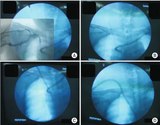
Blood aspiration test confirmed intravascular placement that showed no resistant regurgitation. A hydrophilic guide wire was inserted and a 14.5F 19-cm perm catheter insertion was finished as usual (Fig. 2). The right IJV permcath functioned well. The brachio-jugular AVG created later at the left upper-arm of the patient.
PA-LT
A
B C
D E
0.15 cm
2
1
Fig. 1. (A) Hemodialysis catheter tip malposition induced catheter mal func tion. Both ascending arm venography showed stenosis of the left subclavian vein with col- laterals. Right innominate vein ste no sis also suspected in the ve no gra phy. (B) Doppler ultra- sono gra phy revealed already oc clu ded right IJV. Diameter of right IJV was checked as 1.5 mm.
Lumen of the vein was com ple- te ly collapsed without com pre- s sibility. (C) Diameter of left IJV was about 10 mm with out ste no- sis. (D) Chest X-ray after catheter insertion showed no immediate com pli cations. (E) Ve no graphy show ed not definite steno sis from left IJV to the right atrium.
Sungwoo Cho and Sangchul Yun: Occluded jugular vein cut-down method
268
Annals of Surgical Treatment and Research 2019;97(5):266-269
DISCUSSION
Perm catheter is ideally used with a plan for subsequent per- manent access. The order of preference for the site of placement of dialysis catheters is right IJV, left IJV, external jugular veins, subclavian veins, and femoral veins [2]. Subclavian catheter placement has a high association with stenosis. Subclavian catheter should be performed in the patient whom further arm accesses will not be planned. In this patient, hemodialysis was initially started with the permcath via the right IJV. After primary failure with right and left elbow AVF, the permcath was inserted via the left subclavian vein. When the patient was transferred to our hospital, left subclavian vein stenosis and right IJV occlusion was diagnosed. The right innominate vein also seemed to be slightly stenosed. However, the left IJV was intact. The diameter of the left IJV was about 10 mm. Jugular venography after left subclavian permcath removal also showed intact flow from the left IJV to the right atrium. The next permanent access could be AVG using the left IJV as outflow at the left upper-arm of the patient. The patient needed bridge permcath. Right IJV and left subclavian vein were stenosed after just one catheter procedure in this patient. Other vein sites such as a right subclavian vein or femoral vein would be at high risk of stenosis. The completely occluded right IJV was surgically reopened and used for catheter insertion successfully.
A percutaneous technique for unconventional access has been introduced in the literature [3]. Needle recanalization is a technique of using a needle to go through a chronically
thrombosed venous segment or to artificially create a new tract to the central vein. This technique creates a new tract by needle for catheter insertion. The mean patency of this percutaneous technique is reported as 13 months [4]. Needle recanalization needs stent insertion or balloon dilatation. Sometimes contrast extravasation into mediastinum could be seen [4]. However, open surgical method can create a passageway through the true lumen or subintimal space of a totally occluded IJV. Garrett vascular dilators have a blunt smooth tip. Vein perforation can be prevented with gentle maneuvering under C-arm guide. Even extravasation in cases of vessel tearing is limited due to totally collapsed jugular vein. Furthermore, a ‘cut-down’ method is less expensive than percutaneous needle recanalization. With this technique, a previously exhausted vein can be reused for dialysis. Using a femoral vein for catheter insertion could be avoided.
CONFLICTS OF INTEREST
No potential conflict of interest relevant to this article was reported.
ACKNOWLEDGEMENTS
This work was supported by the Soonchunhyang University Research Fund.
A B
C D
Fig. 2. (A) Garrett vascular dilator was inserted into right inno mi- nate vein through previously oc- clu ded right internal jugular vein.
Ascending arm venography was overlaped on the C-arm ima ge.
This image confirmed the exact position of vascular di lator. (B) Plas tic vein dilator was inserted into the vein. Blood aspiration test showed regur gitation without sig ni ficant regi stant. (C) Tunneled cuffed dual lumen hemodialysis ca the ter was placed. (D) There was no im me diate complication such as hemopneumothorax, ca the ter kin king, catheter mal- po si tion, etc. Immediate cathe ter func tion test with syringe as pi- ra tion sho wed no resistant. Left perm cathe ter removal was done.
Annals of Surgical Treatment and Research 269
REFERENCES
1. Schmidli J, Widmer MK, Basile C, de Donato G, Gallieni M, Gibbons CP, et al.
Editor’s choice - vascular access: 2018 clinical practice guidelines of the Euro- pean Society for Vascular Surgery (ESVS).
Eur J Vasc Endovasc Surg 2018;55:757-818.
2. Vascular Access 2006 Work Group. Clini-
cal practice guidelines for vascular access.
Am J Kidney Dis 2006;48 Suppl 1:S176-247.
3. Rahman S, Kuban JD. Dialysis catheter place ment in patients with exhausted ac- cess. Tech Vasc Interv Radiol 2017;20:65- 74.
4. Funaki B, Zaleski GX, Leef JA, Lorenz JN,
Van Ha T, Rosenblum JD. Radiologic place- ment of tunneled hemodialysis cathe ters in occluded neck, chest, or small thyro- cervical collateral veins in central venous occlusion. Radiology 2001;218:471-6.
Sungwoo Cho and Sangchul Yun: Occluded jugular vein cut-down method