─ 43 ─ eISSN 2287-1683
pISSN 1738-8767
Journal of Trauma and Injury Vol. 28, No. 1, March, 2015
� Case Report �
� Address for Correspondence : Ki Seong Eom, M.D., Ph.D.
Department of Neurosurgery, Wonkwnag University School of Medicine, 344-2 Shinyong-dong, Iksan, Jeonbuk, #570-711, Korea
Tel : 82-63-859-1467, Fax : 82-63-852-2606, E-mail : [email protected] Submitted : March 6, 2015 Revised : March 19, 2015 Accepted : March 19, 2015
양측성 만성 경막하출혈의 자발적 흡수: 증례보고
원광대학교 의과대학, 1원광대학교 의과대학 신경외과학교실
선경웅, 박지민1, 엄기성1
- Abstract -
Bilateral Spontaneous Resolution of Chronic Subdural Hematoma: A Case Report
Gyeongung Seon, Ji-Min Park, M.D.
1, Ki Seong Eom, M.D., Ph.D.
1Wonkwang University School of Medicine, Iksan, Korea
1Department of Neurosurgery, Wonkwang University School of Medicine, Iksan, Korea
Although spontaneous resolution of chronic subdural hematoma (C-SDH) in the elderly has rarely been reported, spontaneous resolution of bilateral C-SDH is very rare. Here, we report the case of a 73-year-old female patient with no significant head trauma history who had a bilateral C-SDH spontaneously resolve despite receiving only conservative treatment. However, because of a lack of detailed knowledge about the mechanisms of resolution, treatment is often limited to surgical interventions that are generally successful, but invasive and prone to recurrence. We review the litera- ture and discuss the possible relation of C-SDH’s spontaneous resolution with its clinical and radiological characteristics.
[ J Trauma Inj 2015; 28: 43-46 ]
Key Words: Chronic subdural hematoma, Elderly, Spontaneous resolution
I.
Introduction
Chronic subdural hematoma (C-SDH) is a rela- tively common condition in old age and is thought to develop when head injury causes trauma to vessels between the arachnoid and dura mater.(1,2) There have been many surgical and nonsurgical tech- niques to treat C-SDH. Burr-hole trephination and
hematoma removal being the widely used effective surgical treatments.(2,3) However, rarely case reports have demonstrated the spontaneous resolu- tion of C-SDH by conservative treatment. Here, we report a spontaneously resolving C-SDH and review the previously reported cases, and their clinical characteristics.
─ 44 ─
- Journal of Trauma and Injury Vol. 28, No. 1 -
II.
Case Report
Four months before her visit to our hospital, a 73- year-old female was admitted to a local clinic for headache and dizziness. Her medical history was significant for diabetes and hypertension, for which
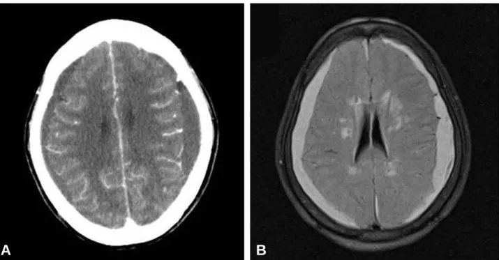
the patient had been taking oral medications for several years. In addition, she was suffering from an unstable angina, and was taking anticoagulants. She had no memory of her head injury. Neurological examination revealed only a decrease of cerebral cognitive functions (Mini-mental state examina- tion-K: 23). Her Glasgow coma scale was 15. Brain computed tomography (CT) scans revealed an iso- dense C-SDH along both cerebral convexities, with- out a significant mass effect (Fig. 1A). Magnetic res- onance imaging (MRI) revealed C-SDH of the bilat- eral frontotemporoparietal areas, with mild brain atrophy (Fig. 1B). She was treated conservatively because she had a stable neurologic status. Her headache and dizziness diminished steadily without any significant medication use. Follow-up CT scans obtained after 2 month showed that the hematoma had disappeared completely (Fig. 2). One year after her initial symptoms developed, the patient is doing well, and no complications in relation to the C-SDH have been observed.
III.
Discussion
C-SDH is a relatively common neurosurgical dis- order that has a high incidence in the geriatric pop- Fig. 1. Brain computed tomography (A) and magnetic resonance imaging (B) show chronic subdural hematoma along both cerebral
convexities without a significant mass effect.
A B
Fig. 2. Follow-up computed tomography image obtained after 2 month showing complete disappearance of the hematoma.
ulation. Pathologically, C-SDH is defined as a per- sistent liquefied old hematoma in the subdural space lasting more than three weeks and encased by a membranous capsule.(3) Generally, surgical methods like burr-hole trephination with closed-system drainage, craniotomy, subduro-peritoneal shunt, or twist-drill drainage are considered. Burr-hole trephination is most used as a first-line treatment because of the simplicity and effectiveness of the procedure.(4-6) However, the reoperation rate after burr-hole trephination is estimated at 5~24%.(7) The natural history and mechanisms of spontaneous hematoma resolution are still controversial. Various theories have been suggested to explain the mecha- nisms of the formation and resolution of C-SDH.
Kawano and Suzuki reported that the modified smooth-muscle cells in the outer membrane might play a role in the resolution of C-SDH, since the cells produce collagen that reinforces the membrane and reduces its fragility.(8) Yamashima et al.(9) described the structure of macrocapillaries, also called ‘sinusoids’, in the outer membrane of sub- dural hematomas. They suggested that the endothe- lial gap junctions of macrocapillaries in the outer membrane play an important role in the leakage of blood, causing hematoma enlargement by microhe- morrhages and increased fibrinolytic activity. The endothelial gap junctions are sometimes bridged by platelets, reducing microhemorrhages and the size of the hematoma. Nakamura et al.(10) reported that ventricular dilatation elevates counter pressure against the subdural hematoma, which may be important in its spontaneous resolution. Lee postu- lated that maturation of the neomembrane and sta- bilization of the neovasculature might result in the spontaneous resolution of hematoma.(11)
Some C-SDHs can be spontaneously resolved by medication. Ambrosetto reported that hematomas disappeared by treatment with adrenocortical hor- mone and 50% glucose, without surgery.(12) Giuffre´
suggested that hormonal factors play an important role in the pathogenesis of subdural hematomas.(13) Glover and Labadie proposed that corticosteroids inhibited the formation of protein-permeable mem- branes, decreasing the size of C-SDH.(14) There are few reports of specific underlying disorders in the
literature of total spontaneous resolution of C-SDH.
Hakan Sec et al.(15) reported the spontaneous dis- appearance of C-SDH in idiopathic thrombocy- topenic purpura despite very low platelet levels.
Takami et al.(16) reported the spontaneous rapid resolution of a large C-SDH in the posterior fossa of a patient with aplastic anemia who was receiving therapeutic platelet and red blood cell infusions periodically. Jukovic´ et al.(17) reported a patient with liver failure, who received conservative treat- ment and showed complete resolution of their C- SDH. Parlato et al.(18) insisted indications for con- servative treatment include being over 70 years of age, worsening mental function, presence of brain atrophy, and absence of clinical and radiological symptoms of increased intracranial pressure are clinical and radiological signs that allow one to choose conservative treatment.
In the present case, the patient had unstable angina and was administered anticoagulants, but had clinically mild symptoms and brain atrophy.
However, no significant mass effect on brain CT correlated with the resolution of her hematoma.
Spontaneous resolution of bilateral C-SDH within 2 month is a very rare event. We think that the clini- cal and radiological characteristics of the hematoma may be associated with its spontaneous resolution.
However, further investigation is required.
REFERENCES
01) Hancock DO. Cerebral collapse associated with chronic sub- dural hematoma in adults. A comparison of two methods of treatment. Lancet 1965; 20: 633-4.
02) Tabaddor K, Shulman K. Definitive treatment of chronic sub- dural hematoma by twist-drill craniotomy and closed-system drainage. J Neurosurg 1977; 46: 220-6.
03) Markwalder TM. Chronic subdural hematomas: a review. J Neurosurg 1981; 54: 637-45.
04) Camel H, Grubb RL. Treatment of chronic subdural hematoma by twist-drill craniotomy with continuous catheter drainage. J Neurosurg 1986; 65: 183-7.
05) Hamilton MG, Frizzell JB, Tranmer BI. Chronic subdural hematoma: The role of craniotomy reevaluated. Neurosurgery 1993; 33: 67-72.
06) Park BA, Lee YK, Kim YT, Moon ES. The risk factor associ- ated with hypertensive intracerebral hemorrhage. J Korean Geriatr Soc 2004; 8: 223-7.
07) Benzel EC, Bridges RM Jr, Hadden TA, Orrison WW. The
─ 45 ─
Gyeongung Seon, et al.: Bilateral Spontaneous Resolution of Chronic Subdural Hematoma: A Case Report
single burr hole technique for the evacuation of non-acute sub- dural hematomas. J Trauma 1994; 36: 190-4.
08) Kawano N, Suzuki K. Presence of smooth-muscle cells in the subdural neomembrane. J Neurosurg 1981; 54: 646-51.
09) Yamashima T, Yamamoto S, Friede RL. The role of endothe- lial gap junctions in the enlargement of chronic subdural hematomas. J Neurosurg 1983; 59: 298-303.
10) Naganuma H, Fukamachi A, Kawakami M, Misumi S, Nakajima H, Wakao T. Spontaneous resolution of chronic subdural hematomas. Neurosurgery 1986; 19: 794-8.
11) Lee KS. Natural history of chronic subdural hematoma. Brain Inj 2004; 18: 351-8.
12) Ambrosetto C. Post-traumatic subdural hematoma: Further observations on nonsurgical treatment. Arch Neurol 1962; 6:
287-92.
13) Giuffré R. Physiopathogenesis of chronic subdural hematomas:
a new look to an old problem. Riv Neurol 1987; 57: 298-304.
14) Glover D, Labadie EL. Physiopathogenesis of subdural
hematomas. Part 2: Inhibition of growth of experimental hematomas with dexamethasone. J Neurosurg 1976; 45: 393-7.
15) Seçkin H, Kazanci A, Yigitkanli K, Simsek S, Kars HZ.
Chronic subdural hematoma in patients with idiopathic throm- bocytopenic purpura: a case report and review of the literature.
Surgical Neurology 2006; 66: 411-4.
16) Takami H, Oshiro N, Hiraoka F, Murao M, Ide T. Rapid reso- lution of a spontaneous large chronic subdural haematoma in the posterior fossa under conservative treatment with platelet administration to aplastic anaemia. Clin Neurol Neurosurg 2013; 115: 2236-9.
17) Jukovic´ M, Kojadinovic´ Z, Popovska B, Till VC. Complete spontaneous resolution of compressive chronic subdural hematoma in a patient with liver failure. Med Glas (Zenica) 2012; 9: 417-20.
18) Ciro Parlato, Antonio Guarracino, Aldo Moraci. Spontaneous resolution of chronic subdural hematoma. surg Neurol 2000;
53: 312-7.
─ 46 ─
- Journal of Trauma and Injury Vol. 28, No. 1 -