MATERIALS AND METHODS Patients’ population
The authors retrospectively reviewed the medical records as well as the pre and postoperative computed tomography (CT) scans of the 182 patients who underwent burr-hole trephination from January 2008 to December 2012. There were 131 male (71.9%) and 51 (28.1%) female in this study, ranging in age from 24 to 94 years old (median age : 68.12 years old). The patients were divided into two groups according to the recurrence of CSDH. The clinical and radiological factors were compared be- tween the recurrence group and the no recurrence group. In this study, 35 patients with bilateral CSDH were excluded in analyz- ing the CT density, the width of hematoma, the degree of air col- lection, the number of burr hole, and the drainage tube.
Clinical and radiological evaluations
The recurrence of CSDH was defined as a subsequent increase in hematoma volume in the subdural space for which reopera- INTRODUCTION
Chronic subdural hematoma (CSDH) is one of the most com- monly encountered entities in neurosurgery practice. The inci- dence rate of CSDH has been reported to be as high as 13.1 cas- es per 100000 inhabitants24). It is common in the elderly, with the highest incidence rate in people older than 70 years17,18,23).
Surgical management is the preferred treatment for CSDH, which includes twist-drill drainage, burr-hole trephination, cra- niotomy with capsulectomy, and subduro-peritoneal shunt.
Among them, burr-hole trephination is widely performed be- cause of its less invasiveness and relative simplicity15). However, the incidence rate of recurrent CSDH is reported to range from 2 to 37%4,7,11,12,14,16,21,22,24). Furthermore, the crucial risk factors are still debatable despite the various elements that may be as- sociated with the recurrence of CSDH. Thus, the goal of this study is to determine the predictors for recurrence of CSDH af- ter burr-hole trephination.
•Received : September 30, 2014 •Revised : January 19, 2015 •Accepted : January 20, 2015
•Address for reprints : El Kim, M.D., Ph.D.
Department of Neurosurgery, Dongsan Medical Center, Keimyung University School of Medicine, 56 Dalseong-ro, Jung-gu, Daegu 700-712, Korea Tel : +82-53-250-7823, Fax : +82-53-250-7356, E-mail : [email protected]
•This is an Open Access article distributed under the terms of the Creative Commons Attribution Non-Commercial License (http://creativecommons.org/licenses/by-nc/3.0) which permits unrestricted non-commercial use, distribution, and reproduction in any medium, provided the original work is properly cited.
Independent Predictors for Recurrence of Chronic Subdural Hematoma
Yoon-Gyo Jung, M.D., Na-Young Jung, M.D., El Kim, M.D., Ph.D.
Department of Neurosurgery, Dongsan Medical Center, Keimyung University School of Medicine, Daegu, Korea
Objective : Chronic subdural hematoma (CSDH) is one of the most frequent problems encountered in neurosurgery. Although burr-hole trephination is widely performed to treat CSDH, the incidence rate of recurrent CSDH is still 2–37%. The goal of this study is to determine the risk factors that affect recurrent CSDH.
Methods : A total of 182 patients were included in this study who underwent burr-hole trephination. The clinical factors and radiographic features between the recurrence and the no recurrence groups were analyzed to find the parameters related to the postoperative recurrence of CSDH.
Results : For the recurrence of CSDH that occurred in 25 patients (13.7%), among various risk factors, pre and postoperative midline displace- ments, which are more than 10 mm (p=0.000), and preoperative hemiparesis (p=0.026) had contributed to recurrent CSDH with statistical signifi- cance by univariate analysis. Unilateral CSDH were more frequently related to recurrent CSDH (16.3%), although it was not a statistical significant result (p=0.052). Furthermore, preoperative midline displacement only had statistical meaning for the recurrence of CSDH by multivariate analysis.
Conclusion : This study indicates that the midline displacement on the preoperative computed tomography scan is the only independent predictor for the recurrence of CSDH.
Key Words : Chronic subdural hematoma · Hemiparesis · Midline displacement · Recurrence.
Clinical Article
pine position and enough fluid was supplied to promote brain expansion. The closed-system drainage tube was usually re- moved after 2–3 days, when the drainage became negligible.
Statistical analysis
A univariate analysis was performed with Pearson’s chi-square test and Student t-test to assess the relationship between each fac- tor and the recurrence of CSDH. A multivariate analysis was per- formed using a logistic regression model. The relationship be- tween the variables and the recurrence of CSDH are presented based on the 95% confidence interval and the odd ratio (OR).
The statistical significance was set at p<0.05.
RESULTS
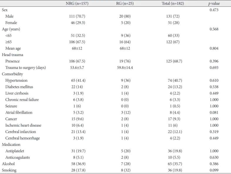
Patients’ characteristics are summarized in Table 1. Reopera- tion was performed in 25 patients (13.7%) because of the symp- tomatic recurrence of CSDH. The demographic data and history of head trauma were not significantly associated with recurrence of CSDH. Underlying medical diseases, antithrombotic usage, and smoking or alcohol consumption were also not related to the recurrence of CSDH.
tion was required because of newly developed symptoms21). Hematoma type was classified into four types according to their density on CT scan : homogeneous, laminar, separate, and trabec- ular type14). Brain atrophy was divided into three stages : none or mild atrophy, definite atrophy, such as dilated sulci, and severe at- rophy, such as widely dilated sulci and subdural space10,17). Air col- lection was categorized into two groups according to the sub- dural air found on immediate postoperative CT scan : none or mild, such as residual air bubble, and definite, such as total re- placement with air17). The midline displacement of the septum pellucidum and pineal body were measured at the level of fora- men of Monro on the CT scans taken before and after surgery.
The cut-off values for the midline displacement were defined based on previous reports10,16,19). Follow-up was for at least 1 year.
Surgical procedure and management
All patients underwent one or two burr-hole trephination with closed-system drainage under general anesthesia. After dural incision, the outer hematoma membrane was opened, and irri- gation was performed with lactated Ringer solution. A silicon tube was inserted into the cavity of the hematoma and connect- ed to a closed drainage system. All patients were kept in the su-
Table 1. Characteristics in 182 patients with chronic subdural hematomas
NRG (n=157) RG (n=25) Total (n=182) p value
Sex 0.473
Male 111 (70.7) 20 (80) 131 (72)
Female 46 (29.3) 5 (20) 51 (28)
Age (years) 0.568
<65 51 (32.5) 9 (36) 60 (33)
≥65 106 (67.5) 16 (64) 122 (67)
Mean age 68±12 68±12 0.804
Head trauma
Presence 106 (67.5) 19 (76) 125 (68.7) 0.396
Trauma to surgery (days) 53.6±5.7 59.8±14.4 0.693
Comorbidity
Hypertension 65 (41.4) 9 (36) 74 (40.7) 0.610
Diabetes mellitus 22 (14) 2 (8) 24 (13.2) 0.538
Liver cirrhosis 3 (1.9) 1 (4) 4 (2.2) 0.449
Chronic renal failure 6 (3.8) 0 (0) 6 (3.3) 1.000
Seizure 1 (6) 0 (0) 1 (0.5) 1.000
Atrial fibrillation 5 (3.2) 3 (12) 8 (4.4) 0.081
Cancer 15 (9.6) 2 (8) 17 (9.3) 1.000
Ischemic heart disease 10 (6.4) 1 (4) 11 (6) 1.000
Cerebral infarction 21 (13.4) 1 (4) 22 (12.1) 0.319
Cerebral hemorrhage 3 (1.9) 1 (4) 4 (2.2) 0.449
Medication
Antiplatelet 31 (19.7) 5 (20) 36 (19.8) 1.000
Anticoagulants 8 (5.1) 2 (8) 10 (5.5) 0.630
Alcohol 58 (36.9) 7 (28) 65 (35.7) 0.386
Smoking 28 (17.8) 8 (32) 36 (19.8) 0.099
NRG : no recurrence group, RG : recurrence group
CSDH plays an important role in the enlargement of CSDHs1). The reported recurrence rates of CSDH range from 2 to 37%, and this study showed a recurrence rate of 13.7%. In the previ- ous study, a few factors for the recurrence of CSDH have been reported, such as advanced age, bleeding tendency, brain atro- phy, alcohol abuse, as well as bilateral CSDH, hematoma density, diabetes mellitus, arachnoid cyst, postoperative posture, postop- erative subdural air accumulation, inflammatory cytokines, and some technical aspects of surgery3,5,13,23). However, the crucial risk factors are debatable until now. In this study, the recurrence of CSDH was correlated with the following variables : 1) mid- line displacement of more than 10 mm and 2) clinical presenta- tion of hemiparesis.
Midline displacement
The authors found that the pre and postoperative midline dis- placement of more than 10 mm were the predictors for the re- currence of CSDH. In the series reported, the recurrence rate was significantly higher when the postoperative midline displace- ment was more than 5 mm compared to less displacement2,19). In patients with CSDH, it is reasonable to evaluate the pre and postoperative midline displacement because it shows that hem- orrhage has filled up the potential space in the cranium and has exerted compression on the brain tissue. Fukuhara and cowork- ers showed that advanced age, brain atrophy, large amount of hematoma, and prolonged compressed parenchyma influenced brain elasticity2,6). Brain with high elastance tends to reexpand poorly, and poor reexpansion of the brain may lead to the per- sistence of postoperative midline displacement2,6). A prolonged postoperative midline displacement may cause impaired adhe- sion between the inner and outer neomembranes, thus facilitat- ing postoperative recurrence2,15).
Hemiparesis
CSDH is asymptomatic in a large number of patients, but it may also cause high intracranial pressure that results in coma.
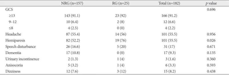
Among these extreme states, nearly every constellation of speech, sensorimotor, neuropsychiatric, or mood disturbances may oc- Clinical manifestations between the recurrence and the no
recurrence group are shown in Table 2. The incidence of recur- rence in patients with hemiparesis (18.8%) was higher than that in those without hemiparesis (7.4%) with statistical significance (p=0.026). However, any other clinical factors were not related to the recurrence of CSDH.
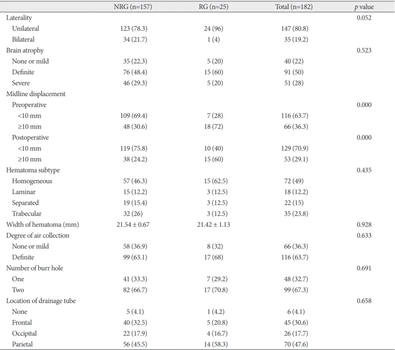
Comparison of radiological factors between the two groups is demonstrated in Table 3. In patients with unilateral CSDH, the incidence of recurrence (16.3%) was higher than that in pa- tients with bilateral CSDH (2.9%). Although it did not get the statistical significance (p=0.052), unilateral CSDH tended to be associated with recurrence of CSDH. In patient with midline displacement of more than 10 mm before surgery, the incidence of recurrence rate (27.3%) was higher than that in patients with midline displacement of less than 10 mm (6.0%). The relation- ship between the preoperative midline displacement and the re- currence of CSDH was statistically significant (p=0.000). Im- mediate postoperative midline displacement of more than 10 mm was also associated with the recurrence of CSDH (31.3%
vs. 7.7%; p=0.000). Other radiological parameters did not have statistical meaning to affect recurrent CSDH.
In summary, univariate analysis clarified that hemiparesis (p=0.026), pre (p=0.000) and postoperative (p=0.000) midline displacement of more than 10 mm were risk factors for recur- rence of CSDH. The preoperative midline displacement of more than 10 mm (OR, 5.7; 95%; p=0.000) was the only independent risk factor for the recurrence of CSDH by multivariate logistic regression analysis (Table 4).
DISCUSSION
CSDH is defined as a watery or xanthochromic fluid collec- tion under dura mater. The mechanism of CSDH is a patho- physiologic process that begins as a local inflammatory reaction of the dura mater to injury or external stimuli, such as blood or CSF. This process causes the neovascularization of the outer membrane of CSDH and vascular hyperperrmeability. Exuda- tion through the microcapillaries in the outer membrane of
Table 2. Relationship between clinical presentation and recurrence of chronic subdural hematoma
NRG (n=157) RG (n=25) Total (n=182) p value
GCS 0.696
≥13 143 (91.1) 23 (92) 166 (91.2)
9–12 10 (6.4) 2 (8) 12 (6.6)
≤8 4 (2.5) 0 (0) 4 (2.2)
Headache 87 (55.4) 14 (56) 101 (55.5) 0.956
Hemiparesis 82 (52.2) 19 (76) 101 (55.5) 0.026
Speech disturbance 26 (16.6) 5 (20) 31 (17) 0.671
Dementia 17 (10.8) 0 (0) 17 (9.3) 0.135
Urinary incontinence 2 (1.3) 1 (4) 3 (1.6) 0.360
Anisocoria 5 (3.2) 1 (4) 6 (3.3) 0.593
Dizziness 12 (7.6) 3 (12) 15 (8.2) 0.438
NRG : no recurrence group, RG : recurrence group, GCS : Glasgow Coma Scale
thalamus8,20). When the hematoma thickness increased beyond spatial compensation, both the superficial and deep brain struc- tures shifted and deformed, and hemiparesis occurred in rela- tion to the degree of midline displacement9). In other words, based on the aggravation of the midline displacement, hemipa- resis occurred.
cur24). In the present study, hemiparesis and headache are the most common preoperative symptoms in CSDH, and the inci- dence of recurrent CSDH in patients with hemiparesis is higher than that in those without hemiparesis. The cause of hemiparesis in CSDH was reported to be the reduced local cerebral blood flow in the rolandic cortex or in deep structures, including the
Table 3. Summary of perioperative CT findings in recurrence group and no recurrence group
NRG (n=157) RG (n=25) Total (n=182) p value
Laterality 0.052
Unilateral 123 (78.3) 24 (96) 147 (80.8)
Bilateral 34 (21.7) 1 (4) 35 (19.2)
Brain atrophy 0.523
None or mild 35 (22.3) 5 (20) 40 (22)
Definite 76 (48.4) 15 (60) 91 (50)
Severe 46 (29.3) 5 (20) 51 (28)
Midline displacement
Preoperative 0.000
<10 mm 109 (69.4) 7 (28) 116 (63.7)
≥10 mm 48 (30.6) 18 (72) 66 (36.3)
Postoperative 0.000
<10 mm 119 (75.8) 10 (40) 129 (70.9)
≥10 mm 38 (24.2) 15 (60) 53 (29.1)
Hematoma subtype 0.435
Homogeneous 57 (46.3) 15 (62.5) 72 (49)
Laminar 15 (12.2) 3 (12.5) 18 (12.2)
Separated 19 (15.4) 3 (12.5) 22 (15)
Trabecular 32 (26) 3 (12.5) 35 (23.8)
Width of hematoma (mm) 21.54 ± 0.67 21.42 ± 1.13 0.928
Degree of air collection 0.633
None or mild 58 (36.9) 8 (32) 66 (36.3)
Definite 99 (63.1) 17 (68) 116 (63.7)
Number of burr hole 0.691
One 41 (33.3) 7 (29.2) 48 (32.7)
Two 82 (66.7) 17 (70.8) 99 (67.3)
Location of drainage tube 0.658
None 5 (4.1) 1 (4.2) 6 (4.1)
Frontal 40 (32.5) 5 (20.8) 45 (30.6)
Occipital 22 (17.9) 4 (16.7) 26 (17.7)
Parietal 56 (45.5) 14 (58.3) 70 (47.6)
NRG : no recurrence group, RG : recurrence group
Table 4. Results of univariate and multivariate analysis of variables related to the recurrence of chronic subdural hematoma
OR (95% CI) p value
Univariate analysis
Hemiparesis 2.896 (1.098–7.639) 0.026
Laterality 0.151 (0.020–1.155) 0.052
Preoperative midline displacement 5.839 (2.288–14.900) 0.000
Postoperative midline displacement 4.697 (1.949–11.320) 0.000
Multivariate analysis
Preoperative midline displacement 5.707 (2.156–15.101) 0.000
OR : odds ratio, CI : confidence interval
chiatry 71 : 741-746, 2001
10. Ko BS, Lee JK, Seo BR, Moon SJ, Kim JH, Kim SH : Clinical analysis of risk factors related to recurrent chronic subdural hematoma. J Korean Neurosurg Soc 43 : 11-15, 2008
11. Kotwica Z, Brzeziński J : Chronic subdural haematoma treated by burr holes and closed system drainage : personal experience in 131 patients.
Br J Neurosurg 5 : 461-465, 1991
12. Kwon TH, Park YK, Lim DJ, Cho TH, Chung YG, Chung HS, et al. : Chronic subdural hematoma : evaluation of the clinical significance of postoperative drainage volume. J Neurosurg 93 : 796-799, 2000 13. Markwalder TM : Chronic subdural hematomas : a review. J Neurosurg
54 : 637-645, 1981
14. Nakaguchi H, Tanishima T, Yoshimasu N : Factors in the natural history of chronic subdural hematomas that influence their postoperative re- currence. J Neurosurg 95 : 256-262, 2001
15. Nakaguchi H, Tanishima T, Yoshimasu N : Relationship between drain- age catheter location and postoperative recurrence of chronic subdural hematoma after burr-hole irrigation and closed-system drainage. J Neurosurg 93 : 791-795, 2000
16. Ohba S, Kinoshita Y, Nakagawa T, Murakami H : The risk factors for re- currence of chronic subdural hematoma. Neurosurg Rev 36 : 145-149;
discussion 149-150, 2013
17. Oishi M, Toyama M, Tamatani S, Kitazawa T, Saito M : Clinical factors of recurrent chronic subdural hematoma. Neurol Med Chir (Tokyo) 41 : 382-386, 2001
18. Okada Y, Akai T, Okamoto K, Iida T, Takata H, Iizuka H : A compara- tive study of the treatment of chronic subdural hematoma--burr hole drainage versus burr hole irrigation. Surg Neurol 57 : 405-409; discus- sion 410, 2002
19. Stanisic M, Lund-Johansen M, Mahesparan R : Treatment of chronic subdural hematoma by burr-hole craniostomy in adults : influence of some factors on postoperative recurrence. Acta Neurochir (Wien) 147 : 1249-1256; discussion 1256-1257, 2005
20. Tanaka A, Yoshinaga S, Kimura M : Xenon-enhanced computed tomo- graphic measurement of cerebral blood flow in patients with chronic subdural hematomas. Neurosurgery 27 : 554-561, 1990
21. Torihashi K, Sadamasa N, Yoshida K, Narumi O, Chin M, Yamagata S : Independent predictors for recurrence of chronic subdural hematoma : a review of 343 consecutive surgical cases. Neurosurgery 63 : 1125- 1129; discussion 1129, 2008
22. Wakai S, Hashimoto K, Watanabe N, Inoh S, Ochiai C, Nagai M : Effi- cacy of closed-system drainage in treating chronic subdural hematoma : a prospective comparative study. Neurosurgery 26 : 771-773, 1990 23. Yamamoto H, Hirashima Y, Hamada H, Hayashi N, Origasa H, Endo S :
Independent predictors of recurrence of chronic subdural hematoma : results of multivariate analysis performed using a logistic regression model. J Neurosurg 98 : 1217-1221, 2003
24. Youmans JR, Winn HR : Youmans Neurological Surgery, ed 6. New York : W.B. Saunders, 2011, pp532-543
Laterality
In previous studies, hematoma laterality was not associated with the recurrence of CSDH5,23). Inconsistent with previous studies, our study show that the incidence of recurrence in pa- tients with unilateral CSDH (16.3%) was higher than that in pa- tients with bilateral CSDH (2.9%). Although it did not get the statistical significance (p=0.052) because of the lack of the num- ber of cases, unilateral CSDH tended to be associated with re- currence of CSDH. As such, further study is necessary to clarify the association between the laterality of hematoma and the re- currence of CSDH.
CONCLUSION
The present study suggested that patients who show hemipa- resis or pre and postoperative midline displacement of more than 10 mm could go through recurrent CSDH and shall un- dergo further surgical management. Therefore, the clinical and radiological surveillance is essential for the patients who have moderate midline displacement or motor weakness.
References
1. Abouzari M, Rashidi A, Rezaii J, Esfandiari K, Asadollahi M, Aleali H, et al. : The role of postoperative patient posture in the recurrence of trau- matic chronic subdural hematoma after burr-hole surgery. Neurosur- gery 61 : 794-797; discussion 797, 2007
2. Chon KH, Lee JM, Koh EJ, Choi HY : Independent predictors for recur- rence of chronic subdural hematoma. Acta Neurochir (Wien) 154 : 1541-1548, 2012
3. El-Kadi H, Miele VJ, Kaufman HH : Prognosis of chronic subdural he- matomas. Neurosurg Clin N Am 11 : 553-567, 2000
4. Ernestus RI, Beldzinski P, Lanfermann H, Klug N : Chronic subdural hematoma : surgical treatment and outcome in 104 patients. Surg Neu- rol 48 : 220-225, 1997
5. Foelholm R, Waltimo O : Epidemiology of chronic subdural haemato- ma. Acta Neurochir (Wien) 32 : 247-250, 1975
6. Fukuhara T, Gotoh M, Asari S, Ohmoto T, Akioka T : The relationship between brain surface elastance and brain reexpansion after evacuation of chronic subdural hematoma. Surg Neurol 45 : 570-574, 1996 7. Hamilton MG, Frizzell JB, Tranmer BI : Chronic subdural hematoma :
the role for craniotomy reevaluated. Neurosurgery 33 : 67-72, 1993 8. Ikeda K, Ito H, Yamashita J : Relation of regional cerebral blood flow to
hemiparesis in chronic subdural hematoma. Surg Neurol 33 : 87-95, 9. Inao S, Kawai T, Kabeya R, Sugimoto T, Yamamoto M, Hata N, et al. : 1990 Relation between brain displacement and local cerebral blood flow in patients with chronic subdural haematoma. J Neurol Neurosurg Psy-