Introduction
C alcified chronic subdural hematoma is realtively com- mon since the first description in 18841,9,10). In the elderly, obsevation is recommened for asymptomatic calcified chronic subdural hematoma without acute or progre- ssive neurological disorders
3,7). However, the incidences of clinically exacerbated due to its rupture into the subcortical white matter is very rare.
Case Report
A 37-year-old male patient having a 15-year seizure history was admitted to hospital, who presented highly frequent seizure recently. The patient had been in difficulties maintaining his normal life because of the cerebral palsy since the childhood and no recent head trauma history was found. When admitted, he was alert and had a little larger head circumference. Laboratory examination revealed no bleeding tendency. In the plain skull film, the left fronto- temporal bone was thickened and an oval-shaped large calcification lesion was observed Brain computed tomography scans demonstrating a hyperdense extracerebral mass with calcified margin at the left frontotemporal lobe and severe brain atrophy, ventricular dilatation and hematoma infer- omedially at the frontal lobe were observed. By performing
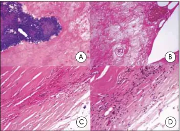
craniotomy, calcified chronic subural hematoma and the membrane were completely eliminated and then intracerebral hematoma was aspirated(Fig. 1). According to the intraope- rative findings, the dura mater was very thickened, the outer membrane was partially calcified, and the mass contents showed a yellowish brown semisolid state. The inner membrane is as thick as, or thicker than, the outer membrane and severely adhered to the arachnoid membrane, which was dissected and removed. On the histopathological examination of the inner membrane of the calcified chronic subdural hematoma, there are hypertrophy due to the calcificaton and highly vascular granulation proliferation with scattered chronic inflammatory cells are also noted(Fig. 2). The seizure was considerably controlled after the operation.
Discussion
C alcification and ossification occurs in 0.8-10% of chronic subdural heamtoma patients5). The calcified chronic subdural hematoma is manifested mainly by seizure, dimentia, mental retardation, growth retardation or headache, but sometimes incidentally found without any symptom. The mechanism of calcification is that poor circulation and absorption in the subdural space and intravascular thrombo- sis
1). Moreover, there is a view that a local factor around the hematoma is involved in the calcification as well as the bilateral chronic subdural hematoma, which develops the calcification only in one side
7). Additionally, abnormal inherent metabolic tendency can play a role in calcification.
However, the mechanism of calcification is still unclear and the periods of calcification are quite different
2). Like the histopathological findings from this present case, the calcified
Calcified Chronic Subdural Hematoma Associated with Intracerebral Hematoma
Jong-Soo Park, M.D.,
1Eun-Ik Son, M.D.,
1Dong-Won Kim, M.D.,
1Sang-Pyo Kim, M.D.
2Departments of Neurosurgery,
1Pathology,
2Keimyung University School of Medicine, Daegu, Korea
Calcified chronic subdural hematoma is relatively common, however, its rupture into the subcortical white matter is very rare. A 37-year-old patient with a large, calcified, chronic subdural hematoma which ruptured intracerebrally forming a frontal lobe hemorrhage is reported. Craniotomy for removal of the hematoma and calcification achieved marked reduction in seizure frequency.
KEY WORDS : Calcified chronic subdural hematoma · Seizure · Intracerebral hematoma · Craniotomy.
VOLUME 34 August, 2003