CASE REPORT
pISSN 1738-2262/eISSN 2093-6729Korean Journal of Spine 9(1):41-43, 2012 www.e-kjs.org
Copyright © 2012 The Korean Spinal Neurosurgery Society 41
Idiopathic Lumbosacral Spinal Chronic Subdural Hematoma
HyunJin Ma, Insoo Kim
Department of Neurosurgery, Dongsan Medical Center, Keimyung University College of Medicine, Daegu, Korea Reports of spontaneous spinal chronic subdural hematoma (SDH) are extremely rare. In the limited cases reported, the origin, symptoms and treatments are diversely revealed. Therefore accurate diagnoses and proper treatments are difficult to determine. We report a 74-year-old male presented with severe low back pain and both buttocks pain. Magnetic resonance imaging showed a diffuse subdural infiltrating mass compress nerve roots at the level of L2-S2. Emergency operation for decompression and excision of mass revealed spinal chronic SDH. His pain was subsided after the operation.
We report a rare case of idiopathic lumbosacral spinal chronic SDH and discuss various causes and treatments.
Key Words: Spinal subdural hematomaㆍSpinal cord compression
●Received: January 8, 2012 ● Revised: March 24, 2012
●Accepted: March 26, 2012
Corresponding Author: Insoo Kim, MD, PhD
Department of Neurosurgery, Dongsan Medical Center, 56 Dalseong-ro, Jung-gu, Daegu 700-712, Korea
Tel: +82-53-250-7730, Fax: +82-53-250-7356 E-mail: [email protected]
INTRODUCTION
Spinal cord hematomas are uncommon. Among spinal cord hematomas, subdural hematoma (SDH) is an even more rare entity. Based on causes, spinal SDH is classified as idiopathic or secondary8,9). Spinal SDH can be developed as a result of anticoagulation therapy, blood dyscrasia, spinal puncture, trauma, spinal anesthesia or vascular malformation1,7). Spinal SDH is classified as acute, subacute, chronic according to the course of clinical symptoms8).
The symptoms of acute spinal SDH begin with severe and sudden onset back pain accompanied by severe radicular pain, and rapid progression to paraplegia within several days. The symptoms of subacute SDH begin with less well localized back, hip and leg pain, gradually progress to paraplegia over a period of 1 week. Reports of spontaneous spinal chronic SDH are extremely rare with only 23 cases5). Spinal chronic SDH begins with the signs and symptoms of progressive spinal cord com- pression occurring over a period of several months to several years, with mild or absent pain. Spontaneous spinal SDH occurs most commonly in the lower thoracic or thoracolumbar lesions and develops during the sixth decade of life5,12). For the best outcomes, early surgical decompression is commonly needed.
We report a case of spontaneous spinal chronic SDH located at lumbosacral region without underlying pathology.
CASE REPORT
A 74-year-old male was presented with low back pain and both buttocks pain for 16 days. The pain in both buttocks had gradually progressed to both legs in the L5 dermatome and the intensity of pain was aggravated during the last 3 days.
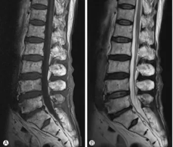
There were no history of trauma, infection or anticoagulant therapy. On admission, his vital signs were stable, and neuro- logical examination revealed normal tone in upper limb and lower limb. Power in extremities were grade 5/5 at all joints in Medical Research Council Muscle Strength Grading Scale2). The Plane X-ray films of lumbar spine showed multiple spur formation and intervertebral space narrowing at L5-S1 and some degenerative change. Lumbar spine magnetic resonance image revealed a diffuse subdural infiltrating mass compress nerve roots at the level of L2-S2. The mass presented an iso- to-hyperintense signal on T1-weighted magnetic resonance imaging (MRI) to the spinal cord, and an iso-to-hypointense signal on T2-weighted MRI (Fig. 1).
As the patient complained of unbearable pain, we under- went an emergency surgical decompression with L4 and L5 laminotomy. There was no obvious extradural hematoma and abnormality but the dura mater was bluish in color and tense.
After durotomy, subdural hematoma with thin membrane was exposed. The hematoma was dark, red wine color and well liquefied, and was evacuated by suction. After normal saline irrigation and bleeding control, watertight dural closure was
HJ Ma, et al.
42 www.e-kjs.org
Fig. 1. T1-weighted image shows an iso-to-hyperintense signal lesion at L2-S2 level (A). T2-weighted image shows an iso-to- hypointense signal lesion at L2-S2 level (B).
Fig. 2. Postoperative T1-weighted (A) and T2-weighted (B) images show no residual hematoma.
performed.
Postoperatively, pain in the buttocks and lower extremities were resolved. Two weeks after operation, the patient had no pain and was discharged. A follow-up MRI study performed 2 months after operation, documented no evidence of residual hematoma (Fig. 2). At 1-year-follow up, there was no postope- rative complication and the patient had no more pain.
DISCUSSION
Spinal SDH is remarkably uncommon, which must be diag- nosed accurately and promptly. In some cases, compression of the spinal cord by spinal SDH could lead to acute para- plegia and requires treatment before irreversible neurological damage occurs.
Based on causes, idiopathic spinal SDH is rare and in secon- dary spinal SDH, the most common causes of spinal SDH are coagulopathies, anticoagulant therapy, spinal trauma, or post iatrogenic circumstance7,9,11). Other various causes in- clude tumors, ruptured vascular malformation including arte- riovenous malformation, or aneurysm of spinal artery5,9,11).
The mechanisms of formation of spinal SDH remain unclear.
Various hypotheses of spinal SDH have been proposed, increa- sed intravascular pressure in subdural space due to increased intrathoracic or intraabdominal pressure resulting from trau- ma, cause a failure in the spinal cord vascular. Another hy- pothesis is migration of hematoma from intracranial subara- chnoid to the spine due to gravity8,13).
MRI provides signal from hematoma changes over time af- ter onset that is similar to those of brain hematoma5,6).
Immediately after attack, hematoma appearance on T1-wei- ghted image is intermediate signal intensity area and on T2- weighted image is low signal intensity. Between 7 to 14 days after attack, hematoma appears as high-to-intermediate signal intensity on T1-weighted image, and an increase in signal in- tensity on T2-weighted image.
In our case, signal intensity was high-to-intermediate in T1- weighted image, and low-to-intermediate image in T2-weigh- ted image. The MRI findings are not typical results of chronic spinal SDH which make preoperative diagnosis difficult. In the operation field, nature of hematoma was much liquefied and was dark, red wine like color. We could suck out hema- toma easily and irrigate hematoma completely.
The treatment of choice is laminectomy and drainage of hematoma4,9,13). Despite of spontaneous remission of intra- spinal hematoma has been reported, early surgical decompre- ssion is commonly required, because of progression of neuro- logical deterioration caused by spinal cord compression, ische- mia, and direct injury to spinal cord. Early surgical decompre- ssion results in a good outcome. The literature showed 80%
improvement in neurological function after surgery1,3,10,13).
CONCLUSION
Spinal chronic SDH is rare but should be considered in patients with sudden back pain, radiating pain to the upper or lower extremities or trunk and motor, sensory, autonomic disturbance. In these patients, prompt radiologic assessments, especially MRI, is required. Good outcome can be achieved with early diagnosis and surgical decompression.
Lumbosacral Spinal Chronic Subdural Hematoma
Korean Journal of Spine 9(1) March 2012 43
REFERENCES
1. Abla AA, Oh MY: Spinal chronic subdural hematoma. Neuro- surg Clin N Am 11:465-471, 2000
2. Amidei C.: Measurement of physiologic responses to mobi- lisation in critically ill adults. Intensive Crit Care Nurs 3:2012 3. Brandt RA: Choronic spinal subdural hematoma. Surg Neurol
13:121-123, 1980
4. Dampeer RA: Spontaneous spinal subdural hematoma. Am J Crit Care 19:191-193, 2010
5. Diyora B, Sharma A, Mamidanna R, Kamat L: Chronic cervico- thoracic spinal subdural hematoma. Neurol Med Chir (Tokyo) 49(7):310-312, 2009
6. Duprez T, Grandin C, Malghem J: MRI monitoring of an acute spinal subdural hematoma with spontaneous resolution. Acta Neurol Belg 95:101-103, 1995
7. Greiner Perth-R, Mohsen Allam Y, Sibermann J, Gahr R:
Traumatic subdural hematoma of the thoraco-lumbar junction
of spinal cord. J Spinal Disord Tech 20(3):239-241, 2007 8. Hamaguchi H, Osawa T, Koyama K, Daito M, Yamazoe S,
Mikami Y, et al: Idiopathic lumbar spinal subdural hematoma.
Orthopedics 31(7):715, 2008
9. Kim MS, Lee CS, Lee SJ, Rhee JJ: Spinal subdural hematoma following intracranial aneurysm surgery: four case reports.
Neuro Med Chir 47(1):22-25, 2007
10. Leber KA, Pendl G, Kogler S, Kammerhuber F, Ebner F: Simul- taneous spinal and intracranial chronic subdural hematoma:
case illustration. J Neurosurg 87:644, 1997
11. Mashiko R, Nohuchi S, Uemura K, Takada T, Matsmura A:
Lumbosacral subdural hematoma: case report. Neurol Med Chir 46(5):258-261, 2006
12. Nam Gu Kang, Rae Seop Lee, Kyu Yong Cho: Spontaneous Spinal Subdural Hematoma: Two Case Reports. Korean J Spine 3(2):177-121, 2006
13. Su YF, Hwang YF, Lee KS, Lin CL, Howng SL, Hwang SL:
Spinal chronic subdural hematoma in a patient with ventriculo- peritoneal shunting after a minor trauma. Spinal Cord 42:717- 719, 2004