Ⅰ. 서 론
두경부에 발생하는 편평세포암종은 재발과 전이의 가능성이 높은 질환이며, 종양의 전이는 암환자에서 사망의 주된 원인이 다. 암환자의 치료에는 악성도에 따라 수술요법, 방사선요법, 항 암화학요법 등이 사용되고 있으며, 암의 조기 진단이나 전이의 가능성을 예측하는 것은 암의 효과적인 조절에 중요하다. 그러 므로 원발종양의 재발과 전이의 가능성을 미리 짐작할 수 있는 예후인자가 있다면 각 환자에서 치료방법을 선택하는데 도움이
될 것이다.
암환자에서 치료계획을 세우거나 예후를 판정하는데 Broders1) 분류에 의한 조직학적 등급과 TNM 병기를 많이 사용하는데, 이 들은 주관적인 경향이 많으며 예후 판정과 일치되지 않은 경우 가 많았다2-6). Jakobsson 등7), Crissman 등8), Bryne 등9) 그리고 Klijanienko 등10)은 좀 더 객관적인 방법으로 다인자 악성등급체 계(multifactorial malignancy grading system)를 도입하였다. 그리고 종양의 두께11), DNA 배수체12,13)를 사용하기도 하고 림프절의 크기 와 경부에서의 범위14), 조직학적 등급, 각화도, 혈관색전3,15)이 이 용되기도 한다. 그 외에 부가적으로 사용되는 지표(parameter)로 는 유사분열지수, DNA 함량, Ki-67, PCNA (proliferating cell nuclear antigen), bromodeoxyuridine 표지지수가 예후 판정의 지표로 사 용되고 있다16-18).
p53 유전자 변이는 두경부의 편평세포암종에서 가장 흔한 유 전자 변이 중의 하나 로서19)p53 단백의 과발현과 관계가 있다.
그 외에도 c-erb-B2 종양유전자단백20,21), Rb유전자22), bcl-2유전23)
두경부 편평세포암종에서 전이와 예후 인자에 대한 연구
서구종∙유선열∙김옥준*∙최홍란*
전남대학교 치과대학 구강악안면외과학교실, 구강병리학교실*, 전남대학교 치의학연구소
Abstract (J. Kor. Oral Maxillofac. Surg. 2002;28:7-15)
유 선 열
501-190, 광주광역시 동구 학1동 8번지 전남대학교 치과대학 구강악안면외과 Sun-Youl Ryu
Dept. Oral & Maxillofacial Surgery, College of Dentistry, Chonnam National Univ.
8, Hak-1-Dong, Dong-Gu, Kwangju, 501-757, Korea Tel : 82-62-220-5439 Fax : 82-62-228-8712 E-mail : [email protected]
CORRELATION OF METASTASIS AND PROGNOSTIC FACTORS IN SQUAMOUS CELL CARCINOMA OF HEAD AND NECK
Gu-Jong Seo, Sun-Youl Ryu, Ok-Joon Kim*, Hong-Ran Choi*
Department of Oral and Maxillofacial Surgery, Department of Oral Pathology*, College of Dentistry, Dental Science Research Institute, Chonnam National University
The present study was carried out to evaluate the correlation of metastasis and prognostic factors in squamous cell carcinoma of head and neck.
Examination was performed on a series of thirty-seven patients who were confirmed to squamous cell carcinoma and its lymphatic metastasis by pathologist. Correlations of metastasis and other factors such as angiogenesis, histologic grading, and p53 expression and ras oncogene were studied.
The depth of tumors was around 1 to 27mm. Twenty cases were more than 10mm deep, of which seventeen cases were shown lym- phatic metastasis. Total score of histologic grading including keratinization, nuclear atypia, growth pattern and intensity of inflammation was ranged from 5 to 10 points. Of these factors, nuclear atypia with intensity of inflammation, and nuclear atypia with growth pattern was correlated with nuclear atypia each. For angiogenesis, number of new-formed vessels were counted 13 to 58 each. Twenty-eight cases were shown to lymphatic metastasis. No correlation with histologic grading and lymphatic metastasis was found. The results of immunohistochemical staining for p53 and ras oncogene revealed that positive cases were 16 and 22, negative for 21 and 15 each.
However, both were not correlated with histologic grading and lymphatic metastasis.
These results were revealed that angiogenesis was not correlated with lymphatic metastasis of squamous cell carcinoma arising in head and neck. Nuclear atypia with intensity of inflammation and dysplasia with growth pattern were correlated with histologic grad- ing, which suggested that more careful and adequate advice is needed for effective treatment.
Key words: Prognostic factors, Metastasis, Squamous cell carcinoma, Head and neck
등이 있다. Zariwala와 Schmid24)는 p53 유전자 변이가 두경부 종양 에서 전이의 증가와 관련된다고 하였고, Shin 등25)은 원거리 전이 와는 관련이 없으나 재발 또는 부분적인 실패의 위험성이 높다 고 하였으며, Ahomadegbe 등19), Bourhis 등26), Somers 등27)은 관련 이 없다고 하였다. Guerry 등28)은 c-erb-B2 와 p53 발현이 전이와 관련이 있는데 그 중에서 c-erb-B2가 유용하며, p53은 발현이 증 가하기는 하지만 통계적 의의는 없다고 하였다.
맥관형성(angiogenesis)은 기존의 혈관에서 새롭게 형성된 모세 혈관을 의미하며 종양세포의 성장과 증식 및 전이에 필수적인
요소29,30)로서 원발종양의 전이 가능성과 관련이 있다고 알려져
있다. 이러한 연관성은 피부의 흑색종31-33)에서 처음 보고되었으 며 이어서 유방암34)과 전립선암35), 두경부암종36), 초기의 비소세포 폐암종(non small-cell lung carcinoma)37)에서도 맥관형성과 전이와 의 관계를 증명하는 많은 연구결과가 발표되었다.
그러나 두경부암종에서는 다른 부위의 암종과는 달리 연구결 과가 상반되는 경우가 많다. 맥관형성은 Gasparini 등36), Mikami 등38), Williams 등39)에 의하면 종양의 전이와 관련이 있고 Zatterstom 등40)에 의하면 방사선치료의 민감성과 관련이 있다고 하였다. 그러나 Dray 등41), Gleich 등42,43)그리고 Leedy 등44)은 맥관 형성이 종양의 전이 또는 방사선치료의 민감성 등과 관련이 없 다고 하였다.
이에 본 연구에서는 두경부의 편평세포암종의 전이와 예후인 자와의 관계를 알아보고자, 편평세포암종으로 치료받은 환자 중 에서 림프절 전이 여부가 확인된 37례를 대상으로 조직학적 등 급과 p53과 ras 종양유전자의 발현 그리고 맥관형성과 전이와의 관계를 관찰하였다.
Ⅱ. 재료 및 방법 1. 연구재료
1995년 1월부터 1999년 6월까지 전남대학교병원에 내원하여 두경부암종으로 진단한 환자 중에서 조직의 크기가 1cm 이상으 로 맥관형성을 측정할 수 있는 58례를 선택하고, 다시 이 중에서 림프절 전이 여부를 확인할 수 있는 37례를 대상으로 하였다.
2. 광학현미경적 관찰
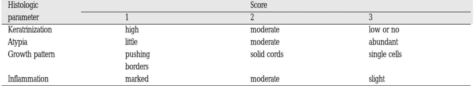
광학현미경하에서 먼저 종양의 깊이를 측정하였다. 조직절편 에서 조직 소견의 등급을 정하기 위하여 Crissman 등8), Bryne 등9) 과 Klijanienko 등10)의 등급체계(Table 1~3)를 참고하여 이들이 공 통적으로 사용한 각화도(keratinization), 핵의 비정형(atypia), 성장 양상(growth pattern), 염증세포 분포 정도에 따라 각각 1�3점으 로 점수를 산정하였다(Table 4).
3. 면역조직화학염색
3um 두께로 박절하여 Probe-On 슬라이드에 부착시켜 건조시킨 다음 검색에 사용하였고, 염색의 전 과정은 Probe-On 슬라이드 를 맞대어 생기는 capillary gap action 원리를 이용하여 개발된 Microprobe Immuno/DNA 염색기(Fisher�)를 이용하여 시행하였 다. 파라핀 절편이 부착된 슬라이드는 탈파라핀과 함수과정을 거쳐 조직항원이 잘 노출될 수 있도록 1×Automation buffer(Biomeda)에 3분간 부치시켰고 1×Automation buffer를 제 거한 후 CD34 (QBEnd/10, BioGenex), 변이형 p53 유전자(BP53.12, Zymed(San Francisco. CA)), 그리고 ras 종양유전자(Santa Cruz Biotechnology) 산물에 대한 항체를 항체 희석액으로 1 : 50으로 희석하여 20분간 부치시킨 후 완충액으로 수세하였다. 일차항체 의 검출을 위한 이차항체는 biotin이 부착된 anti-mouse IgG를 이 용하여 10분간 부치시킨 후 완충액으로 수세하고, avidin-alkaline phosphatase에 10분간 작용시켰고 alkaline phosphatase의 발색은 완충액으로 수세를 한 후 Fast Red TR salt를 이용하여 발색시켰으 며 헤마톡실린으로 대조염색을 시행하고 Crystal mount(Fisher�) 로 봉입한 후 양성반응을 광학현미경으로 검색하였다. 염색의 전 과정에 있어서 부치온도는 45℃로 하였으며 음성대조군은 일 차항체 대신 항체 희석액을 부치시켜 실험에 이용하였다.
p53
유전자와 ras 종양유전자에 대한 면역조직화학 염색을 시 행하여 대략 100개의 세포중 10개 이상의 세포에서 양성이면 양 성 반응으로 판정하였다.4. 맥관형성 측정
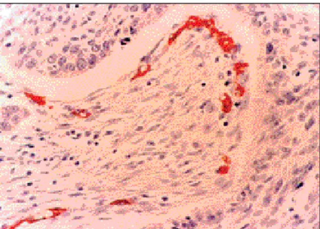
미세혈관의 밀도는 환자의 결과에 대한 지식이 없는 상태에서 측정하였다. 저배율에서 가장 빈도가 높은 부위를 선택하였는데 일반적으로 종양의 주위에서 가장 높게 분포하였다. 400배의 배 율에서 4 부위를 선택하여 사진촬영을 한 후 인화하여 미세혈관 의 수를 계산하였다. 400배의 배율에서 측정 가능한 부위는 대략 0.2mm2(0.1885mm2per field) 정도이다.
한 개의 미세혈관은 적색으로 CD34에 면역염색된 내피세포군 으로서 인접한 미세혈관, 종양세포, 다른 결합조직 요소와 분리 되어 있어야 한다. 내강(lumens)은 있을 수도 있지만 없어도 되고 내강에 적혈구가 없어도 된다. 경화부위(sclerotic area), 염증부위, 정상조직과 인접한 부위의 미세혈관은 제외하였다.
5. 통계 처리
각 인자 간의 상관관계를 Pearson product moment correlation과 회귀분석법으로 검사하였다.
Table 1.Grading scheme for squamous cell carcinoma (Cited from Crissman et al, 1984)
Histologic Score
parameter 1 2 3 4
Cytoplasmic High degree, Moderate, 20-50% Poor, 5-20% No evidence keratinization well-formed of cells, attempts of cells with of
keratinizaton
pearls at pear formation suggestion of keratinization
Nuclear Few enlarged 50-70% mature considerable Anaplastic
differentiation nuclei, >75% appearing nuclei nuclear pleom- tumor
mature appearing orphism, 25%
mature appearing
Mitoses, 0-1 2-3 4-5 >5
average number/
HPF
Inflammatory Continuous rim Patchy rim Occasional patch none response
Vascular/lymphatic Not identified Not identified Not identified Identified invasion
Pattern of Pushing borders Solid cords Thin irregular Single cells invasion cords
Table 2.Grading scheme for squamous cell carcinoma (Cited from Bryne et al, 1992) Morphological Score
feature 1 2 3 4
Degree of Highly keratiniza- Moderately Minimal keratinizat- No keratini- keratinization tion (>50% of keratinized ion (5-20% of the zation (0-5%
the cells) (20-50% of the cells) cells) of the cells)
Nuclear Little nuclear Moderately Abundant nuclear Extreme
polymorphism polymorphism abundant nu- polymorphism nuclear poly- (>75% mature clear polymor- (25- 50% mature morphism
cells) phism (50-75% cells) (0-25%
mature cells) mature cells)
Number of
mitoses (high 0-1 2-3 4-5 >5
power field)*
Pattern of Pushing, well- Infiltrating, soild Small groups Marked and
invasion delineated cords, bands and or cords of widespread
infiltrating borders or strands infiltrating cellular dissocia- cells (n>15) tion in small
groups and/or in single cells(n<15)
Lymphoplasma- Marked Moderate Slight None
cytic infiltration
Ⅲ. 결 과
연령 분포는 30대에서 70대(최고 80세)까지이며, 40대�60대가 32명으로 대부분을 차지하였다. 성별 분포는 남자가 29명, 여자 가 8명으로 남자가 더 많았다(Table 5).
종양의 깊이는 1�27mm로 다양하였다. 종양의 깊이가 10mm 이상인 증례는 37례 중 20례로서 이 중에서 17례가 림프절 전이 를 보여 종양의 깊이와 림프절 전이 간에 상관성을 보였다. 종양 세포의 각화도, 핵의 비정형 정도, 성장 양상 그리고 염증세포 분
포 정도를 합한 조직학적 등급은 총점 12점에서 5�10점의 분포 를 보였다. 조직학적 등급의 4가지 인자 중에서 핵의 비정형과 염증세포 분포 정도는 조직학적 등급과 높은 상관관계(R=0.727) (Fig. 1)를 보였다. 그리고 핵의 비정형과 성장 양상은 조직학적 등급과 다소 높은 상관관계(R=0.599)(Fig. 2)을 보였다.
맥관형성은 13�58개의 분포(Fig. 3)를 보였으며 조직학적 등 급, 림프절 전이와의 상관관계는 없었다. 림프절 전이는 28례에 서 관찰되었고 전이된 림프절은 1�12개까지 나타났다. 조직학 적 등급은 맥관형성과 림프절 전이와 상관관계가 없었다.
Table 3.Grading scheme for squamous cell carcinoma (Cited from Klijanienko et al, 1995) Differentiation
Keratinization Nuclear atypia Growth pattern
Intensity of inflammatory infiltrates Desmoplasia
Presence of tumor cell emboli in the peripheral microvessels (scored from 1 to 4)(for emboli, present=4, not found=1)
Table 4.Histologic parameters and score used in this study
Histologic Score
parameter 1 2 3
Keratrinization high moderate low or no
Atypia little moderate abundant
Growth pattern pushing solid cords single cells
borders
Inflammation marked moderate slight
Fig. 1. Correlation of atypia and inflammation by histologic grade
Fig. 2. Correlation of atypia and growth pattern by histologic grade
p53과 ras 종양유전자의 면역조직화학 염색에서 각각 양성 반
응은 16례와 22례에서, 음성반응은 21례와 15례에서 나타났다 (Fig. 4, 5). p53은 음성 반응이 양성 반응에 비해 많았고 ras는 양 성 반응이 음성 반응에 비해 높게 나타났으며 조직학적 등급과 림프절 전이와의 상관성은 없었다.Ⅳ. 고 찰
각 암환자에서 적절한 치료방법을 선택할 수 있는 임상적 진행 상태과 유사한 정확한 평가방법이 필요하고 종양의 생물학적 양 상을 미리 예측하기 위해 병리학자들은 등급(grading)을 산정한 다. 암종의 양적인 등급은 Broders1)가 처음으로 정상 편평세포와 닮은 정도에 따라 4등급으로 분류하는 방법을 제안하였으며, 그 Table 5. General characteristics of patients
Case Age Sex Depth Kera Atypia Growth Infla Grade Ves/x400 p53 ras LN(No.)
1 60 M 4 1 3 2 2 8 20 - - 3
2 56 M 9 1 1 1 2 5 17 - - -
3 49 M 3 3 2 1 1 7 13 + - 2
4 61 M 15 3 1 1 2 7 22 + - 10
5 58 F 5 1 1 2 1 5 25 - - 3
6 65 M 12 1 2 3 1 7 26 - + 10
7 44 F 13 1 2 3 3 9 42 - - 1
8 66 M 8 2 2 1 3 8 35 + - -
9 62 M 20 2 2 1 3 8 46 - + 4
10 62 M 18 2 2 2 1 7 24 - + 4
11 74 M 20 3 1 2 3 9 32 - + 5
12 47 M 7 1 1 2 1 5 15 - + -
13 32 M 5 1 1 3 2 7 26 - - 4
14 70 M 3 1 1 2 3 7 37 + + 1
15 72 F 5 3 1 2 3 9 32 - - 1
16 72 F 2 2 1 3 3 9 48 + + 1
17 50 M 10 1 3 3 3 10 33 + + 1
18 65 M 12 1 2 3 3 8 58 - - 2
19 55 M 11 2 3 3 2 10 54 - - 1
20 55 M 16 3 1 3 3 10 37 - - -
21 67 F 8 1 1 2 1 5 42 + + 1
22 43 M 5 2 1 2 2 7 38 + + -
23 56 M 8 1 2 2 2 7 30 - + 1
24 80 F 13 2 2 2 2 8 40 + + 2
25 56 M 8 1 2 2 2 7 30 + + 2
26 69 M 13 1 1 2 2 6 30 + + -
27 53 M 12 3 1 3 3 10 21 - + 1
28 53 M 18 3 1 3 3 10 49 + - 1
29 61 M 6 3 2 2 2 9 48 + - -
30 47 M 8 1 2 3 3 9 25 - + 1
31 63 M 13 1 2 2 2 7 35 + + 3
32 63 M 10 3 3 2 1 9 53 - + 8
33 60 M 16 2 1 3 2 8 18 - + -
34 56 F 1 3 1 1 1 6 35 - - -
35 43 F 27 2 1 3 1 7 40 + + 2
36 49 M 15 1 1 2 3 7 25 - + 5
37 46 M 10 1 1 2 3 7 57 + + 1
Abbreviations : M; Male, F; Female, Kera; keratinization, Infla; inflammation, Ves; number of vessel, +; positive, -; negative, LN(No.); number of lymph node metastasis.
후 많은 병리학자들은 고도, 중도 및 저도의 3등급으로 분류하여 사용하였다. 또한 종양의 크기, 림프절 전이와 원격전이를 평가 한 TNM 분류법4-6)이 있다. Broders 분류에 의한 조직학적 등급은 예후를 판정하는데 많이 이용되고 있지만 치료계획을 세우는데 는 적당하지 않았다3). 이는 주관적이며 예후와 일치하지 않은 경 우2,3)가 많아 객관적인 방법이 도입되었다8,45-50).
Crissman 등8)이 사용한 등급체계는 Jakobsson 등7)의 방법을 변 형시켜 각화도, 핵분화, 분열세포 숫자, 염증반응, 혈관이나 림프 관의 침윤, 종양세포의 침윤양상 등 6가지 인자들을 각각 1~4점 으로 점수를 매겨서 사용하였다(Table 1). Bryne 등9)은 Crissman 등8)의 등급체계를 변형하여 각화도, 핵의 다형성, 세포분열, 종 양세포의 침윤양상, 염증세포(임파구, 형질세포)의 침윤정도를 각각 1~4점씩 총 20점으로 판정하였다(Table 2). Klijanienko 등10) 은 Crissman 등8)의 등급체계를 변형하여 7가지의 조직학적 지표 를 사용하였는데 분화도, 각화도, 핵의 비정형, 성장 양상, 염증 세포의 정도, 결합조직 형성(desmoplasia), 종양세포 색전 등의 지 표를 사용하여 각각 1~4점으로 분류하였다(Table 3).
그 외에도 부가적으로 유사분열지수, DNA 함량, Ki-67, PCNA,
(level)이 전이를 예측하는데 가장 좋다고 하였다. Roland 등3)와 Janot 등15)은 조직학적 등급, 각화도, 혈관색전이 관련이 있다고 하였다.
본 연구에서는 비교적 객관적인 방법이라고 할 수 있는 Bryne 등9), Crissman 등8), Klijanienko 등10)의 조직학적 등급체계를 이용하 였다. 이들이 사용한 측정 인자는 서로 유사한 듯 하지만 조금씩 달라서, 본 연구에서는 이들의 공통점인 종양세포의 각화도, 핵 의 비정형, 성장 양상, 염증세포 분포 정도를 예후인자로 택하고 각 인자 당 1~3점으로 분류하여 측정하였다. 또 예후와의 관계를 알아보기 위하여 일반적으로 환자의 생존율을 조사하고 있으나, 본 연구에서는 암환자의 예후에 치명적인 림프절 전이와 예후인 자와의 관계를 조사하였다.
본 연구결과에서 종양세포의 각화도, 비정형 정도, 성장 양상 그리고 염증세포 분포 정도를 합한 조직학적 등급은 총점 12점 에서 5~10점의 분포를 보였다. 조직학적 등급체계의 4가지 인자 중에서 비정형과 염증세포 분포 정도 그리고 비정형과 성장 양 상은 조직학적 등급과 상관성을 보였으나, 조직학적 등급과 림 프절 전이와는 상관관계가 없었다.
Fig. 3. Immnohistochemical staining for CD34 in squamous cell carcinoma
Fig. 5. Immunohistochemical staining for ras in squamous cell carcinoma.
Fig. 4. Immunohistochemical staining for p53 in squamous cell carcinoma
이는 1~27mm 정도로 아주 다양하였으며, 종양의 깊이와 림프절 전이는 관계가 없었다. 종양의 깊이가 10mm 이상인 증례는 37례 중 20례였으며 이 중에서 17례가 림프절 전이를 보여 종양의 깊 이가 깊을수록 전이가 많이 됨을 알 수 있었다.
맥관형성은 전이 뿐만 아니라 고형성 종양(solid tumor)의 성장 에 필수적이다30,51). 또한 맥관형성은 종양형성의 초기에 일어나
며31,51)종양의 진행과 전이를 촉진시킨다52,53)고 알려져 있다. 본 연
구에서는 종양환자의 예후를 추측할 수 있는 유용한 예후 인자 를 찾아내고자 최근 많은 연구자들이 연구하고 있는 맥관형성을 측정하였다. 맥관형성의 계수방법은 다양하여 혈관의 직경과 둘 레, 숫자를 측정하는 컴퓨터를 이용한 분석법(computerized vas- cular analysis), 현미경에 부착된 eyepiece graticule을 이용하는 Chalkey count54), 관찰자의 주관에 따라 염색 정도에 따라 판단하 는 분석법(subjective visual vascular grading system) 등이 있다52,55). 미세혈관을 측정하는 종양의 넓이는 일반적으로 200~400배 (0.155~0.848mm2) 범위를 이용하고 있다. 본 연구에서는 혈관에 대한 면역조직화학염색 후 400배 시야에서 4부위에 대해 사진촬 영을 하였으며 이는 측정의 재생성을 도모하기 위함이었다.
종양의 중앙부는 맥관형성이 낮으며, 특히 큰 종양에서는 괴사 가 나타나기도 한다. 맥관형성이 왕성한 부위는 종양성장의 선 행구간(leading edge)과 관련이 있어서 종양세포의 증식과 전파 의 가능성을 제공한다. 이러한 소혈관은 결체조직으로 둘러싸여 있는데 이는 기존 혈관에서 생성되었음을 의미한다56). 일반적으 로 종양의 가장자리에서 혈관이 풍부하므로 맥관형성을 측정하 고자 할 때는 종양의 가장자리를 이용한다. 본 연구에서도 종양 의 가장자리에서 맥관형성을 측정하였으며, 저배율에서 혈관 분 포가 가장 풍부한 부위를 찾은 후 400배의 배율에서 4 부위를 선 택하였다.
본 연구에서는 CD3458)를 이용하여 혈관에 대한 면역조직화학 염색을 시행하여 관찰하였으며 섬유아세포, 일부 종양세포 혹은 형질세포와 교차반응의 가능성59)을 피하기 위하여 경화부위 (sclerotic area), 염증부위, 정상조직과 인접한 부위의 미세혈관은 제외하였다. 또한 미세혈관은 갈색으로 면역염색된 내피세포군 으로서 인접한 미세혈관, 종양세포, 다른 결합조직요소와 분리 되어 있어야 하며 내강의 유무는 중요하지 않았다.
종양의 맥관형성을 정량적으로 측정한 연구에서 피부암33,60),
유방암34,52,57,61-63), 전립선암35), 초기의 폐암(비소세포암종)37)그리고
두경부의 암종36)에서 혈관분포가 전이의 가능성과 관계가 있다 고 보고되었다. 그러나 두경부암종에서는 다른 부위의 암종과 달리 연구결과가 상반되는 경우가 많았다. Mikami 등38)과 Gasparini 등36)은 발생부위와 종양의 병기가 다양한 증례를 대상 으로 한 연구에서 미세혈관 밀도와 종양의 전이가 관련이 있다 고 하였고, Dray 등45)은 발생부위는 한정시키지 않고 종양의 병기 를 T1~T3, N0, M0로 한정한 연구에서 맥관형성과 관련이 없다고 하였다. Williams 등39)은 T1~T3의 구강종양에서 관련이 있다고 하 였고, Leedy 등44)은 T1과 T2 설종양에서는 관련이 없다고 하였으 며 이는 혀가 유방과는 다르게 혈관이 매우 풍부하므로 새로운 혈관이 필요하지 않으며 또한 Factor Ⅷ을 사용하였는데 염색이
잘되지 않았기 때문이라고 하였다.
본 연구결과에서 맥관형성은 13�58개의 분포를 보였으며 조 직학적 등급과 림프절 전이와의 상관관계는 없었다. Gleich 등42,43) 은 조기의 T1 구강 편평세포암종과 T2�T4의 구강 편평세포암종 에서 맥관형성과 종양의 전이 가능성과의 연관성을 찾지 못했는 데 그 이유를 다음과 같이 설명했다. 첫째, 조기 암종은 성장에 있어서 미세혈관 의존성이 낮다. 둘째, 두경부 암종에서 미세혈 관 밀도를 측정하는데 부적절하며 이는 기존 혈관과 새로 형성 된 혈관과의 감별이 어렵고 두경부 자체가 혈관이 풍부한 부위 이므로 종양의 성장에 신생혈관의 의존도가 상대적으로 낮다.
셋째, 커다란 종양에서 부분적인 미세혈관 밀도로 전체종양을 판단할 수 없다. 넷째, CD31 면역염색방법이 모든 미세혈관을 나 타내는데 적절하지 않다고 하였다. 이 밖에도 두경부 종양의 유 전자 변이로 인해 무산소 환경에서도 성장할 수 있도록 하므로 신생혈관에 대한 의존도가 낮다고 하였다. 이러한 현상으로 Graeber 등64)은 동물실험에서 p53 유전자 변이가 된 종양세포가 무산소 부위에서도 성장할 수 있음을 관찰하였다.
p53
유전자 변이는 두경부 편평세포암종에서 가장 흔한 유전 자 변이 중의 하나로서19)p53
단백의 과발현과 관계가 있다. 그밖 에 c-erb-B220,21)종양유전자단백, Rb 유전자22), bcl-2유전자23)등이 있다. 그러나 예후와의 관계는 결과가 일정하지 않다19,25,26). Sauter 는 설암에서 p53의 발현과 생존율의 증가가 관련이 있다고 하였 고 Zariwala와 Schmid24)도 p53 유전자 변이가 두경부 종양에서 전 이의 증가와 관계가 있다고 하였다. Guerry 등28)은 c-erb-B2와p53
발현이 전이와 관련이 있는데 그 중에서 c-erb-B2가 유용하 다고 하였다. 즉 p53 발현이 증가하기는 하였으나 통계적 의의는 없었다. 그러나 Ahomadeghe 등19), Bourhis 등26), Somers 등27)은 p53 유전자 변이가 관련이 없다고 하였고, Shin 등25)은 원격전이와는 관련이 없으나 재발 또는 부분적인 실패의 위험성이 높다고 하 였다.본 연구결과에서 p53과 ras 종양유전자의 면역조직화학염색 결과 각각 양성 반응은 6례와 22례에서, 음성 반응은 21례와 15례 에서 나타났다. p53은 음성 반응이 양성 반응에 비해 많았고 ras 는 양성 반응이 음성 반응에 비해 높게 나타났으며 조직학적 등 급과 림프절 전이와의 상관성은 없었다.
본 연구결과로 미루어 볼 때 두경부 편평세포암종은 맥관형성 과 관련이 없거나 맥관형성이 많은 인자 중의 하나일 것이라고 생각되었다. 그러나 종양세포의 비정형과 염증세포 분포 정도 그리고 핵의 비정형과 성장 양상은 조직학적 등급과 상관성을 보이고 종양의 깊이와 림프절 전이는 상관성이 있으므로 진단시 에 유의하여야 하며 임상의들이 예후 판정시에 고려하여야 할 것으로 사료된다. 또한 환자의 생존율을 높이기 위하여 유용한 예후 인자를 찾는 것이 중요하다고 사료된다.
Ⅴ. 결 론
본 연구에서는 두경부 편평세포암종의 전이와 예후 인자와의 관계를 알아보고자, 편평세포암종으로 치료받은 환자 중에서 림
프절 전이 여부가 확인된 37례를 대상으로 조직학적 등급과 p53 과 ras 종양유전자의 발현 그리고 맥관형성과 전이와의 관계를 관찰하였다.
종양의 깊이는 1~27mm였고, 10mm 이상이 20례였으며 이 중 17례에서 림프절 전이를 보여 종양의 깊이와 림프절 전이 간에 상관성을 보였다. 종양세포의 각화도, 핵의 비정형 정도, 성장 양 상 및 염증세포 분포 정도를 합한 조직학적 등급은 총점 12점에 서 5~10점의 분포를 보였다. 조직학적 등급의 4가지 인자 중에서 비정형 정도와 염증세포 분포 정도 그리고 비정형 정도와 성장 양상은 조직학적 등급과 상관성을 보였다. 맥관형성은 13~58개 의 분포를 보였으며 조직학적 등급과 림프절 전이와의 상관관계 는 없었다. 림프절 전이는 28례에서 관찰되었으며 전이의 숫자 는 1~10까지였다. 조직학적 등급은 맥관형성과 림프절 전이와 상관관계가 없었다. p53과 ras 종양유전자의 면역조직화학염색 상 양성반응은 각각 16례와 22례였으며 이들과 조직학적 등급 및 림프절 전이와의 상관성은 없었다.
이상의 결과로 두경부 편평세포암종에서 맥관형성은 림프절 의 전이와 상관관계를 보이지 않았으나 종양세포의 비정형 정도 와 염증세포 분포정도 그리고 비정형 정도와 성장 양상은 조직 학적 등급과 상관성을 보이므로 진단시에 유의하여야 함을 시사 하였다.
참고문헌
1. Broders AC : Carcinoma; grading and practical application. Arch Pathol 2:376-381, 1926.
2. Ensley JF, Crissman J, Kish J, Jacobs J, Weaver A, Kinzie J : The impact of conventional morphologic analysis on response rates and survival in patients with advanced head and neck cancers treated initially with cisplatin-containing combination chemotherapy.
Cancer 57:711-717, 1986.
3. Roland JJ, Caslin AW, Nash J, Stell PM : Value of grading squamous cell cardinoma of the head and neck. Head Neck 14:224-229, 1992.
4. Platz H, Fries R, Hudec M, Tjoa AM, Wagner RR : The prognostic relevance of various factors at the time of the first admission of the patient. J Max Fac Surg 11:3-12, 1983.
5. Frierson HF, Cooper PH : Prognostic factors in squamous cell carci- noma of the lower lip. Hum Pathol 17:346-354, 1986.
6. Sugarbaker PH : Cololectal cancer. In: Devita VT, Hellman S, Rosenberg SA, eds. Cancer; Principles and Practice of Oncology.
Philadelphia, Lippincott. 1985. pp795-884.
7. Jakobsson PA : Histologic grading of malignancy and prognosis in glottic carcinoma of the larynx. Workshop No. 14. In: Alberti PW, Bryce DP, eds. Centennial Conference on Laryngeal Cancer. New York, Appleton-Century-Crofts, 1976. pp847-852.
8. Crissman JD, Liu WY, Gluckman JL, Cummings G. Prognostic value of histopathologic parameters in squmous cell carcinoma of the oropharynx. Cancer 54:2995-3001, 1984.
9. Bryne M, Koppang HS, Lilleng R, Kjerheim A : Malignancy grading of the deep invasive margins of oral squamous cell carcinomas has high prognostic value. J Pathol 166:375-381, 1992.
10. Klijanienko J, El-naggar AK, de Braud F, Rodriguez-Peralto JL, Rodriguez R, Itzhaki M, Russo A, Janot F, Luboinski B, Cvitkovic E.
Tumor vascularization, mitotic index, histopathologic grade, and DNA ploidy in the assessment of 114 head and neck squamous cell
confined to the tongue and floor of mouth. Am J Surg 152:345-350, 1986.
12. Cooke LD, Cooke TG, Forester G : Flow cytometric analysis of DNA content in squamous carcinoma of the tongue. Clin Otolaryngol 19:131-134, 1994.
13. Kennedy JC, El-Badawy N, DeRose P. Comparison of cell prolifera- tion in breast carcinoma using image analysis (Ki-67) and flow cyro- metric systems. Anal Quant Cytol Histol 14:304-311, 1992.
14. Mamelle G, Pampurik J, Luboinski B, Lancar R, Lusinchi A, Bosq J.
Lymph nodes prognosis factors in head and neck squamous cell carcinoma. Am J Surg 168:494-498, 1994.
15. Janot F, Klijanienko J, Russo A, Mamet JP, De Braud F, El-Naggar AK, Pignon JP, Luboinski B, Cvitkovic E. Prognostic value of clinocopathological parameters in head and neck squamous cell carcinoma: a prospective analysis. Br Cancer 73:531-538, 1996.
16. Kearslay JH, Furlong KL, Cooke RA, Waters MJ. An immunohisto- chemical assessment of cellular proliferation markers in head and neck squamous cell cancers. Br J Cancer 61:821-7, 1990.
17. Shingaki S, Suzuki I, Nakajima T, Kawasaki T. Evaluation of histopathologic parameters in predicting cervical lymph node metas- tasis of oral and oropharyngeal carcinomas. Oral Surg Oral Med Oral Pathol 66:683-8, 1988.
18. Tytor M, Frazen G, Olafsson J. DNA ploidy in oral cavity carcinomas with special reference to prognosis. Head Neck 11:257-63, 1989.
19. Ahomadegbe JC, Barrois M, Fogel S, Le Bihan ML, Douc-rasy S, Duvillard P, Armand JP, Riou G : High incidence of p53 alter- ations(mutation, deletion, overexpression) in head and neck prima- ry tumors and metastases; absence of correlation with clinical out- come. Frequent protein overexpression in normal epithelium and in early non-invasive lesions. Oncogene 10:1217-1227, 1995.
20. Mizutani T, Onda M, Tokunaga A, Yamanaka N, Sugisaki Y.
Relationship of c-erb-B2 protein expression and gene amplification to invasion and metastasis in human gastric cancer. Cancer 72:2083- 2088, 1993.
21. Slamon DJ, Clark GM, Wong SG, Levin WJ, Ullrich A, McGuire WL.
Human breast cancer: correlation of relapse and survival with amplification of the HER-2/neu oncogene. Science 235:177-182, 1987.
22. Logothetis C, Xu H, Ro J, Hue S, Sahin A, Ordonez N, Benedict W.
Altered expression of retinoblastoma protein and known prognostic variables in locally advanced bladder cancer. J Natl Cancer Inst 84:1256-1261, 1992.
23. Pezzella Turley H, Kuzu I, Tungekar MF, Dunnill MS, Pierce CB, Harris A, Gatter KC, Masson DY. Bcl-2 protein in non-small-cell lung carcinoma. N Engl J Med 329:690-694, 1993.
24. Zariwala M, Schmid S. P53 gene mutations in oropharyngeal carci- nomas: a comparison of solitary and multiple primary tumors and lymph node metastasis. Int J Cancer 56:807-811, 1994.
25. Shin DM, Lee SL, Lippman SM, Lee JJ, Tu NZ, Choi G, Heyne K, Shin HJC, Ro JY, Goepfert H, Hong WK, Hittelman WN. p53 expres- sion; predicting recurrence and second primary tumors in head and neck squamous cell carcinoma. J Natl Cancer Inst 88:519-529, 1996.
26. Bourhis J, Bosq J, Wilson GD, Bressac B, Talbot M, Leridant AM, Dendale R, Janin N, Armand JP, Luboinski B, Malaise EP, Wibault P, Eschwege F : Correlation between p53 gene expression and tumor- cell proliferation in oropharyngeal cancer. Int Cancer 57:458-462, 1994.
27. Somers K, Merrick A, Lopez M, Incognito L, Schechter G, Casey G.
Frequent p53 mutations in head and neck cancer. Cancer Res 52:5997-6000, 1992.
28. Guerry M, Vabre L, Talbot M, Mamelle G, Leridant AM, Hill C, Bosq J, Luboinski B, Janot F. Prognostic value of histological and biologi- cal markers in pharyngeal squamous cell carcinoma : a case-control study. Br J Cancer 77(11):1932-1936, 1998.
29. Folkman J, Watson K, Ingber D, Hanahan D. Induction of angiogen-
dependent. J Natl Cancer Inst 82:4-6, 1990.
31. Herlyn M, Clark WH, Rodeck U. Biology of tumor progression in human melanocytes. Lab Invest 56:461-474, 1987.
32. Srivastava A, Laidler P, Hughes LE, Woodcock J, Shedden EJ.
Neovascularization in human cutaneous melanoma; a quantitative morphological and Doppler ultrasound study. Eur J Cancer Clin Oncol 22:1205-9, 1986.
33. Srivastava A, Laidler P, Davies RP, Horgan K, Hughes LE. The prog- nostic significance of tumor vascularity in intermediatethickness (0:76-4.0mm thick) skin melanoma. Am J Pathol 133:419-423, 1988.
34. Weidner N, Folkman J, Pozza F. Tumour angiogenesis; a new signif- icant and independent prognostic indicator in early-stage breast car- cinoma. J Natl Cancer Inst 84:1875-1887, 1992.
35. Weidner N, Carroll PR, Flax J, Blumenfeld W, Folkman J. Tumor angiogenesis correlates with metastasis in invasive prostate carcino- ma. Am J Pathol 143:401-409, 1993.
36. Gasparini G, Weidner N, Maluta S. Intratumoral microvessel density and p53 protein; correlation with metastasis in head-and-neck squa- mous-cell carcinoma. Int J Cancer 55:739-744, 1993.
37. Macchiarini P, Fontanini G, hardin MJ, Squartini F, Angeletti CA.
Relation of neovascularisation to metastasis of non-small-cell lung cancer. Lancet 340:145-146, 1991.
38. Mikami Y, Tsukuda M, Mochimatsu I. Angiogenesis in head and neck tumor. Nippon Jibiinkoka Gakkai Kaiho(J Oto-RhinoLaryngol Soc Japan) 96:645-650, 1993.
39. Williams JK, Carlson GW, Cohen C, Derose PB, Hunter S, Jurkiewicz MJ. Tumor angiogenesis as a prognostic factor in oral cavity tumors.
Am J Surg 168:373-380, 1994.
40. Zatterstrom UK, Brun E, Willen R, Kjellen E, Wennerberg J. Tumor angiogenesis and prognosis in squamous cell carcinoma of the head and neck. Head Neck 17:312-318, 1995.
41. Dray TG, Hardin NJ, Sofferman RA. Angiogenesis as a prognostic marker in early head and neck cancer. Ann Otol Rhinol Laryngol 104:724-729, 1995.
42. Gleich LL, Biddinger PW, Pavelic ZP, gluckman JL. Tumor angio- genesis in T1 oral cavity squamous cell carcinoma: role in predict- ing tumor aggressiveness. Head Neck 18:343-346, 1996.
43. Gleich LL, Biddinger PW, Duperier FD, Gluckman JL. Tumor angio- genesis as a prognostic indicator in T2-T4 oral cavity squamous cell carcinoma: A clinical-pathologic correlation. Head Neck 19:276-280, 1997.
44. Leedy DA, Trune Dr, Kronz JD, Weidner N, Cohen JI. Tumor angio- genesis, the p53 antigen, and cervical metastasis in squamous cell carcinoma of the tongue. Otolaryngol Head Neck Surg 111:417-422, 1994.
45. Anneroth G, Hansen LS, Silverman S : Malignancy grading in oral squamous cell carcinoma: I. Squamous cell carcinoma of the tongue and floor of the mouth: histologic grading in the clinical evaluation.
J Oral Pathol 15:162-8, 1986.
46. Barona de Guzman R, Martorell MA, Basterra J, Armengot M, Alvarez-Valeds R, Garin L : Prognostic value of histopathological parameter in 51 supraglottic squamous cell carcinomas.
Laryngoscope 103:538-40, 1993.
47. Bryne M, Koppang HS, Lilleng R, Stene T, Bang, Dabelsteen E. New malignancy grading is a better prognostic indicator than Broders’
grading in oral squamous cell carcinomas. J Oral Pathol Med 18:432- 437, 1989.
48. Bryne M, Nielsen K, Koppang HS, Dabelsteen E. Reproducibility of two malignancy grading systems of high prognostic value for oral cancer patients. J Oral Pathol Med 20:369-372, 1991a.
49. Bryne M. Prognostic value of various cellular and molecular features for oral squamous cell carcinomas: a review. J Oral Pathol Med 20:413-420, 1991b.
50. Zatterstrom UK, Wennerberg, Ewers SB, Willen R, Attewell R.
Prognostic factors in head and neck Cancer: histologic grading, DNA ploidy, and nodal status. Head Neck 13:477-87, 1991.
51. Bouck N : Tumor angiogenesis: The role of oncogenes and tumor suppressor genes. Cancer Cells 2:179-85, 1990.
52. Weidner N, Semple JP, Welch WR, Folkman J. Tumor angiogenesis and metastasis-correlation in invasive breast carcinoma. N Engl J Med 324:1-8, 1991.
53. Weinstat-Saslow D, Steeg PS. Angiogenesis and colonization in the tumor metastatic process: basic and applied advances. FASEB J 8:401-7, 1994.
54. Chalkey H. Method for the quantitative morphloical analysis of tis- sues. J Natl Cancer Inst 4:47-53, 1943.
55. Fox SB, Leek RD, Weekes MP, Whitehouse RM, Gatter KC, Harrist AL. Quantitation and prognostic value of breast cancer angiogene- sis: comparison of microvessel density, chalkley count, and comput- er image analysis. J Pathol 177:275-283, 1995.
56. Albo D, Granick MS, Hhala N, Atkinson B, Solomon MP : The rela- tionship of angiogenesis to biological activity in human squamous cell carcinoma of the head and neck. Ann Plast Surg 32:588-594, 1994.
57. Bosari S, Lee AKC, De Lellis RA : Microvessel quantitation and prog- nosis in invasive breast carcinoma. Hum Pathol 23:755-761, 1992.
58. Simpson J, Ahn C, Battifora H, Esteban J. Vascular surface area as a prognostic indicator in invasive breast carcinoma. Lab Invest 70:22A(Abstract)
59. Traweek ST, Kandalaft PL, Mehta P. The human hematopoietic progenitor cell antigen (CD34) in vascular neoplasia. Am J Clin Pathol 96:25-31, 1991.
60. Vacca A, Ribatti D, Roncali L. Melanocyte tumor progression is asso- ciated with changes in angiogenesis and expression of the 67-kilo- dalton laminin receptor. Cancer 72:455-461, 1993.
61. Gasparini G, Weidner N, Bevilacqua P. Tumor microvessel density, p53 expression, tumor size, and peritumoral lymphatic vessel inva- sion are relevant prognostic markers in node-negative breast carci- noma. J Clin Oncol 12:454-466, 1994.
62. Horak Er, Leek R, Klenk N. Angiogenesis assessed by platele/endothelial cell adhesion molecule antibodies, as indicator of node metastases and survival in breast cancer. Lancet 340:1120- 1124, 1992.
63. Toi M, Kashitani J, Tominaga T. Tumor angiogenesis is an indepen- dent prognostic indicator in primary breast carcinoma. Int J Cancer 55:371-374, 1993.
64. Graeber TG, Osmanian C, Jacks T. Hypoxia-mediated selection of cells with diminished apoptotic potential in solid tumors. Nature 379:88-91, 1996.