K.-H. Bae
Hongcheon Institute of Medicinal Herb, 101 Yeonbong-ri, Hongcheon-eup, Hongcheon, Gangwon, 250-930, Korea K.-H. Yoo ・ E.-S. Yoon ( )
Department of Biology, College of Nature Sciences, Kongju National University, Kongju 314-701, Korea
e-mail: [email protected] H.-B. Lee
Division of Forest Resources, College of Forest and Environmental Sciences, Kangwon National University, Chuncheon, Kangwon-do 200-701, Korea
Callus induction and plant regeneration of Iris dichotoma Pall. in endangered species
Kee-Hwa Bae ・ Kyoung-Hwa Yoo ・ Hak-Bong Lee ・ Eui-Soo Yoon
Received: 23 August 2012 / Accepted: 1 September 2012
ⓒKorean Society for Plant Biotechnology
Abstract Iris dichotoma Pall. is an important endangered
plant belonging to the family Iridaceae. A method was developed for the rapid micropropagation of I. dichotoma through plant regeneration from leaf, rhizome, and root explant-derived calli. Leaf, rhizome, and root segments were cultured on Murashige and Skoog (MS) medium supple- mented with 2,4-dichlorophenoxy acetic acid (2,4-D; 0-3.0 mg・L
-1) for callus induction. Callus production was highest at 1.0 mg・L
-12,4-D, where 73.8% and 45.5% of cultured rhizome and root cuttings, respectively, produced calli. The viable calli were maintained at an induced concentration of 2,4-D (3.0 mg・L
-1). They were then transferred to MS medium supplemented with various concentrations of 2,4-D (0-3.0 mg・L
-1) in combination with 6-benzyladenine (BA: 0, 1.0 and 3.0 mg・L
-1) for adventitious shoot regeneration. The addition of a low concentration of 2,4-D into BA-containing medium significantly increased the frequency of shoot regeneration in leaf, rhizome, and root-derived calli. The highest number of adventitious shoots (26.4 per callus) formed at 0.5 mg・L
-12,4-D and 1.0 mg/l BA. For rooting of the shoots, half- strength MS medium supplemented with different concentrations of indole 3-butyric acid (IBA) 0-3.0 mg・L
-1was tested. The optimal results were observed using half-strength MS medium supplemented with 1.0 mg・L
-1IBA, on which 98% of the regenerated shoots developed roots with an average of 3.5 roots per shoot within 45 days.
The plantlets raised in vitro were acclimatized and transferred to soil with 95% success. This in vitro propagation protocol will be useful for conservation and mass propagation of this endangered plant.
Keywords acclimatization, callus, in vitro, Iris dichotoma,
proliferation
Introduction
The genus Iris includes over 300 species, spread mostly across the northern temperate zone (Schulze 1988). Iris (=Pogoniris) comprises rhizomatous irises with bearded outer tepals (Mathew 1981). Four genuses and 31 species of Iris are distributed across South Korea. Five native species are critically endangered: Iris dichotoma, I. koreana, and I. odaesanensis. The distribution area of I. dichotoma is limited to Daecheong-do and the islands of Korea. Popu- lations are generally small, and some have disappeared or declined. For this reason, the South Korean government has designated the species as “Threatened to extinct: the first grade (I) for preservation” (Lee and Choi 2006). Lee and Choi (2006) treated it as critically endangered at the regional level according to the IUCN (2001) Red List criteria (Gärdenfors et al. 2001).
The natural mode of reproduction in the I. dichotoma species limits the large-scale production of valuable plants (Wang et al. 1999). Reproduction of this species using seeds is rarely carried out due to problems with germination, low seed production, capacity for cross-pollination, and the long juvenile period in plant development. Furthermore, similar to vegetative reproduction, the time required to obtain sufficient quantities of planting stock is 4-5 years. On the other hand, the cell and tissue culture method has become popular in iris reproduction, since it considerably increases plant multiplication factors (Shimazu et al. 1997; Wang et
DOI:http://dx.doi.org/10.5010/JPB.2012.39.3.182Research Article
al. 1999a; Shibli and Ajlouni 2000) and improves the quality of valuable planting stock (Baruch and Quak 1966; Mielke and Anderson 1989). The developed clonal reproduction techniques are used as an alternative method of conserving rare iris species (Radojevic and Subotic. 1992; Shibli and Ajlouni 2000).
In vitro propagation of the tissue of monocotyledons is complicated by their low regenerative capacity compared to dicotyledons (Kamo et al. 1990; Wang and Nguyen 1990). Analysis of the regenerative capacity in some orna- mental monocotyledons has demonstrated that it is lower in Iridaceae than in Amaryllidaceae, Araceae, and Liliaceae (Hussey 1975). In this paper, it is shown that the selection of organ or tissue as an explant is important in the develop- ment of plant reproduction in callus culture. Tissues of generative organs have been used in most studies of iris regeneration in vitro. For instance, flower tissues were used for direct regeneration of I. ensata (Ichihashi and Kato 1986; Kawase et al. 1991). The perianth tube and upper ovary proved to be the most convenient for microreproduction of I. ensata cultivars (Kawase et al. 1995). The patterns of shoot and organogenic callus formation in young stem culture were studied in cultivars and wild type I. ensata (Yabuya et al. 1991).
Numerous studies have demonstrated that the hormonal composition of the medium is the most important factor for the in vitro regeneration of irises (Radojevic et al.
1987; Laublin et al. 1991; Radojevic and Subotic 1992;
Gozu et al. 1993; Jehan et al. 1994; Shimizu et al. 1996;
Wang et al. 1999b). The morphogenetic capacity of explant tissues can be induced by varying the composition of phytohormones in the medium for the production of organogenic calluses and differentiation of accessory buds via organogenesis or embryoidogenesis. In view of its decorativeness and rarity in nature, an efficient propagation method for I. adriatica should be established. As is common in irises, propagation by splitting rhizomes is slow (Hussey 1975; Jehan et al. 1994); thus, micropropagation in vitro might be the method of choice. Various iris species have been propagated through organogenesis or somatic embry- ogenesis, using explants from the leaf base (Gozu et al.
1993; Shibli and Ajlouni 2000), mature zygotic embryos (Radojević and Subotić 1992; Boltenkov et al. 2004), ovary sections (Laublin and Cappadocia 1992), and root sections (Laublin et al. 1991).
Propagation of I. dichotoma via in vitro plant regeneration has not been reported. In this study, we undertook to develop a protocol for regeneration from leaf, rhizome, and root explant sections as a method for efficient propagation in
vitro for this species. Those techniques would help signifi- cantly in the multiplication and conservation of this endemic Iris species.
Materials and methods
Plant materials and culture conditions
Seeds of I. dichotoma were scarified by immersion in 70%
EtOH for 1 minute and then sterilized with commercial bleach 1% (v/v) (5% of sodium hypochlorite) with a few drops of Tween-20 (Sigma, USA) for 30 minute. The seeds were washed five times in sterile water and placed into petri dishes containing 20 ml solid hormone-free 1/3 MS medium (Murashige and Skoog 1962) under cool white fluorescent lights (56 μmol・m
-2・s
-1) on a 16 h photoperiod or in the dark at 25℃. The germination test was carried out on triplicates of 30 seeds.
Effect of explant type and growth regulators on callus induction
Half strength MS basal medium was used to induce callus induction and growth. Two-week-old seedlings were used as explant source. Explants (5~10 mm in length) from the leaf, rhizome and root were placed on the media containing 0, 0.1, 0.5, 1.0, and 3.0 mg・L
-12,4-dichlorophenoxy acetic acid (2,4-D) or 0, 0.1, 0.5, 1.0, and 3.0 mg・L
-1a-naphthalene acetic acid (NAA) supplemented with sucrose (30 g・L
-1) and solidified with agar (8 g・L
-1); the pH of the medium was adjusted to 5.7. The explants, 60 for each explant type, were incubated under cool white fluorescent lights on a 16 h photoperiod at 25℃ and subcultured on fresh media every 4 weeks.
Adventitious shoot induction and multiplication
Calli subcultured on the same medium for two generations,
were used to induce adventitious shoots. They were trans-
ferred onto 1/2 MS medium supplemented with sucrose
(30 mg・L
-1) and solidified with agar (8 g・L
-1), pH 5.7,
added with 0.1, 0.5, 1.0, and 3.0 mg・L
-12,4-D and 1.0, 3.0
mg・L
-1BA. Calli were maintained under cool white fluor-
escent lights (56 μmol・m
-2・s
-1) on a 16 h photoperiod at
25℃. After 8 weeks, shoot induction and multiplication
were evaluated and expressed as shooting frequency and
number of shoots per callus. The experiments were performed
on 20 calli for each callus line, and repeated twice.
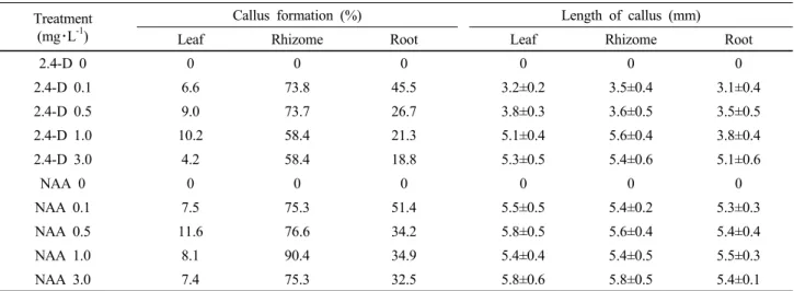
Table 1. Effect of 2,4-D and NAA on callus formation and its length derived from root, rhizome and leaf explants of I. dichotoma on 1/2 MS medium including 30 g・L-1 sucrose after 4 weeks of culture
Treatment (mg・L-1)
Callus formation (%) Length of callus (mm)
Leaf Rhizome Root Leaf Rhizome Root
2.4-D 0 0 0 0 0 0 0
2.4-D 0.1 6.6 73.8 45.5 3.2±0.2 3.5±0.4 3.1±0.4
2.4-D 0.5 9.0 73.7 26.7 3.8±0.3 3.6±0.5 3.5±0.5
2.4-D 1.0 10.2 58.4 21.3 5.1±0.4 5.6±0.4 3.8±0.4
2.4-D 3.0 4.2 58.4 18.8 5.3±0.5 5.4±0.6 5.1±0.6
NAA 0 0 0 0 0 0 0
NAA 0.1 7.5 75.3 51.4 5.5±0.5 5.4±0.2 5.3±0.3
NAA 0.5 11.6 76.6 34.2 5.8±0.5 5.6±0.4 5.4±0.4
NAA 1.0 8.1 90.4 34.9 5.4±0.4 5.4±0.5 5.5±0.3
NAA 3.0 7.4 75.3 32.5 5.8±0.6 5.8±0.5 5.4±0.1
*Data are the means ± SD, of three experiments. Different alphabetical letters are significantly different according to Duncun’s multiple range test at α ≤ 0.05.
Root induction
For root induction, individual shoots of 3-4 cm in length, were placed into a test tube containing 1/2 MS medium (10 ml) supplemented with sucrose (30 g・L
-1) and solidified with agar (8 g・L
-1); pH 5.7. The auxin indole-3-butyric acid (IBA) was assayed at 0, 0.5, 1.0, and 3.0 mg・L
-1. After 4 weeks, rooting was evaluated and expressed as rooting frequency, root number, and the longest root length per plantlet.
Acclimatization
Well-developed, plantlets were selected and removed from the culture tubes. The roots were cleared of medium with water to prevent contamination. The plantlets were potted in sterilized universal planting soil and transferred to a greenhouse for acclimatization. Pots were shielded with plastic covers to initially maintain the plants at high humidity, and the plantlets were acclimatized by gradually opening the covers. After 1 month, they were completely uncovered and hardened to the greenhouse.
Statistical analysis
All data were analyzed using analysis of variance (ANOVA), and expressed as means ± standard error (SE). Each experi- ment comprised three replications with at least 200 seeds per replication. To examine significant differences among the treatments, multiple comparison tests were then performed by Duncan’s multiple range test at α ≤ 0.05 (SAS).
Results and discussion
Effects of explant type and growth regulators on callus induction
In I. dichotoma, callus formation varied significantly depend- ing on explant type (Table 1). Rhizome part the earliest signs of callus formation after 4 weeks of culture, while leaves and roots started to generate callus from cut surfaces after 5 weeks of culture. Calli were compact, globular, and yellowish on 1/2MS medium with 1.0 mg・L
-12,4-D (Fig.
1C), and globular and dark yellowish on 1/2MS medium with 1.0 mg・L
-1NAA (Fig. 1B); the control exhibited no response (Fig. 1A). The rhizomes showed 90.4% callus for- mation after 8 weeks. However, root and leaves exhibited a significantly lower callus induction with 8.1 and 39.4%
callus formation respectively (Table 1). Conversely, a passaged I. ensata culture was obtained from the globular callus formed after the development of the embryos at the stem base induced by 2.0 mg・L
-1NAA and 0.5 mg/l BA (Boltenkov et al. 2004). Callus induction in monocots requires long time for its initiation (Geier 1986). It was reported that the induction of callus was difficult and the proliferation of initiated callus was very slow and somehow difficult to maintain compared to other iris species (Zheng et al., 1998;
Luciani et al., 2006). The highest callus size was achieved
when 3.0 mg・L
-1NAA on 1/2MS medium (5.8 mm) was
used in comparison with 0.1 mg・L
-12,4-D on 1/2MS medium
(3.2 mm) (Table 1). Callus formation from plates also varied
significantly depending on plant growth regulators and their
combinations (Table 1). The formation of morphogenic callus
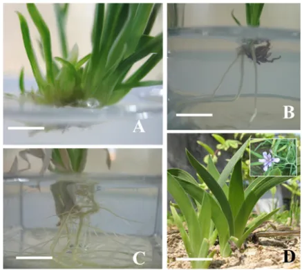
Fig. 1. Plant regeneration from callus derived from in vitro cultured explant types of I. dichotoma. A: Rhizome part on control medium after 4 weeks culture, B: Rhizome part on 1/2 MS medium including 1.0 mg・L-1 NAA after 8 weeks culture, C: Rhizome part on 1/2 MS medium including 1.0 mg・L-1 2,4-D after 8 weeks culture, D: Callus regeneration on 1/2 MS medium with 0.5 mg・L-1 BA and 1.0 mg・L-1 2,4-D after 8weeks of culture, E: Rooting form callus on 1/2 MS medium without plant growth regulators after 8 weeks of culture, F: In vitro shoots formed
Table 2. Effect of 2,4-D with BA on adventitious shoot formation and its length derived from explants of root, rhizome and leaf in I. dichotoma on 1/2 MS medium including 30 g・L-1 sucrose after 4 weeks of culture
Treatment (mg・L-1) Adventitious shoot formation (%) Length of shoot (mm)
2,4-D BA Leaf Rhizome Root Leaf Rhizome Root
0 0 0 0 0 0 0 0
0.1
1.0
98 98 100 12.2±1.2 13.5±1.4 13.1±1.4
0.5 100 100 100 13.8±1.3 13.6±1.5 13.5±1.5
1.0 100 100 100 15.1±1.4 15.6±1.4 13.8±1.4
3.0 100 100 100 15.3±1.5 15.4±1.6 15.1±1.6
0.1
3.0
100 100 100 13.2±1.2 13.5±1.4 13.1±1.4
0.5 100 100 100 13.8±1.3 13.6±1.5 13.5±1.5
1.0 100 100 100 15.1±1.4 15.6±1.4 13.8±1.4
3.0 100 100 100 15.3±1.5 15.4±1.6 15.1±1.6
*Data are the means ± SD, of three experiments. Different alphabetical letters are significantly different according to Duncun’s multiple range test at α ≤ 0.05.
in a culture of I. pumila (Radojevic et al. 1987), I. pseudacorus, and I. virginica embryos also required 2,4-D. This con- centration was also reported to be the optimal concentration for callus formation compared to other iris species (Myers and Simon 1999; Luciani et al. 2006).
Plantlet production and soil transfer
The highest callus growth rate was obtained in either 0.5 or 1.0 mg・L
-1BA and 1.0 mg・L
-12,4-D, while BA con- centration higher than 2.0 mg・L
-1inhibited callus pro-
liferation and promoted shoot formation. However, the best callus growth was obtained in the presence of 1.0 mg・L
-12,4-D, indicating that 2,4-D played a key role in callus proliferation (data not shown). In this sense, our results showed that 2,4-D not only induced callus initiation, but also maintained callus proliferation and agreed with previous results observed in iris callus culture (Boltenkov et al. 2004).
Proliferated compact calli were transferred to 1/2MS medium
supplemented with different BA and 2,4-D concentrations
under light conditions to investigate their potential for shoot
regeneration (Table 2). After 4 weeks of culture, most of
Table 3. Effect of 2,4-D and NAA on callus formation and its length derived from explants of root, rhizome and leaf in I. dichotoma on 1/2 MS medium including 30 g・L-1 sucrose after 4 weeks of culture
Treatment (mg・L-1) No. of root Length of root (mm)
IBA 0 3.1±1.2*d 4.4±0.4d
IBA 0.5 4.1±1.8bc 8.1±1.0b
IBA 1.0 6.1±0.9a 12.8±1.2a
IBA 3.0 4.7±1.1b 7.9±0.3c
*Data are the means ± SD, of three experiments. Different alphabetical letters are significantly different according to Duncun’s multiple range test at α ≤ 0.05.
Fig. 2. Plant regeneration from callus derived from I. dichotoma A: Shoot multiplication on 1/2 MS medium without plant growth regulators after 8 weeks of culture B: Rooting on 1/2 MS medium without plant growth regulators after 8 weeks of culture C: Rooting on 1/2 MS medium with 1.0 mg・L-1 IBA after 8 weeks of culture D: Plants survived ex vitro (Flowering of plantlets transferred to soil)
the compact calli started to turn to light green (Fig. 1D), and control was rooting from calli after 4 weeks of culture (Fig. 1E). Most of the calli at the early stage developed many yellowish green globular structures. Calli formed numerous shoots when they were cultured on 1/2MS medium supplemented with different concentrations of BA and 2,4-D (Table 2) (Fig. 1F). BA plays a key role in shoot regeneration in vitro (Ayabe et al. 1995; Ayabe and Sumi 1998; Guo et al. 2005; Xu et al. 2008). In the present experiment, BA induced shoot regeneration at the rate of 100% when cultured on medium with 1.0 or 3.0 mg・L
-1BA, although the highest BA concentration at 3.0 mg・L
-1appeared to show a suppressive effect on shoot differentiation (Table 2).
These results agreed with the reports of Barandiaran et al.
(1999) and Luciani et al. (2006), where BA could induce
shoot regeneration from calli, but were different from the observations of Myers and Simon (1999), who found that BA alone did not induce shoot regeneration. Regenerated shoot-derived calli were transferred to 1/2 MS medium with 0, 0.5, 1.0, and 3.0 mg・L
-1IBA to investigate the root induction into plantlets. After 4 weeks of culture, more than 90% root induction into plantlets was observed, with well-developed leaves and roots in all media (Fig. 2B-C).
The length of roots of plantlets was the longest in the 1/2
MS medium with 1.0 mg・L
-1IBA (Table 2). Shoot regen-
eration was enhanced by the combinations of BA and 2,4-D
compared with the use of control. Root formation varied
with different NAA concentrations. In this sense, root for-
mation increased with higher NAA concentrations and callus
growth was suppressed after rooting under light conditions.
However, those calli regenerated shoots also produced roots on growth regulator-free medium (data not shown). When the converted plantlets were acclimated in artificial soil and then transferred to greenhouse, about 96.4% of the plants survived 2 months after transfer (Fig. 2D) and pro- duced flowers after maturing (Fig. 2D).
We conclusively established high-frequency plant regen- eration via callus induction in I. dichotoma. This protocol can be applied to mass propagation of this endangered endemic species and can be applied to molecular breeding by genetic transformation in I. dichotoma.
References
Ayabe M, Sumi S (1998) Establishment of a novel tissue culture method, stem-disc culture and its practical application to micropropagation of garlic (Allium sativum L.). Plant Cell Rep 17:773-779
Ayabe M, Taniguchi K, Sumi SI (1995) Regeneration of whole plants from protoplasts isolated from tissue cultured shoot primordia of garlic (Allium sativum L.). Plant Cell Rep 15:
17-21
Bae KH, Kwon HK, Choi YE (2009) In vitro germination and plantlet conversion from the culture of fully mature seeds of Cypripedium guttatum Swartz. Prog Ornament Plant 9:
160-165
Barandiaran X, Martin N, Rodriguez-conde MF, di-Pietro A, Martin J (1999b) An efficient method for callus culture and shoot regeneration of garlic (Allium sativum L). Hort Science 34:348-349
Baruch ER, Quak F (1966) Virus-free plants of Iris “Wedgewood”
obtained by meristem culture. Neth J Plant Pathol 72:
270-273
Boltenkov EV, Rybin VG, Zarembo EV (2004) Cultivation of Iris ensata Thunb. callus tissue Prikl Biokhim Mikrobiol 40:244-251
Gärdenfors U, Hilton-Taylor C, Mace GM, Rodríguez JP (2001) The application of IUCN Red List criteria at regional levels.
Conser Biology 15:1206-1212
Geier T (1986) Anthurium. In: Evans, PV, Sharp DA, Bajal YPS (Eds) Handbook of Plant Cell Culture (vol 5) Collier Macmillan/Macmillan New York/London pp. 228-252 Gozu Y, Yokoyama M, Nakamura M (1993) In vitro pro-
pagation of Iris pallida, Plant Cell Rep 13:12-16 Guo DP, Zhu ZJ, Hu XX, Zheng SJ (2005) Effect of cytokinins
on shoot regeneration from cotyledon and leaf segment of stem mustard (Brassica juncea var. tsatsai). Plant Cell Tissue Organ Cult 83:123-127
Hussey G (1975) Totipotency in tissue explants and callus of some members of the Liliaceae, Iridaceae and Amaryllidaceae.
J Exp Bot 26:253-262
Ichihashi S, Kato S (1986) Clonal propagation of Iris kaempferi by means of flower organ culture. Bull Aichi Univ Edu
35:135-143
Jehan H, Courtois D, Ehret C, Lerch K, Petiard V (1994) Plant regeneration of Iris pallida Lam. and Iris germanica L. via somatic embryogenesis from leaves, apces and young flowers.
Plant Cell Rep 13:671-675
Kawase K, Mizutani H, Yoshioka M, Fukuda S (1991) Pro- pagation of Iris by tissue culture. 2. Culture of flower organs.
J Jpn Soc Hort Sci 60:436-437
Kawase K, Mizutani H, Yoshioka M, Fukuda S (1995) Shoot formation on floral organs of Japanese Iris in vitro. J Jpn Soc Hort Sci 64:143-148
Laublin G, Saini HS, Cappadocia M (1991) In vitro plant regeneration via somatic embryogenesis from root culture of some rhizomatous Irises Plant Cell Tiss Org Cul 27:
15-21
Lee JS, Choi BH (2006) Distribution and red data of wild orchids in the Korean Peninsula. Kor J Plant Taxon 36:
335-360
Laublin G, Cappadocia M (1992) In vitro ovary culture of some apogon garden Irises (Iris pseudacorus L, I. setosa Pall, I. versicolor L.). Bot Acta 105:319-322
Luciani GF, Mary AK, Pellegrini C, Curvetto NR (2006) Effects of explants and growth regulators in garlic callus formation and plant regeneration. Plant Cell Tissue Organ Cult 87:
139-143
Mathew B (1981) The Iris. B. T. Batsford Ltd., London. MITIĆ B. 2002. Iris adriatica (Iridaceae), a new species from Dalmatia (Croatia). Phyton 42:305-313
Mielke KA, Anderson WC (1989) In vitro bulblet formation in Dutch Iris. Hortscience 24:1028-1031
Murashige T, Skoog F (1962) A revised medium for rapid growth and bioassays with tobacco tissue cultures. Physiol Plant 15:473-497
Myers JM, Simon PW (1999) Regeneration of garlic callus as affected by clonal variation, plant growth regulators and culture conditions over time. Plant Cell Rep 19:32-36 Radojevic L, Subotic A (1992) Plant regeneration of Iris setosa
Pall. through somatic Embryogenesis and organogenesis. J Plant Physiol 139:690-696
Radojevic L, Sokic O, Tucic B (1987) Somatic embryogenesis in tissue culture of Iris (Iris pumila L.). Acta Hort 212:
719-723
SAS (2001) SAS/STAT User’s guide (8.02) SAS Institute Inc.
Cary NC USA
Schulze W (1988) Wild-Iris für den Garten, Fortschritt Erfurt, Jena
Sears RG, Deckard EL (1982) Tissue culture variability in wheat:
callus induction and plant regeneration. Crop Science 22:
546-550
Shibli RA, Ajlouni MM (2000) Somatic embryogenesis in the endemic black Iris. Plant Cell Tiss Org Cult 61:15-21 Shimizu K, Nagaike H, Yabuya T, Adachi T (1997) Plant
regeneration from suspension culture of Iris germanica.
Plant Cell Tissue and Organ Culture 50:27-31
Shimizu K, Yabuya T, Adachi T (1996) Plant regeneration from
protoplasts of Iris germanica L. Euphytica 89:223-227 Wang WC, Nguyen HT (1990) A novel approach for efficient
plant regeneration from long-term suspension culture of Wheat. Plant Cell Rep 8:639-642
Wang Y, Jeknic Z, Ernst RC, Chen THH (1999) Efficient plant regeneration from suspension-cultured cells of tall bearded Iris. Hortscience 34:730-735
Wang Y, Jeknic Z, Ernst RC, Chen THH (1999) Improved plant regeneration from suspension-cultured cells of Iris germanica L. ‘Skating Party’. Hortscience 34:1271-1276 Xu Z, Um YC, Kim CH, Lu G, Guo DP, Liu HL, Bah AA,
Mao A (2008) Effect of plant growth regulators, temperature and sucrose on shoot proliferation from the stem disc of Chinese jiaotou (Allium chinense) and in vitro bulblet for- mation. Acta Physiol Plant 40:521-528
Yabuya T, Ikeda Y, Adachi T (1991) In vitro propagation of Japanese garden Iris, Iris ensata Thunb. Euphytica 57:77-81 Zheng S, Henken B, Sofiari E, Jacobsen E, Krens FA, Kik C
(1998) Factors influencing induction, propagation and regen- eration of mature zygotic embryo-derived callus from Allium cepa. Plant Cell Tissue Organ Cult 53:99-105