K. H. Yoo ・ E. S. Yoon ( )
Department of Biology, College of Nature Sciences, Kongju National University, Kongju 314-701, Korea
e-mail: [email protected] K. H. Bae
Hongcheon Institute of Medicinal Herb, 101 Yeonbong-ri, Hongcheon-eup, Hongcheon, Gangwon, 250-930, Korea National Institute of Biological Resources, 42 Hwangyeong-ro, Seo-gu, Incheon, 404-708, Korea
M. H. Lee
Plant Quarantine Technology Center, Suwon 443-440, Korea J. H. Jeong
Department of Health Food Processing, Jeonnam Provincial College, Damyang, Jeollanam-do 517-802, Korea
Y. E. Choi
Division of Forest Resources, College of Forest and Environmental Sciences, Kangwon National University, Chunchon, Kangwon-do 200-701, Korea
Plant regeneration from callus of Iris odaesanensis Y. N. Lee native to Korea via organogenesis
Kee-Hwa Bae ・ Kyoung-Hwa Yoo ・ Mi-Hyun Lee ・ Jae-Hun Jeong ・ Yong-Eui Choi ・ Eui-Soo Yoon
Received: 12 September 2013 / Accepted: 23 September 2013
ⓒKorean Society for Plant Biotechnology
Abstract Iris odaesanensis Y. N. Lee. is an important endangered and native plant belonging to the family Iridaceae in Korea. This study describes a method for rapid micropropagation of this species via from leaf, rhizome and root explants derived calli. Leaf, rhizome and root explants were cultured on Murashige and Skoog (MS) medium supplemented with 2,4-dichlorophenoxy acetic acid (2,4-D) for callus induction. Rhizome explants yielded calli at a frequency of 72% when cultured at 1.0 mg/l 2,4-D. Calli were maintained at 1.0 mg/l 2,4-D. These calli were transferred to MS medium supplemented with 0, 0.5, 1.0, and 2.0 mg/l 2,4-D in combination with 0, 0.5, 1.0, and 3.0 mg/l BA for adventitious shoot induction. The highest number of adventitious shoot (228.9 per petri-dish) were formed at 1.0 mg/l 2,4-D and 1.0 mg/l BA. WPM medium was the best to convert calli into plantlets, where up to 98.2%
of calli were regenerated into plantlets. This in vitro propagation protocol should be useful for conservation of this endangered plant.
Keywords Iris odaesanensis Y. N. Lee, Callus, In vitro, organogenesis, Proliferation
Introduction
The genus Iris is a perennial herbaceous plant, which includes about 300 species of flowering plants with showy flowers. The genus is widely distributed from temperate zone to the subarctic zone in the Northern hemisphere (Schulze 1988; Shibata 1998). These plants are commonly planted in gardens, and widely used in floral arrangement for ornamental purposes.
Iris odaesanensis is native to South Korea, and called yellow spot iris or yellow flag. It is firstly collected in Mt.
Omi, Gyeongsangbuk-do in 1963, and that was described as Iris koreana for. albiflora (Lee and Lee, 1964). Lee (1974) found the same species in both Mt. Odae and Hoenggye-ri in Gangwon province but named it as 'Iris odaesanensis Y. N. Lee'. I. odaesanensis is a rare wild perennial herbaceous plant which is subjected to strict protection as an endemic Iridaceae. The populations size declines rapidly. For this reason, the Ministry of Environment (MEV) has designated the species as ‘Threatened to extinct:
the first grade (Ⅱ) for preservation’ (Lee and Choi 2006).
Also, this species reproduction by seeds is rarely used due to poor germination, low seed production, capacity by cross-pollination, and long juvenile period in plant development.
At the same time, similar to vegetative reproduction, as long as 4-5 years are required to obtain sufficient quantities of planting stock.
Plant tissue culture is a powerful alternative technique for conservation and propagation of plants, especially for those that are rare and difficult to propagate by conventional methods (Shimazu et al. 1997; Wang et al. 1999a; Shibli and Ajlouni 2000) and improves the quality of valuable planting stock (Baruch and Quak 1966; Mielke and Anderson 1989).
DOI:http://dx.doi.org/10.5010/JPB.2013.40.3.163 Research Article
The developed techniques for clonal reproduction are used as an alternative way to conserve rare iris species (Radojevic and Subotic 1992; Shibli and Ajlouni 2000).
In vitro propagation of monocotyledons plants is more complicated because of their low regenerative capacity compared to dicotyledons plants (Kawase et al. 1991; Wang and Nguyen 1990). Analysis of the regenerative capacity in some ornamental monocotyledons demonstrates that it is lower in Iridaceae than in Amaryllidaceae, Araceae, and Liliaceae (Hussey 1975). These studies showed that the selection of organ or tissue as an explant is important in development of plant reproduction through callus cultures.
Accordingly, tissues of reproductive organs were used in most studies of iris regeneration in vitro. For instance, flower tissues were used for direct regeneration of Iris ensata. (Ichihashi and Kato 1986; Kawase et al. 1991).
The perianth tube and upper ovary proved to be the most convenient for microreproduction of I. ensata cultivars (Kawase et al. 1995). The patterns of shoot and organogenic callus formation in young stem culture were studied in cultivars and wild-type I. ensata (Yabuya et al. 1991).
Numerous studies demonstrated that the hormonal com- position of the medium is the most important factor for in vitro regeneration of irises (Radojevic et al. 1987; Laublin and Cappadocia 1992; Radojevic and Subotic 1992; Gozu et al. 1993; Jehan et al. 1994; Shimizu et al. 1996; Wang et al. 1999b). Various iris species have been propagated through organogenesis or somatic embryogenesis, using explants from the leaf base (Gozu et al. 1993; Shibli and Ajlouni 2000), mature zygotic embryos (Radojević and Subotić 1992; Boltenkov et al. 2004), ovary sections and root sections (Laublin and Cappadocia 1992), but, so far, no studies have been reported on callus derived organogenesis in I. odaesanensis.
Therefore, in the present study, for the first time, we report the establishment of a high frequency plant regeneration system via organogenesis in I. odaesanensis.
Materials and Methods
Plant materials and culture conditions
Mature seeds of I. odaesanensis were collected from Mt.
Odae National Park in Korea, and sown on seed beds prepared in a greenhouse at the Kongju National University.
Mature seeds of I. odaesanensis were scarified by immersion in 70% EtOH for 1 min and then sterilized with commercial
bleach 1% (v/v) (5% of sodium hypochlorite) with a few drops of Tween-20 (Sigma, USA) for 30 min. The seeds were washed 5 times in sterile water and placed into petri dishes containing hormone-free 1/3 MS solid medium (Murashige and Skoog 1962) under cool white fluorescent lights (30 μmolm-2s-1) on a 16 h photoperiod or in the dark at 25℃.
Callus induction
After cutting the leaf, rhizome, and root segments into 10 mm in sizes, they were cultured on MS medium supplemented with 0, 0.5, 1.0, and 3.0 mg/l 2,4-dichlorophenoxy acetic acid (2,4-D) and a-naphtalene acetic acid (NAA). All media were supplemented with 30 g/l sucrose and solidified with 8.0 g/l plant agar, and then adjusted to 5.8 pH before autoclaving at 121℃ for 20 min. The culture room was maintained at 25 ± 1℃ in darkness. The frequency of callus induction was evaluated after 12 weeks of culture.
Adventitious shoot induction
Calli subcultured on the same medium for two generations were used for inducing adventitious shoot. Calli were transferred onto MS medium supplemented with sucrose (30 g/l) and solidified with agar (8 g/l); pH 5.7; added with 0, 0.5, 1.0, and 2.0 mg/l2,4-D and 0, 0.5, 1.0, 3.0 mg/l BA. Calli were maintained under cool white fluorescent lights (30μmolm-2s-1) on a 16 h photoperiod at 25℃. After 8 weeks, adventitious shoot induction was evaluated and expressed as shooting frequency and number of adventitious shoot per callus. The experiments were performed on 20 calli for each callus line, and repeated five time.
Plantlet conversion
Adventitious shoots were transferred to WPM (Lloyd and McCown 1980), half-strength WPM, one-third strength WPM medium and MS, half-strength MS, and one-third strength MS medium for the growth of plantlets. The culture room was maintained at 25 ± 1℃ with a 16 h photoperiod under 30μmolm-2s-1cool white fluorescent light.
Plantlet conversion rate was evaluated by counting plantlets with well-developed leaves and roots after 4 weeks of culture. Plantlet height was evaluated by measuring average length of shoots and roots after 4 weeks of culture. A total of four germinated embryos were transferred onto each plastic square culture vessel (7.2 cm × 7.2 cm × 10 cm).
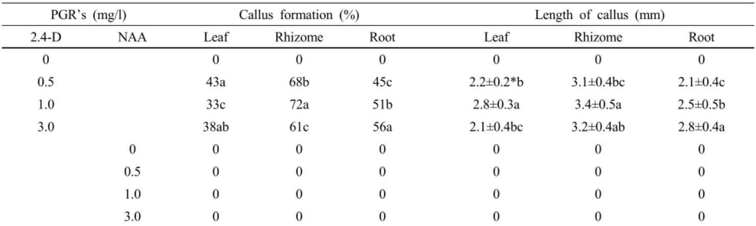
Table 1 Effect of 2,4-D and NAA on callus and adventitious root formation from root, rhizome and leaf explants of I. odaesanensis.
MS medium containing 30 g/l sucrose was used. Date were collected after 12 weeks of culture
PGR’s (mg/l) Callus formation (%) Length of callus (mm)
2.4-D NAA Leaf Rhizome Root Leaf Rhizome Root
0 0 0 0 0 0 0
0.5 43a 68b 45c 2.2±0.2*b 3.1±0.4bc 2.1±0.4c
1.0 33c 72a 51b 2.8±0.3a 3.4±0.5a 2.5±0.5b
3.0 38ab 61c 56a 2.1±0.4bc 3.2±0.4ab 2.8±0.4a
0 0 0 0 0 0 0
0.5 0 0 0 0 0 0
1.0 0 0 0 0 0 0
3.0 0 0 0 0 0 0
*aData are the means ± SD, of five time experiments. Different alphabetical letters are significantly different according to Duncun's multiple range test at P〈 0.05.
Fig. 1 Plant regeneration from callus derived from various explants in I. odaesanensis. A: leaf explants on MS medium supplemented with 1.0 mg/l 2,4-D after 12 weeks culture (scale bar indicates 10 mm), B: Root explant at 1.0 mg/l 2,4-D after 12 weeks culture (scale bar indicates 10 mm), C: Rhizome explant on at 1.0 mg/l 2,4-D after 12 weeks culture (scale bar indicates 10 mm), D:
Adventitious root induction from root explant at 1.0 mg/l NAA after 12 weeks of culture (scale bar indicates 15 mm) Each experiment was performed five times.
Statistical analysis
All data were analyzed using ANOVA and expressed as means ± standard error (SE). To examine significant differences among the treatments, multiple comparison tests were then performed by Duncan’s multiple range test at p ≤ 0.05 (SAS 2001).
Results and Discussion
Effects of explant type, culture media and growth regulators on callus induction
Callus formation varied significantly depending on kind of explant of I. odaesanesis (Table 1). Rhizome explant formed callus after 4 weeks of culture, but leaves and roots generated callus from cut surfaces after 6 weeks of culture. Calli of
Fig. 2 Plant regeneration from callus derived from rhizome explants in I. odaesanensis, A: Proliferation of callus on MS medium with 1.0 mg/l 2,4-D and 1.0 mg/l BA after 8 weeks of culture (scale bar indicates 0.2 mm), B: Shoot bud initiation on MS medium without plant growth regulators after 4 weeks of culture (scale bar indicates 0.8 mm), C: Proliferation of shoots on MS medium after 4 weeks of culture (scale bar indicates 10 mm), D: Plantlet with well-developed leaves and roots grown on MS medium after 4 weeks of culture (scale bar indicates 45 mm)
leaf, root and rhizome (Fig. 1A-C) were compact, globular and yellowish on MS medium with 1.0 mg/l 2,4-D. Yellowish adventitious root were induced from leaf, rhizome and root explant on MS medium with 1.0 mg/l NAA after 12 weeks of culture (Fig. 1D), but control (non-treated 2,4-D or NAA) did not form callus. Rhizome explant showed callus formation at a frequency of 72% after 12 weeks of culture (Table 1). However, roots and leaves exhibited a significantly lower callus induction with 51 and 33% callus formation respectively (Table 1). Conversely, I. ensata culture was obtained from the globular callus formed after the development of the embryos at the stem base induced by 2 mg/l NAA and 0.5 mg/l BA (Boltenkov et al. 2004). It was reported that the induction of callus was difficult and the proliferation of initiated callus was very slow and somehow difficult to maintain compared to other iris species (Zheng et al. 1998; Luciani et al. 2006). The highest callus size was achieved when 1.0 mg/L 2,4-D was
supplemented to MS medium (Table 1). Callus formation from plates also varied significantly depending on plant growth regulators and their combinations (Table 1). The formation of morphogenic callus in a culture of I. pumila (Radojevic et al. 1987), I. pseudacorus, and I. virginica embryos also required 2,4-D.
Adventitious shoot induction and in vitro plantlet production
For determination of adventitious shoot induction from callus, both types (compact and friable ones) were transferred to MS medium supplemented with 2,4-D and BA and placed under illumination. After 20 to 25 days of culture, only the compact calli turned greenish (partially green) and several adventitious shoot regenerated on the calli (Fig . 2A). The differentiated multiple shoots were divided and transplanted onto the same medium(Fig. 2B). The highest adventitious shoot induction rate was obtained in 1.0 mg/l
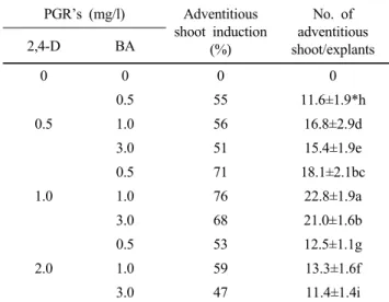
Table 2 Effect of 2,4-D in combination with BA on adventitious shoot production from rhizome derived callus segments of I.
odaesanensis. MS medium containing 30 g/l sucrose was used.
Date were collected after 12 weeks of culture PGR’s (mg/l) Adventitious
shoot induction (%)
No. of adventitious shoot/explants
2,4-D BA
0 0 0 0
0.5
0.5 55 11.6±1.9*h
1.0 56 16.8±2.9d
3.0 51 15.4±1.9e
1.0
0.5 71 18.1±2.1bc
1.0 76 22.8±1.9a
3.0 68 21.0±1.6b
2.0
0.5 53 12.5±1.1g
1.0 59 13.3±1.6f
3.0 47 11.4±1.4i
*aData are the means ± SD, of five time experiments. Different alphabetical letters are significantly different according to Duncun's multiple range test at P〈 0.05.
Table 3 Effect of various kinds of medium on conversion of adventitious shoot of I. odaesanensis into plantlest. Medium containing 30 g/l sucrose was used. Data were collected after 4 weeks of culture
Various kinds of medium
Conversion into plantlet (%)
Length of shoot (cm)
Length of root (cm)
WPM 91.4 8.9±2.8*d 4.7±1.8*f
1/2WPM 98.2 11.4±0.8c 5.6±2.1e
1/3WPM 94.1 12.2±2.1b 6.2±1.9b
MS 96.6 13.8±3.1a 6.4±1.3a
1/2MS 94.9 12.8±1.4b 5.9±2.3bc
1/3MS 93.6 11.9±4.8c 5.8±4.5cd
*aData are the means ± SD, of five time experiments. Different alphabetical letters are significantly different according to Duncun's multiple range test at P〈 0.05.
2,4-D and 1.0 mg/l BA (Table 2). Proliferated compact calli were transferred to 1/2MS medium supplemented with different BA and 2,4-D concentrations under light conditions to investigate their potential for shoot elongation (Table 3). After 4 weeks of culture, most of the compact calli started to turn to light green (Fig. 2A). Most of the callus at the early stage of shoot development had many yellowish green globular structures. Calli formed numerous shoots when they were cultured on MS medium supplemented with different concentrations of BA and 2,4-D (Table 2) (Fig. 2A). Adventitious shoot of I. odaesanensis were transferred to various media (WPM, 1/2WPM, 1/3WPM,
MS, 1/2MS, and 1/3MS) to investigate the conversion into plantlets. After 4 weeks of culture, more than 90% of the adventitious shoot converted into plantlets with well-developed leaves and roots in all media (Fig. 2C, D). However, there was a remarkable difference on the growth of plantlets among the six media (Table 3). The length of shoots and roots of plantlets was the longest on MS medium. Therefore, MS medium was the most effective for growth in I.
odaesanensis. BA plays a key role in shoot regeneration in vitro (Ayabe et al. 1995; Ayabe et al. 1998; Guo et al.
2005; Xu et al. 2008). In the present experiment, BA enabled shoot regeneration at a frequency of up to 100%
when rhizome explants were cultured on medium with 1.0 or 3.0 mg/l BA, although BA at a level of 3.0 mg/l showed a suppressive effect on shoot differentiation (Table 2). These results agree with the reports of Barandiaran et al. (1999) and Luciani et al. (2006), where BA could induce shoot regeneration from callus, but were different from the observations of Myers and Simon (1999) who found that BA alone did not induce shoot regeneration.
In conclusion, we established a system for high frequency plant regeneration via callus induction in I. odaesanensis.
This protocol can be applied to mass propagation and molecular breeding by genetic transformation in this endangered endemic species.
References
Ayabe M, Sumi S (1998) Establishment of a novel tissue culture method, stem-disc culture and its practical application to micropropagation of garlic (Allium sativum L.). Plant Cell Rep 17:773-779
Ayabe M, Taniguchi K, Sumi SI (1995) Regeneration of whole plants from protoplasts isolated from tissue cultured shoot primordia of garlic (Allium sativum L.). Plant Cell Rep 15:17-21
Barandiaran X, Martin N, Rodriguez-conde MF, di-Pietro A, Martin J (1999) An efficient method for callus culture and shoot regeneration of garlic (Allium sativum L). Hort Science 34:348-349
Baruch ER, Quak F (1966) Virus-free plants of Iris “Wedgewood”
obtained by meristem culture. Neth. J. Plant Pathol, 72:270-273 Boltenkov EV, Rybin VG, Zarembo EV (2004) Cultivation of Iris
ensata Thunb. callus tissue, Prikl. Biokhim Mikrobiol 40:244-251
Gozu Y, Yokoyama M, Nakamura M (1993) In vitro propagation of Iris pallida Plant Cell Rep 13:12-16
Guo DP, Zhu ZJ, Hu XX, Zheng SJ (2005) Effect of cytokinins on shoot regeneration from cotyledon and leaf segment of stem mustard (Brassica juncea var. tsatsai). Plant Cell Tissue Organ Cult 83:123-127
Hussey G (1975) Totipotency in tissue explants and callus of some members of the Liliaceae, Iridaceae and Amaryllidaceae. J Exp Bot 26:253-262
Ichihashi S, Kato S (1986) Clonal propagation of Iris kaempferi by means of flower organ culture. Bull Aichi Univ Edu 35:135-143
Jehan H, Courtois D, Ehret C, Lerch K, Petiard V (1994) Plant regeneration of Iris pallida Lam. and Iris germanica L. via somatic embryogenesis from leaves, apecs and young flowers. Plant Cell Rep 13:671-675
Kawase K, Mizutani H, Yoshioka M, Fukuda S (1991) Propagation of Iris by tissue culture. 2. Culture of flower organs. J Jpn Soc Hort Sci 60:436-437
Kawase K, Mizutani H, Yoshioka M, Fukuda S (1995) Shoot formation on floral organs of Japanese Irisin vitro. J Jpn Soc Hort Sci 64:143-148
Lee YN (1974) New taxa on Korean flora (1). Korean Jour Botany 17(1):33-35
Lee JS, Choi BH (2006) Distribution and red data of wild orchids in the Korean Peninsula. Kor J Plant Taxon 36:335-360 Lee WJ, Lee WH (1964) Report on study of a new species of
Iridaceae plants Seoul Agricultural College 1-4
Laublin G, Cappadocia M (1992) In vitro ovary culture of some apogon garden Irises (Iris pseudacorus L, I. setosa Pall, I.
versicolor L.). Bot Acta 105:319-322
Lloyd E, McCown B (1980) Commercially feasible micropropagation of mountain laurel (Kalmia latifolia) by use of shoot tip culture. Comb Proc Int Plant Prop Soc 30:421-427
Luciani GF, Mary AK, Pellegrini C, Curvetto NR (2006) Effects of explants and growth regulators in garlic callus formation and plant regeneration. Plant Cell Tissue Organ Cult 87:139-143 Mielke KA, Anderson WC (1989) In vitro bulblet formation in
Dutch Iris. Hortscience 24:1028-1031
Murashige T, Skoog F (1962) A revised medium for rapid growth and bioassays with tobacco tissue cultures. Physiol Plant 15:473-497
Myers JM, Simon PW (1999) Regeneration of garlic callus as affected by clonal variation, plant growth regulators and
culture conditions over time. Plant Cell Rep 19:32-36 Radojevic L, Subotic A (1992) Plant regeneration of Iris setosa
Pall. through somatic Embryogenesis and organogenesis. J Plant Physiol 139:690-696
Radojevic L, Sokic O, Tucic B (1987) Somatic embryogenesis in tissue culture of Iris (Iris pumila L.). Acta Hort 212:719-723 SAS (2001) SAS/STAT User’s guide (8.02) SAS Institute Inc.
Cary NC USA
Schulze W (1988) Wild-Iris für den Garten Fortschritt Erfurt Jena Shibata K (1998) A cyclopedia of useful plants and plant products.
Hokuryukan Tokyo pp 514-519
Shibli RA, Ajlouni MM (2000) Somatic embryogenesis in the endemic black Iris. Plant Cell Tiss Org Cult 61:15-21 Shimizu K, Nagaike H, Yabuya T, Adachi T (1997) Plant
regeneration from suspension culture of Iris germanica. Plant Cell Tissue and Organ Culture 50:27-31
Shimizu K, Yabuya T, Adachi T (1996) Plant regeneration from protoplasts of Iris germanica L. Euphytica 89:223-227 Wang WC, Nguyen HT (1990) A novel approach for efficient plant
regeneration from long-term suspension culture of Wheat.
Plant Cell Rep 8:639-642
Wang Y, Jeknic Z, Ernst RC, Chen THH (1999a) Efficient plant regeneration from suspension-cultured cells of tall bearded Iris. Hortscience 34:730-735
Wang Y, Jeknic Z, Ernst RC, Chen THH (1999b) Improved plant regeneration from suspension-cultured cells of Iris germanica L. ‘Skating Party’. Hortscience 34:1271-1276
Xu Z, Um YC, Kim CH, Lu G, Guo DP, Liu HL, Bah AA, Mao A (2008) Effect of plant growth regulators, temperature and sucrose on shoot proliferation from the stem disc of Chinese jiaotou (Allium chinense) and in vitro bulblet formation. Acta Physiol Plant 40:521-528
Yabuya T, Ikeda Y, Adachi T (1991) In vitro propagation of Japanese garden Iris, Iris ensata Thunb. Euphytica 57:77-81 Zheng S, Henken B, Sofiari E, Jacobsen E, Krens FA, Kik C (1998)
Factors influencing induction, propagation and regeneration of mature zygotic embryo-derived callus from Allium cepa.
Plant Cell Tissue Organ Cult 53:99-105