■투 고 : 2009년 11월 27일, 수 정 : 2009년 12월 18일, 채 택 : 2009년 12월 21일
■교신저자 : 백정한, 대구시 수성구 상동 165 대구한의대학교 부속 대구한방병원 소아과 (Tel : 053-770-2128, E-mail : [email protected])
蒲公英의 염증성 사이토카인 발현 및 조절에 관한 연구
노경호․백정한
대구한의대학교 한의과대학 소아과학교실
Abstract
Inhibitory Effect of Taraxaci Herba Extract (THE) on Pro-inflammatory Mediatory
Noh Kyung Ho, Baek Jung Han
Department of pediatrics, College of Oriental Medicine, Daegu Haany University
ObjectivesThe purpose of this study is to find out the effect of
Taraxaci Herba Extract
(THE), LPS, on pro-inflammatory mediatoryMethods
After the treatment of
Taraxaci Herba
MeOH extract dissolved in EMEM for 1 hour prior to the addition of LPS (1 μg/ml), cell viability was measured by MTT assay, Nitric Oxide production was monitored by measuring the nitrite content in culture medium. And levels of cytokine and PGE2 were analyzed by sandwich immunoassays.Results
THE inhibited the production of nitrite and nitrate (0.03 and 0.1 mg/ml), TNF-α, (0.03 and 0.1 mg/ml), IL-1β(0.03 and 0.1 mg/ml), IL-6 (0.01, 0.03 and 0.1 mg/ml) and PGE2(0.03 and 0.1 mg/ml) activated with LPS. In Raw 264.7 cells activated with lipopolysaccharide.
Conclusions
According to the results above,
Taraxaci Herba
can produce anti-inflammatory effect, which may play a role in adjunctive therapy in Gram-negative bacterial infections.Key words: Taraxaci Herba
, LPS, TNF-α, IL-1β, IL-6, PGE2Ⅰ. 緖 論
NO (nitric oxide) 생성 저해제는 septic shock, 다발성 경화증, 류마티스 관절염과 같은 각종 염증성 질환의 염증반응조절제로서의 가능성 에 관한 연구가 활발하게 이루어지고 있는데, 최근에는 이러한 조절제를 찾기 위해 苦楝皮
1), 玄蔘
2), 龍膽草
3)등의 한약재와 鹿茸藥鍼
4), 紅 花子藥鍼
5), 紫何車藥鍼
6)등의 藥鍼 및 托裏消 毒飮
7), 龍膽瀉肝湯
8), 加味淸上防風湯
9)등의 방제에 대하여 많은 연구가 진행되고 있다.
蒲公英 (Taraxaci Herba)은 국화과 (Compositae) 에 속한 다년생 초본인 민들레의 전초를 건조 한 것으로 taraxasterol, cholin, ilulin 및 pectin 등 을 함유한다. 또한, 蒲公英은 性寒하며, 味苦 甘 하고, 入肝胃經하며, 淸熱解毒 消腫散結 利 尿通淋 , 緩下의 효능이 있어 상기도염과 폐렴, 급성간염, 급성담낭염, 요료감염, 십이지장궤 양 등에 치료효과가 있다
10).
최근 蒲公英에 관한 연구동향으로는 박 등
11)의 蒲公英 추출물이 자궁내막증 유발 白鼠에 미치는 영향, 임 등
12)의 膣炎에 대한 蒲公英의 效果 , 백
13)의 포공영의 자유라디칼 소거 및 간 세포 보호활성, 이 등
14)의 蒲公英에 의한 肝芽 細胞腫 의 細胞 自滅死 誘導 效果, 손 등
15)의 포공영약침액의 발암과정 blocking agent로서 의 활성, 하
16)의 蒲公英 藥鍼이 Rat의 Adjuvant 關節炎 에 미치는 影響, 박 등
17)의 蒲公英 煎湯 液 을 이용한 카드뮴 독성 解毒 효과연구, 이 등
18)의 포공영의 항위염 작용, 김 등
19)의 蒲公 英 水抽出物 이 鎭痛,抗炎作用에 미치는 影響 이외에, 치면세균막 및 치은염에 미치는 영향에 관한 연구
20)등이 실험적으로 규명되어 왔다.
Gram-negative 박테리아의 세포벽 구성성분 인 LPS (lipopolysaccharide)는 인지질, 다당류 및 소량의 단백질로 구성되며, 염증반응을 유발
하는 유력한 인자로 대식세포의 TLR (Toll-Like Receptor)과 결합하여 다양한 cytokine을 생성시 키므로 염증반응 연구에서 빈용되는 실험모 델이며
21), 대식세포는 염증 반응시에 IL (inter- leukin), TNF-α (tumor necrosis factor-α)와 같은 cytokine을 생산하고, COX-2 (Cyclooxygenase-2) 를 활성화시켜 PG (Prostaglandin)를 생산하여 감염초기의 생체 방어에 중요한 역할을 하는 세포로 알려져 있다
22-3).
대식세포가 탐식된 이물질을 분해시킬 때 생성되는 IL-1β (interleukin-1β), TNF-α 및 NO 는 숙주에 치명적인 결과를 초래할 수 있는 것 으로 보고되고 있다
22-7).
이러한 염증반응은 bacteria의 제거에 유리하 게 작용하지만, 통제범위를 넘어선 과도한 염 증반응은 TNF-α, IL-1 (interleukin-1) 및 IL-6 (interleukin-6) 등의 염증매개 cytokine을 다량 생산하게 되고, 이러한 염증매개물질들은 조 직손상이나, 장기의 부전을 유발한다
28).
이에 저자는 본 연구에서 蒲公英의 염증성 질환에 대한 효과를 실험적으로 확인하기 위 해 蒲公英을 RAW 264.7 세포에 전처리한 후, LPS로 유도한 NO 생성과 세포생존율 및 TNF- α , IL-1β (interleukin-1β), IL-6, PGE
2(Prostaglandin E
2)등의 염증매개물질 생성에 미치는 영향을 살펴보았다.
Ⅱ. 材料 및 方法
1. 추출물의 제조
蒲公英 추출물( Taraxaci Herba Extracts, THE)
은 蒲公英( Taraxaci Herba ) 300g을 MeOH 3L에
넣고 4일간 추출한 다음 추출물을 거어즈로 1
차 여과하고 3000×g에서 3분간 원심분리하고,
상층액만을 취하여 0.2μm filter (Nalgene, New
York, NY, USA)로 여과하였다. 이를 rotary evaporator (EYELA, Tokyo, Japan)로 동결건조하 고 사용 때까지 -20℃에서 보관하였다. THE의 수율은 12.65%였으며 실험에서 THE는 EMEM (Eagle's minimum essential medium)에 녹여 사용 하였다.
2. 세포배양
Murine macrophage cell line인 Raw 264.7 cells 은 한국세포주연구재단 (서울)에서 구입하였으 며, Dulbecco’s modified Eagle’s medium (DMEM) 에 10% fetal bovine serum (FBS), 100 U/ml penicillin 및 100 μg/ml streptomycin을 혼합한 배지를 사용하여 37℃, 5% CO
2incubator에서 배양하였다. 실험과정의 모든 cells은 80~90%
의 confluency에서 실험하였고, 20 passages를 넘 기지 않은 cell만 사용하였다.
3. 시약
LPS (Escherichia coli 026:B6; Difco, Detroit, MI, U.S.A.) 와 3-(4,5-dimethylthiazol-2-yl)-2,5- diphenyltetrazoleum (MTT)은 Sigma (St. Louis, MO, USA)에서 구입하였고, fetal bovine serum (FBS) 과 antibiotics는 Gibco/BRL (Eggenstein, Germany)로부터 구입하였으며, Antibody는 BD Bioscience (USA), Cayman (USA), Zymed (USA) 에서 구입하였고, NC paper는 Schleicher &
Schuell (USA)에서 구입하였다. TNF-α, IL-1β와 IL-6의 ELISA Kit는 Pierce endogen (Rockford, IL, USA)에서 구입하였으며, PGE
2의 ELISA Kit 는 RnD Systems(Minneapolis, MN, USA)에서 구 입하였다.
4. NO 생성량 측정
Raw 264.7 세포주로부터 생성된 nitric oxide (NO)의 양은 세포 배양액 중에 존재하는 NO
2-의 형태로서 Griess 시약을 이용하여 측정하였 다. 세포배양 상등액 100 ㎕와 Griess시약 (1%
sulfanilamide in 5% phosphoric acid + 1% α- naphthylamide in H
2O) 100 ㎕를 혼합하여 96well plates에서 10분 동안 반응시킨 후 540 nm에서 Titertek Multiskan Automatic ELISA microplate reader (Model MCC/340, Huntsville, AL)로 흡광도를 측정하였다. NO
2-의 농도는 sodium nitrate를 희석하여 흡광도를 측정하여 표준 곡선을 얻었다.
5. 세포 생존율 측정
Raw 264.7 cells을 96 well plate에 5×10
4cells/
well로 분주한 다음 THE를 농도별로 처치하여 세포의 생존율을 구하였다. 세포에 0.01 - 0.1 mg/ml의 농도로 THE를 처치하고 37℃, 5%
CO2에서 배양하였다. 배양후 생존세포에 MTT (0.5 mg/ml)를 4시간 처치한 후 배지를 제거하 고 생성된 formazan crystals을 DMSO에 녹여 Titertek Multiskan Automatic ELISA microplate reader (Model MCC/340, Huntsville, AL)로 540 nm에서 흡광도를 측정하였다. 세포생존율은 control cell에 대한 백분율로 나타내었다. [i.e.
viability (% control) = 100×/(absorbance of treated sample)/(absorbance of control)].
6. Cytokine의 측정
Cytokine을 측정하기 위하여 6-well plate에
cells (1×10
6/ml)을 분주하고 THE를 농도별로 처
치한 다음, 1시간 후에 LPS를 처치하였다. LPS
처치후 6-12시간에 배지를 수거하여 cytokine
을 측정하였다. 수거된 배지는 측정전까지 -7
0℃에서 보관하였다. TNF-α, IL-1β와 IL-6
는 ELISA Kit (Pierce endogen, Rockford, IL,
USA)를 사용하여 측정하였으며, 실험의 방법
은 manufacturer's instruction에 따랐다.
0 0.5 1 1.5 2 2.5 3 3.5 4
CON LPS CSE 0.01 CSE 0.03 CSE 0.1 Nitric Oxide Production (Fold Increase)
NO
**
##
##
Fig. 1. Inhibition of NO production by Taraxaci Herba Extract(THE) in Raw 264.7 cells .
Raw 264.7 cells were treated with various concentrations of THE dissolved in EMEM for 1hr prior to the addition of LPS(1㎍/㎖), and the cells were further incubated for 18hr.
Control cells were incubated with vehicle alone.
The concentrations of nitrite and nitrate in culture medium were monitored as described in the Methods section.
Data represent the mean±SD with three separate experiments(*: significant as compared to control, **P<0.01, #:
significant as compared to LPS alone, ##P<0.01).
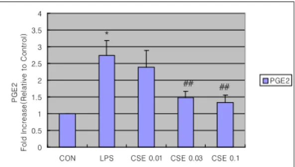
7. PGE
2의 측정
THE를 농도별로 처치하고, 1시간 후에 LPS 를 처치한 후, 배양된 배지를 E-tube에 모아 서 ELISA Kit (RnD Systems, Minneapolis, MN, USA)를 이용한 PGE
2측정에 사용하였다. PGE
2와 link된 acetylcholinesterase가 도포되어 있는 plate에 standard와 배양 배지를 첨가하여 4℃
에서 18시간 동안 반응시켰다. 18시간 후에 standard solution과 배지를 제거하고 Kit에 포 함되어 있는 wash buffer로 5회 반복 세척하였 다. 200 ul의 substrate reagent를 첨가하여 37°C 에서 1시간 동안 반응시킨 후, stop solution을 첨가하여 반응을 종결시켰다. PGE
2의 함량은 405 nm에서 흡광도를 측정하여 standard curve 에 준하여 결정하였다.
8. 통계적 검증
실험 결과는 mean±SD로 나타내었으며, 처 치군 간의 유의성은 one way analysis of varience (ANOVA)로 검정한 후 Newman - Keuls test로 검정하였다. 통계적 유의성 검정은 p <0.05 또 는 p <0.01로 하였다.
Ⅲ. 結 果
1. THE가 LPS로 유도된 Raw cell의 NO production에 미치는 영향
THE의 NO 생성억제정도를 관찰하기 위하 여 THE를 0.01, 003, 0.1 ㎎/㎖의 농도로 Raw 264.7 cell에 처리하여 생성되는 NO의 양을 측 정하였다.
NO의 양은 처치를 하지 않은 control에 대 한 비율로 나타내었다.
LPS군에서는 control군에 비교하여 NO의 생 성량이 유의성(P<0.01) 있게 증가하였으며, THE 0.01 ㎎/㎖를 처치한 실험군에서는 LPS군에 비 해 유의성이 없었으나, THE 0.03, 0.1 ㎎/㎖를 처치한 실험군에서는 LPS군에 비해 유의성 (P<0.01) 있게 NO의 생성을 억제하였다(Fig. 1).
2. THE가 Raw cell의 생존율에 미치는 영향
THE 0.01, 0.03, 0.10 mg/ml의 농도에서 LPS
로 유도된 NO 생성의 감소가 THE의 세포독
성으로 인한 것인지를 관찰하기 위하여 THE
0.01, 0.03, 0.10 mg/ml의 농도로 처리하고 24
0.8 0.85 0.9 0.95 1 1.05
CON LPS CSE 0.01 CSE 0.03 CSE 0.1 Cell Viability (% of Control)
viability
**
Fig. 2. Effects of Taraxaci Herba Extract(THE) on the cell viability in LPS stimulated Raw 264.7 cells.
The cell viability was measured after indicated time.
Data represent the mean±SD with three separate experiments.
0 2 4 6 8 10 12 14
CON LPS CSE 0.01 CSE 0.03 CSE 0.1 TNF-α Fold Increase(Relative to Control)
TNF-α
**
##
#
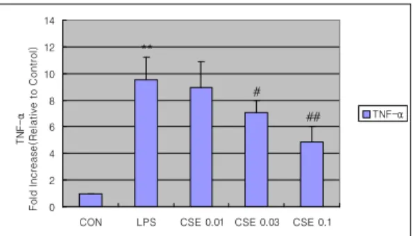
Fig. 3. The effect of Taraxaci Herba Extract(THE) on LPS-inducible TNF-α production.
Production of TNF-α was measured in the medium of Raw 264.7 cells cultured with LPS(1㎍/㎖) in the presence or absence of THE for 18hr.
The amount of TNF-α was measured by immunoassays as described in Section 2.
Data represent the mean±SD with three separate experiments(*: significant as compared to control, **P<0.01,
#: significant as compared to LPS alone, ##P<0.01).
시간 후 MTT assay를 실시하여 세포생존율을 측정하였다. LPS 단독 처리군과 THE 0.01, 0.03, 0.10 mg/ml 군에서는 control군에 비교하 여 유의한 세포독성을 나타내지 않았다(Fig. 2).
3. THE가 LPS로 유도된 Raw cell의 TNF-α에 미치는 영향
LPS로 유도된 RAW 264.7 세포에서 THE의 TNF-α 생성 저해능을 관찰하기 위하여 THE를 0.01, 0.03, 0.10 mg/ml의 농도로 세포에 처리 하고 18시간 후 ELISA kit를 사용하여 TNF-α 생성 정도를 측정하였다.
LPS는 전염증성 cytokine인 TNF-α의 생성을 유의성 있게 증가시켰고, THE 0.03, 0.1 ㎎/㎖
를 처치한 실험군에서는 LPS로 유도된 TNF-α 를 LPS단독 처리군에 비해 각각 유의성(P<
0.05 또는 P<0.01) 있게 감소시켰다(Fig. 3).
4. THE가 LPS로 유도된 Raw cell의 IL-1β에 미치는 영향
LPS로 유도된 RAW 264.7 세포에서 THE의
IL-1β 생성 저해능을 관찰하기 위하여 THE를
0.01, 0.03, 0.10 mg/ml의 농도로 세포에 처리
하고 18시간 후 ELISA kit를 사용하여 IL-1β 생
0 2 4 6 8 10 12
CON LPS CSE 0.01 CSE 0.03 CSE 0.1 IL-6 Fold Increase(Relative to Control)
IL-6
**
# ##
Fig. 5. The effect of Taraxaci Herba Extract(THE) on LPS-inducible IL-6 production.
Production of IL-6 was measured in the medium of Raw 264.7 cells cultured with LPS(1㎍/㎖) in the presence or absence of THE for 6hr.
The amount of IL-6 was measured by immunoassays as described in Section 2.
Data represent the mean±SD with three separate experiments(*: significant as compared to control, **P<0.01).
0 0.5 1 1.5 2 2.5
CON LPS CSE 0.01 CSE 0.03 CSE 0.1 IL-1β Fold Increase(Relative to Control)
IL-1β
**
#
## ##
Fig. 4. The effect of Taraxaci Herba Extract(THE) on LPS-inducible IL-1β production.
Production of IL-1β was measured in the medium of Raw 264.7 cells cultured with LPS(1㎍/㎖) in the presence or absence of THE for 12hr.
The amount of IL-1β was measured by immunoassays as described in Section 2.
Data represent the mean±SD with three separate experiments(*: significant as compared to control, **P<0.01,
#: significant as compared to LPS alone, ##P<0.01).
0 0.5 1 1.5 2 2.5 3 3.5 4
CON LPS CSE 0.01 CSE 0.03 CSE 0.1 PGE2 Fold Increase(Relative to Control)
PGE2
*
## ##
Fig. 6. The effect of Taraxaci Herba Extract(THE) on LPS-inducible PGE2 production