서론
피지샘 암종(sebaceous gland carcinoma)은 피지샘의 부 속 상피(adnexal epithelium)에서 발생하는 암으로, 흔하 지는 않지만 매우 공격적인 성향을 지니고 있어 전이와 관련된 사망률(metastasis-related mortality)이 높게는 40%
까지 보고되기도 한다.1) 피지샘 암종은 눈 주위 영역
(periocular region)에 호발하는데 가장 흔하게 발생하는 부위는 안검(eyelid)이며, 두 번째 호발하는 부위는 이하 선(parotid gland)으로 알려져 있다.2) 주로 6-70대에서 호 발하며, 여성에서 조금 더 많이 발생하는 것으로 알려져 있다. 통증이 없고 천천히 자라는 경우가 많아 임상적으 로 양성종양과 큰 차이가 없어 늦게 진단되는 경우도 많다.3) 경부 림프절 전이나 이하선 주변 림프절로 전이 가 있을 경우 5년 생존율이 5-60% 정도로 매우 낮아지는 위험한 암종이다.4) 저자들은 안검 종양의 광범위 절제술 을 받고 피지샘 암종으로 진단받은 환자에서 수술 당시 관찰되던 이하선 종양이 2년 뒤에 경부 림프절 전이를 동반한 피지샘 암종으로 진단된 1예를 경험하였기에 문 헌 고찰과 함께 보고하는 바이다.
대한두경부종양학회지, 제36권 제2호, 2020. pp.21-25 Korean Journal of Head & Neck Oncology, Vol.36, No.2
https://doi.org/10.21593/kjhno/2020.36.2.21 ISSN 1229-5183(Print) / ISSN 2586-2553(Online)
이하선으로 전이된 안검의 피지샘 암종 1예
채희성1⋅양희준1⋅백승원1⋅김지훈2+
연세대학교 원주의과대학 이비인후과학 교실1, 국민건강보험 일산병원 이비인후과2
A Case of Parotid Metastasis from Sebaceous Carcinoma of the Eyelids
Hee Sung Chae, MD1, Hui Joon Yang, MD1, Seung Won Paik, MD1, Ji-Hoon Kim, MD2+
Department of Otorhinolaryngology-Head and Neck Surgery, Yonsei University Wonju College of Medicine, Wonju, Korea1, Department of Otorhinolaryngology, National Health Insurance Service Ilsan Hospital, Goyang, Korea2
= Abstract =
Sebaceous carcinoma is a relatively rare and aggressive malignant tumor. Periocular area (especially eyelid) is the most common lesion to occur, and the most common extraocular lesion is the parotid gland. Because the lesion also mimic other benign inflammatory diseases, this leads to delayed diagnosis or misdiagnosis. Here, we report a 58-year-old male patient who presented with a non-tender painless left parotid mass after wide excision of sebaceous carcinoma in the left eyelid two years ago. When he was diagnosed with sebaceous carcinoma of left eyelid, there was a small left parotid tumor on the computed tomography. But no further examination and treatment were performed. Two years later, physical examination revealed growing parotid tumor and multiple neck nodes on the left side. After radical parotidectomy and neck dissection, histological examination showed a sebaceous carcinoma and neck node metastasis. Considering the aggressiveness of sebaceous carcinoma, further evaluation for parotid glands should be considered when sebaceous carcinoma of the eyelid was discovered.
Postoperative chemoradiotherapy was performed for disease control. Follow up after two years, and computed tomography showed no sign of recurrence.
Key Words : Sebaceous carcinoma⋅Parotid gland metastasis
Received Revised Accepted
: April 8, 2020 : July 24, 2020 : July 28, 2020
+Corresponding author: Ji-Hoon Kim, MD
Department of Otorhinolaryngology, National Health Insurance Service Ilsan Hospital 100, Ilsan-ro, Ilsandong-gu, Goyang-si, Gyeonggi-do, Korea
Tel: +82-31-900-0253, Fax: +82-31-900-3366 E-mail: [email protected]
증례
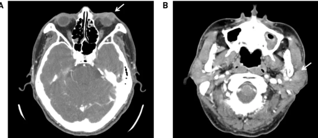
기저질환이 없는 58세 남자 환자가 수개월 전부터 점차 커지기 시작한 좌측 이하선의 무통성 종물을 주소로 내원 하였다. 신체검사 상 좌측 이하선 부위에 약 2 x 3 cm 크기의 단단하고 주변 조직과 고정된 양상의 종물이 촉 진되었다. 좌측 경부 level II에는 1 x 1 cm 가량의 림프절 비대가 관찰되었고 안면마비는 관찰되지 않았다. 환자 는 2년 전 좌측 하안검 부위에 생긴 종물로 본원 안과에 내원한 과거력이 있었다. 당시 시행한 안와 컴퓨터 단층 촬영 검사(orbital CT)에서 1.1 x 0.9 cm 크기의 비균질성 의 조영 증강을 보이는 결절성 병변이 좌측 하안검 부위 에 관찰되었으며, 원뿔 내외 공간(extra & intraconal space) 으로의 침범은 보이지 않았다(Fig. 1A). 안검 종물과 함께 좌측 이하선에 1cm 가량의 약하게 조영 증강되는 종물이 관찰되고 있으나, 당시에는 이하선 종양에 대한 추가 검 사는 진행되지 않았다(Fig. 1B). 본원 안과에서 하안검 종물에 대해서 국소마취하에 절제 생검술(excisional bi- opsy)을 시행하였으며, 조직병리학적 검사 상 미분화된 양 상의 악성 종양이 의심되는 결과가 보고되어 추가 수술이 필요한 상황이었다. 이후 환자는 연고지 관계상 타 병원에 내원하여 국소마취하에 외안각(lateral canthus)을 포함한 하안검 가측 일부에 대해 추가적인 광범위 절제술을 시행 받았으며 최종 조직병리 검사 상 최장 길이는 0.7 cm, 두께 는 0.4 cm인 피지샘 암종으로 확인되었다. 5 mm 이상의 절제연을 확보하였고, 신경 주위 침윤(perineural invasion), 림프 혈관 침윤(lymphovascular invasion)은 관찰되지 않 았다. 이후 환자는 추가 치료 없이 수술을 시행 받은 병원 에서 추적 관찰을 하였다.
환자는 안검 종양을 수술한 지 2년이 지나 이하선의 종물을 주소로 본원 외래에 내원하였다. 내원 당일 종물
의 정확한 조직학적 평가를 위해 세침 흡인 세포 검사를 시행하였으며, 병리과로부터 선낭 암종(adenoid cystic car- cinoma) 혹은 전이성 암종(metastatic carcinoma)이 의심된 다는 소견을 받았다. 종양의 분화가 좋지 않아 세침 흡인 검사만으로는 정확하게 평가가 어려워 이하선 종물에 대한 병리학적 확진을 위하여 수술적 제거를 계획하였 다. 수술 전 시행한 경부 컴퓨터 단층 촬영 검사(neck CT)에서는 약 3 x 2.2 cm 크기의 주변과 경계가 명확하지 않으며 조영이 증강되는 타원형의 종물이 좌측 이하선 천엽에서 기시하여 심엽까지 일부 확장되어 있는 것을 관찰할 수 있었다(Fig. 2A). 또한, 좌측 경부 level II 구역 에도 전이를 의심할만한 림프절들이 관찰되었다(Fig.
2B, 2C). 양전자 방출 단층 촬영 검사(PET-CT)에서는 표 준화 섭취 계수(standard uptake value, SUV) 8.6의 고대사 (hypermetabolic) 종물이 좌측 이하선에서 관찰되었으며 (Fig, 2D), level II, III의 경부 림프절도 전이가 의심되는 상황이었다(Fig. 2E, 2F). 타 장기로의 원격 전이는 관찰 되지 않았다(Fig. 2G). 침샘암 및 경부 림프절 전이 의심 하에 이하선 전절제술 및 제2형 변형 근치적 경부 절제술 을 시행하였다(Fig. 3). 수술 당시 이하선 종물은 안면신 경의 기시부와는 분리가 되었으나, 저작근과 안면신경 의 가지에 유착되어 있어 경부 가지(cervical branch)와 변연 하악 가지(marginal mandible branch)를 제외한 안면 신경의 분지들은 희생되었다. 이하선의 심엽은 이복근 (digastric muscle), 경상 설골근(stylohyoid muscle), 그리 고 경상돌기(styloid process)로부터 박리하여 분리시켜 제거하였다. 전반적으로 종물이 주변의 근육과 유착되 어 있어 근육을 일부 포함하여 적절한 변연을 확보하며 종물을 제거하였다. 수술 후 10일째 배액관 제거 후 특이 합병증 없이 퇴원하였다. 제거된 이하선 종양은 3.2 x 3.0 x 1.7 cm의 크기로 최종 조직병리소견 상 H&E 염색 A
B
Fig. 1. Preoperative imaging evaluation of the eyelid mass. (A) Enhanced CT scan showed heterogeneous mass in the left lower eyelid (white arrow). (B) A small mass of the left parotid gland was incidentally observed at the time of diagnosis (white arrow).
에서 거품 세포질(foamy cytoplasm) 양상의 공포성 세포 질(vacuolated cytoplasm)을 가진 세포가 기저양 세포(ba- saloid cell)로 둘러싸인 모습이 보이며 세포 간의 경계가 불분명하게 관찰되었다. 또한 표피층까지의 파제트병 모양의 성장(pagetoid growth)이 보이며 주위로 괴사 소견 이 관찰되었다(Fig. 4A, 4B). 면역조직화학검사 상 피지 세포에서 특징적으로 양성반응을 보이는 epithelial mem- brane antigen (EMA), androgen receptor (AR) 염색에서 양성반응을 보여 최종적으로 피지샘 암종으로 진단하였 다(Fig. 4C, 4D). 상기 이하선 암종은 절제연이 양성이었으
며, 고분화성 종양으로 샘외 침범(extraglandular extension) 이 관찰되었고 신경 주위 침윤, 림프 혈관 침윤이 모두 관찰되었다. 경부 림프절 조직은 8개에서 전이성 암종이 확인되었으며, level II의 4개의 림프절에서 피막외 침범 (extranodal extension)이 확인되었다. 환자의 병리 결과를 종합하여 pT2N2bM0로 AJCC 병기 IVA로 최종 진단하 였다. 수술 후 다학제 진료를 통해 시스플라틴을 기반으 로 한 동시 항암방사선치료를 진행하였다. 6차례에 걸쳐 항암제를 투여하였고, 33회에 걸쳐 좌측 경부 림프절 부 위에 61.05Gy, 이하선 부위에 69.96Gy의 방사선치료를 A
B
C
D
E
F
G
Fig. 2. Preoperative imaging studies. (A-C) Enhanced CT scan showed a 3.0 x 1.6 x 2.2 cm sized ill-defined lobulated mass in the left parotid gland and multiple enlarged lymph nodes. (D-G) Axial fused image of PET-CT scan showed intense FDG uptakes at the left parotid gland and level II lymph nodes. No distant metastases were revealed.
A
B
Fig. 3. Intraoperative findings and resected specimens. (A) Image shows the surgical site after left radical parotidectomy and neck dissection. (B) 3.2 x 3.0 x 1.7 cm sized firm, irregular surfaced parotid gland mass and left level II-V lymph nodes.
A B C D
Fig. 4. Microscopic and immunohistochemistry findings. (A) H&E staining shows the tumor lobules composed of large foamy cells surrounded by basaloid cells (x 200). (B) Numerous cells with sebaceous differentiation is shown (x 200). (C) Diffuse positive for epithelial membrane antigen (EMA) is shown (x200). (D) Diffuse positive for androgen receptor (AR) is shown (x200).
시행하였다. 환자는 항암방사선치료 종료 후 2년까지 영 상학적 검사에서 재발 소견은 관찰되지 않고 있다. 안면 마비는 House-Brackmann grade 5 수준으로 관찰되어 보 존적인 치료를 유지하고 있으며, 외래에서 재발 여부를 지속적으로 추적 관찰할 예정이다.
고찰
피지샘 암종은 1-5% 가량의 빈도로 발생하는 비교적 드문 악성 종양으로, 안검에 생기는 악성 종양 중 기저 세포암(basal cell carcinoma)과 편평 세포암(squamous cell carcinoma)에 이어 3번째로 많이 발생하는 것으로 알려 져 있다.5) 피지샘 암종은 초기에는 임상적으로 안검 결 막염 같은 염증성 질환과 유사하게 보이는 경우가 많아 진단이 늦어지는 경우가 많다.6,7) 노령, 여성, 아시아인, 두경부 영역에 방사선 치료를 받은 과거력, Muir-Torre syndrome의 유전적 소인, 가족성 망막모세포종(familial retinoblastoma) 등이 현재까지 알려진 발병의 위험인자
이다.8-11) 피지샘 암종은 세포 이형성 정도에 따라 고분화
(well-differentiated), 중분화(moderate-differentiated), 저분 화(poorly-differentiated)로 분류하긴 하지만, 조직학적으 로도 다양한 표현형을 지니기 때문에 병리 진단이 쉽지 않다. 세포형태학적으로 피지샘 암종은 표피층으로의 파제트병 모양 전파(pagetoid spread)를 동반한 소엽상 성 장(lobular growth)과 공포성 세포질(vacuolated cytoplasm) 이 정중앙에 위치한 다각형의 세포(polygonal cell)들이 군집을 이루거나 산재한 형태로 나타난다.12) 현미경적으 로는 다공포성의 투명 세포질(multivacuolated clear cyto- plasm)을 동반한 세포 소엽(cell lobule)이 관찰되며, 핵-세 포질 비(nucleo-cytoplasmic ratio)가 증가하고, 응집된 염 색질이 나타난다.13) 면역조직화학 염색에서는 종양 세포 에서 피지(sebaceous) 혹은 선상(glandular) 분화가 동시 에 확인되기도 한다.14) 또한, EMA, AR, anti-epithelial an- tigen (BerEP4), anti-adipophilin (ADP), p53, Ki-67등의 면 역조직화학 염색을 통해 편평 세포암이나 기저 세포암과 감별할 수 있다. 감별진단이 필요한 기저 세포암의 경우 EMA에서 음성반응을 보이며, 편평 세포암의 경우 AR에 서 음성반응을 보이게 된다.15)
피지샘 암종의 치료는 5mm 이상의 충분한 절제연을 확보하는 수술적 절제가 원칙이다. 국소 림프절 전이가 있을 경우에는 원발 부위의 절제와 경부 림프절 절제술 을 함께 시행하며, 수술 후 방사선 치료를 권고하고 있
다.6,16,17) 일반적으로 피지샘 암종의 경우 신경 주위 침윤
이 약 20% 정도에서 관찰되나 림프 혈관 침윤은 드문
것으로 알려져 있는데, 본 환자의 경우 신경 주위 침윤과 림프 혈관 침윤이 모두 관찰 되었다. 일반적으로 피지샘 암종의 치료에 항암치료를 적용하지 않고 있으나, Jung 등은 재발한 전이성 피지샘 암종 환자에서 5-플루오로우 라실과 시스플라틴의 병합 요법을 이용하여 치료한 사례 를 보고한 바가 있다.18) 수술 후 동시 항암방사선치료가 방사선치료 단독에 비해 생존율에 이익을 주는 지에 대 한 연구는 아직까지 부족하다. 하지만 본 증례의 경우 환자의 전신 상태가 양호하였고 치료에 적극적이었으며, 이하선 종양의 조직학적 소견을 고려하여 의료진의 상의 하에 동시 함암방사선치료를 하기로 결정하였다. 수술 후 동시 항암방사선치료의 효과를 증명하기 위해서는 많은 수의 환자를 대상으로 추가 연구가 필요할 것으로 생각된다.
본 증례의 경우 타 병원에서 수술 당시 안검 종물의 평가를 위해 시행한 안와 컴퓨터 단층 촬영 검사에서 안검 종물과 함께 좌측 이하선에 1cm 가량의 약하게 조 영 증강되는 종물이 관찰되었다(Fig. 1B). 2년 후에 동일 한 좌측 이하선 부위에 발생한 피지샘 암종은 안검의 원발 병소와 병리학적으로 동일하다는 점과 암종이 발생 한 시기의 시간적 선후관계를 고려하였을 때, 안검의 피 지샘 암종이 발견되었을 당시에도 전이의 가능성에 대한 충분한 평가가 필요하였을 것으로 생각된다. 피지샘 암 종의 공격성을 고려하였을 때 이하선 종물에 대한 적극 적인 치료가 고려되어야 했으나 원발 부위만 치료하고 종료하였던 점이 다소 아쉬운 부분으로 생각된다. 피지 샘 암종의 경우 인종별로 임상 양상 및 전이 패턴이 다소 차이가 있으며, 종양 자체가 흔하지 않기 때문에 전이에 대한 진단 및 치료가 늦어지는 경우가 있을 수 있다. Park 등이 29명의 한국인을 대상으로 눈 주위에서 발생한 피 지샘 암종 환자들의 임상병리학적 분석 결과를 보고한 논문에서도 1명에서만 경부 전이가 확인되었다.19) 이와 같이 한국인의 경우 피지샘 암종의 전이 양상이 기존에 서양인에서 보고된 바와 다소 차이를 보이고 있기 때문 에 안검 주위의 악성 종양으로 진단된 환자에서 이하선 및 주위 림프절 영역에 대해 전이를 간과할 가능성이 있을 수 있다. 저자들은 상기 증례를 통해 안검에 발생한 악성 종양의 경우에 두경부 영역으로의 전이 여부를 보 다 적극적으로 평가할 필요가 있다는 것을 확인하였다.
추후 피지샘 암종의 국소 전이에 대한 정확한 진단과 치료 지침을 제시하기 위해서는 더 많은 증례 분석이 필요할 것으로 생각된다.
References
1) Kaliki S, Ayyar A, Dave TV, Ali MJ, Mishra DK, Naik MN.
Sebaceous gland carcinoma of the eyelid: clinicopathological features and outcome in Asian Indians. Eye (Lond). 2015;29:
958-963.
2) Mighell AJ, Stassen LF, Soames JV. Sebaceous carcinoma of the parotid gland. Dentomaxillofac Radiol. 1996;25:51-53.
3) Gnepp DR, Brannon R. Sebaceous neoplasms of salivary gland origin. Report of 21 cases. Cancer. 1984;53:2155-2170.
4) Dasgupta T, Wilson LD, Yu JB. A retrospective review of 1349 cases of sebaceous carcinoma. Cancer. 2009;115:158-165.
5) Cook BE Jr, Bartley GB. Treatment options and future prospects for the management of eyelid malignancies: an evidence-based update. Ophthalmology. 2001;108:2088-2121.
6) Husain A, Blumenschein G, Esmaeli B. Treatment and outcomes for metastatic sebaceous cell carcinoma of the eyelid. Int J Dermatol. 2008;47:276-279.
7) Leibovitch I, Selva D, Huilgol S, Davis G, Dodd T, James CL.
Intraepithelial sebaceous carcinoma of the eyelid misdiagnosed as Bowen's disease. J Cutan Pathol. 2006;33:303-308.
8) Shields JA, Demirci H, Marr BP, Eagle RC Jr, Shields CL.
Sebaceous carcinoma of the ocular region: a review. Surv Ophthalmol. 2005;50:103-122.
9) Snow SN, Larson PO, Lucarelli MJ, Lemke BN, Madjar DD.
Sebaceous carcinoma of the eyelids treated by mohs micro- graphic surgery: report of nine cases with review of the literature.
Dermatol Surg. 2002;28:623-631.
10) Kivelä T, Asko-Seljavaara S, Pihkala U, Hovi L, Heikkonen J.
Sebaceous carcinoma of the eyelid associated with retinoblastoma.
Ophthalmology. 2001;108:1124-1128.
11) Stockl FA, Dolmetsch AM, Codère F, Burnier MN Jr. Sebaceous carcinoma of the eyelid in an immunocompromised patient with Muir-Torre syndrome. Can J Ophthalmol. 1995;30:324-326.
12) Stern RC, Liu K, Dodd LG. Cytomorphologic features of seba- ceous carcinoma on fine needle aspiration. Acta Cytol. 2000;44:
760-764.
13) Jain P, Nanda A, Handa U, Bal A, Mohan H, Gupta SK. FNA di- agnosis of recurrent sebaceous carcinoma. Diagn Cytopathol.
2006;34:124-126.
14) Marnouche el A, Maghous A, Kadiri S. Sebaceous carcinoma of the parotid gland: a case report and review of the literature. J Med Case Rep. 2016;10:174.
15) Hasebe T, Mukai K, Yamaguchi N. Prognostic value of im- munohistochemical staining for proliferating cell nuclear anti- gen, p53, and c-erbB-2 in sebaceous gland carcinoma and sweat gland carcinoma: comparison with histopathological parameter.
Mod Pathol. 1994;7:37-43.
16) MacFarlane JK, Viloria JB, Palmer JD. Sebaceous cell carcino- ma of the parotid gland. Am J Surg. 1975;130:499-501.
17) Callahan EF, Appert DL, Roenigk RK, Bartley GB. Sebaceous carcinoma of the eyelid: a review of 14 cases. Dermatol Surg.
2004;30:1164-1168.
18) Jung YH, Woo IS, Kim MY, Han CW, Rha EY. Palliative 5-fluo- rouracil and cisplatin chemotherapy in recurrent metastatic se- baceous carcinoma: Case report and literature review. Asia Pac J Clin Oncol. 2016;12:e189-193.
19) Park SK, Park J, Kim HU, Yun SK. Sebaceous Carcinoma:
Clinicopathologic Analysis of 29 Cases in a Tertiary Hospital in Korea. J Korean Med Sci. 2017;32:1351-1359.