• 검색 결과가 없습니다.

Inhibitory Effects of Camellia sinensis Extract on the Development of Atopic Dermatitis-like Lesions in NC/Nga Mice

N/A
N/A
Protected

Academic year: 2021

Share "Inhibitory Effects of Camellia sinensis Extract on the Development of Atopic Dermatitis-like Lesions in NC/Nga Mice"

Copied!
11
0
0

로드 중.... (전체 텍스트 보기)

전체 글

(1)

Inhibitory Effects of Camellia sinensis Extract on the Development of Atopic Dermatitis-like Lesions in NC/Nga Mice

1

Tae Hong Kim

2

⋅Si Young Ha

2

⋅Jae-Kyung Yang

2,†

ABSTRACT

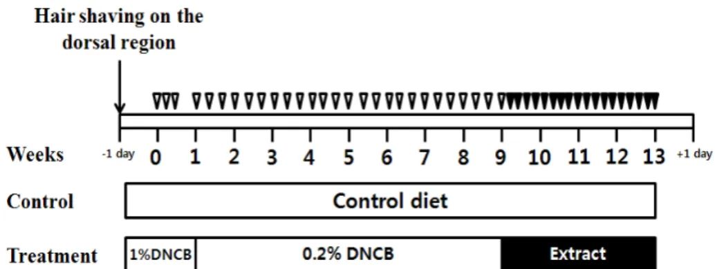
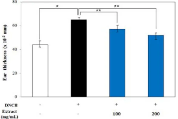
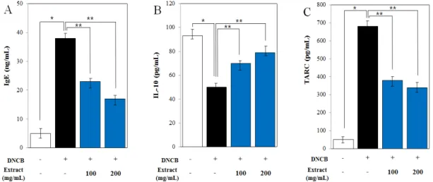
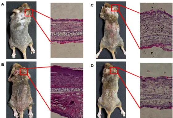
Atopic dermatitis (AD) syndrome is one of the most common and severe skin diseases in Korea; a large population has this disease. We examined the effects of the extract from the leaf and sprig of Camellia sinensis on the develop- ment of AD by using NC mice as a model of atopic dermatitis. Oral administration of the extract to NC/Nga mice treated with 2,4‐dinitrochlorobenzene (DNCB) inhibited the development of AD-like skin lesions as shown by a sig- nificant decrease in the skin symptoms of the disease and a decrease in ear thickness and levels of immunoglobulin E (IgE) and thymus-and activation-regulated chemokine (TARC) level in the skin. Administration of the extract mark- edly suppressed the DNCB-induced mRNA expression of interleukin 4 (IL-4) and tumor necrosis factor α (TNF-α).

The findings suggest that transdermal application of the extract may modulate in the skin of NC/Nga mice. The ex- tract was effective for the prevention and treatment of AD.

Keywords :

atopic dermatitis, NC/Nga mice, IgE, Camellia sinensis

1 Date Received February 13, 2014, Date Accepted April 1, 2014

2 Department of Environmental Materials Science, Institute of Agriculture & Life Science, Gyeongsang National University, Jinju, 660-701, South Korea

Corresponding author : Jae-Kyung Yang (e-mail: [email protected])

1. INTRODUCTION

The incidence of AD has recently increased in in- dustrialized countries, and although the onset of AD is usually during early infancy and childhood, it can also occur in adulthood (Cooper, 1994; Rudikoff and Lebwohl, 1998). Although topical steroids, emol- lients, and oral anti-histamines are used as the first-line therapy for AD, many patients are still con- cerned about the long-term use of these agents (Furue et al. 2003). Currently, treatment and pre- vention of AD through dietary intervention is receiv- ing considerable attention. Recent studies indicate

that intake of diet containing lactic acid bacteria and natural polyphenols such as astragalin and genistein suppresses AD-like skin lesions (Wakabayashi et al.

2008; Kotani et al. 2000). The prevalence of AD has increased progressively by 2 to 3 fold during the past three decades in industrialized countries with a lifetime prevalence of 10-20% in children and 1-3%

in adults (Leung and Bieber, 2003; Schultz and

Hanifin, 2002). Topical glucocorticoids are used for

the treatment of AD as important and effective

remedies. However, these agents can be used only

for a short period of time and in limited skin re-

gions, because long-term use of glucocorticoids caus-

(2)

es a variety of potential side effects (Barnetson and White, 1992). Therefore, the use of complementary or alternative medicine (CAM), a main alternative resource being phytotherapy (use of natural extracts as therapeutic agents), has become increasingly pop- ular for the treatment of AD, and CAM has been used to overcome the shortcomings of conventional therapy (Boneberger et al. 2010). Thus, because of an increase in the use of CAM in AD patients, fur- ther studies are required to establish their efficacy, safety, and therapeutic use (Boneberger et al. 2010).

C. sinensis, a tree from which a beverage com- monly used in Asian countries is obtained, contains compounds having strong antioxidant capacities. It is a significant source of polyphenols, including (-)-epi- gallocatechin gallate (EGCG), (-)-epigallocatechin, (-)-epicatechin gallate, and (-)-epicatechin. These pol- yphenols have recently attracted attention as bio- active agents with anticancer, antidiabetic, antiviral, antimalarial, hepatoprotective, neuroprotective, and cardioprotective effects (Adhami et al. 2007; Noonan et al. 2007). However, little is known about the ben- eficial effects of C. sinensis extract on AD (Lee et al. 2006; Rowe et al. 2007).

Polyphenols obtained from C. sinensis, referred to type of catechins, are present in the leaves of the tea plant. Several in vitro (Trompezinski et al. 2003;

Huang et al. 2005) and in vivo (Afaq et al. 2003;

Hsu et al. 2007) studies have shown that a group of polyphenolic catechins, their major constituent being EGCG, exhibits multiple biological effects on the skin. Trompezinski et al. (Trompezinski et al. 2003) have shown that EGCG inhibits, in a dose-dependent manner, the upregulation of both vascular endothelial grow th factor and IL-8 in T N F-α stim ulated keratinocytes. Afaq et al. (Afaq et al. 2003) have shown that topical application of green tea poly- phenol to SKH-1 hairless mice before ultraviolet-B exposure significantly decreased skin edema and in-

filtration of leukocytes via modulations in the mi- togen-activated protein kinase and nuclear factor kap- pa B signaling pathways. These findings have sug- gested that EGCG may be a novel therapeutic agent for various inflammatory skin diseases such as AD.

Kim et al. (Kim et al. 2012) showed that bath ther- apy with green tea extract is an effective, safe, and non-steroidal therapy for the treatment of patients with AD associated with Malassezia sympodialis.

To date, several animal models have been devel- oped for studies on AD (Yang et al. 2013; Turner et al. 2013; Yoon et al. 2011). The AD model of NC/Nga mice is clinically and histologically very similar to human AD, when mice are raised under conventional conditions (Matsuoka et al. 2003).

NC/Nga mice originated from the Japanese fancy mice at Nagoya University (Japan) in 1957, and they were established as an inbred strain for creating an animal model of human AD. NC/Nga mice raised in air-uncontrolled conventional circumstances sponta- neously develop AD-like skin lesions with a mark- edly elevated serum level of IgE, whereas NC/Nga mice maintained in specific pathogen-free (SPF) con- ditions do not show clinical signs of AD or IgE hy- perproduction (Matsuda et al. 1997). However, a va- riety of antigens can be involved in the development of AD-like skin lesions in NC/Nga mice under con- ventional conditions, which makes it difficult to ana- lyze the pathogenesis of AD-like skin lesions (Shiohara et al. 2004).

The objective of this study was to evaluate the ef-

fects of administration of C. sinensis extract on the

progress of AD-like skin lesions in NC/Nga mice by

continuous application of the DNCB. C. sinensis ex-

tract showed excellent anti-AD activity and thus

could be possibly used to treat and prevent AD. This

study may further provide insights on the potential

of C. sinensis extract as an integral part of a novel

therapeutic modality for AD in humans or animals.

(3)

2. MATERIALS and METHODS 2.1. Preparation of C. sinensis Extracts

For this study, the leaves and branches of C. si- nensis were collected from the Hadong green tea in- stitute (Hwagae-myeon, Hadong-gun, Gyeongnam-do, Korea, 35°N and 127°E) in Hadong, Korea. The leaves and branches were cut using a pair of pruning shears. The leaves and branches were spread out on a large flat surface to dry. To ensure heterogeneity among samples, all leaves were stored at room tem- perature until a constant fresh weight was obtained before use.

The leaves and branches were pretreated to per- form a steaming treatment. Steaming of leaves and branches was performed in an autoclave set at a de- sired temperature of 121℃ for 30 min.

The extracts of pretreated leaves and branches were prepared under two conditions of hot water and ethanol treatment. The hot water extract of pretreated leaves and branches was exactly weighed (50 g) in an Erlenmeyer flask, 1000 mℓ distilled water was added to the flask, and the extract was autoclaved at the desired temperature of 121℃ for 30 min. The ethanol extract of pre-treated leaves and branches was exactly weighed (50 g) in an Erlenmeyer flask, 1000 mℓ of 80% ethanol solution was added into the flask, and the extract was placed in a shaking in- cubator at 25℃ for 48 h.

Then, the extracts were filtered through a glass filter (2G3). The hot water and ethanol extracts were concentrated in a rotary evaporator under reduced pressure at 40℃ and then freeze-dried. Both extracts were stored at 4℃ for subsequent analyses.

The yield from dried hot water and ethanol ex- tracts was approximately 19.7% and 26.1% (w/w), respectively. The hot water extract and ethanol ex- tract were mixed (1:1, w/w) for the experiment.

2.2. Experimental Animals and Diets Preparation

Specific pathogen-free female 5-week-old NC/Nga mice (25 ± 2 g) were purchased from SLC, Inc.

(Shizuoka, Japan). Animals were maintained for 1 week before the start of the experiments. They were housed in an air‐conditioned animal room with a temperature of 23 ± 2℃ and a humidity of 50 ± 10%. Mice were provided with solid feed (CRF‐1, Oriental Yeast, Tokyo, Japan).

2.3. Chemicals and Treatment in NC/Nga Mice Test

2,4-dinitrochlorobenzene (DNCB) was purchased from Sigma-Aldrich (Milwaukee, WI, USA). The ex- perimental schedule for the preparation of AD-like skin lesion in NC/Nga mice is summarized in Fig. 1.

The mice were divided into four groups (n = 5 per group). To induce AD-like immunologic and skin lesions, DNCB was applied onto the dorsal skin and ears. After complete removal of hair on the dor- sal surface of within an area of approximately 8 cm

2

, 200 µℓ of 1% DNCB solution (dissolved in a 3:1 mixture of acetone and olive oil) was applied on their dorsal skin and ears for three consecutive days fors ensitization. Four days after sensitization, the dorsal skin and ears were challenged with 200 µℓ of 0.2% DNCB solution three times per week for eight weeks. After inducing AD, the extract was applied to the dorsal skin and ears of the mice six times per week for four weeks.

A lotion without the extract was applied to the

dorsal skin and ears of the control and DNCB-treat-

ed mice. Animals were killed 64 days after the first

application of DNCB. Blood was collected from the

vena cava and the right ear was removed and used

for histopathological examination.

(4)

Fig. 1. Experimental schedule for the induction of AD-like lesion in NC/Nga mice. Deltas indicate the application of DNCB.

2.4. Body Weights and Ear Thickness of NC/Nga Mice

The body weights of the NC/Nga mice were monitored once a week during the experimental period. Ear thickness was measured with a micro- meter (Mitutoyo, Kawasaki, Japan) on the day of sacrifice. The right ear was removed from each mouse and fixed in 10% formalin solution. It was subsequently embedded in paraffin, sectioned (4 μm), and stained with hematoxylin and eosin.

2.5. Quantitation of Total IgE, IL-10, and TARC (ELISA assay)

Enzyme-linked immunosorbent assay (ELISA) kits for IgE, IL-10, and TARC were obtained from R&D Systems (Minneapolis, MN, USA) and BD Biosciences (San Diego, CA, USA). Cytokines in serum level were measured by sandwich ELISA using the OptEIA Set mouse IgE kit from BD Biosciences, the DuoSet mouse IL-10, and the mouse CCL17/TARC kits from R&D Systems, according to the manu- facturer’s instructions.

Cytokine concentrations were calculated using a linear regression equation obtained from standard ab-

sorbance values.

2.6. RT-PCR Analysis

Total RNA from the spleen was isolated with Trizol (Molecular Research Center Inc., Cincinnati, USA) according to the manufacturer’s instruction.

cDNA was synthesized from total RNA (2 g) using oligo (dT) 18 primers (0.5 g) and the Prime RT Premix kit (Daejeon, Korea) according to the manu- facturer’s instruction. The primers used in this study were as follows: IL-4 (forward) 5’- AGT TGT CAT CCT GCT CTT CTT T -3’, (reverse) 5’- GAC TGG GAC TCA TTC ATG GTG C -3’; TNF-α (forward) 5’- CAC CAC GCT CTT CTG TCT ACT GAA C -3’, (reverse) 5’- CCG GAC TCC GTG ATG TCT AAG TAC T -3’; and GAPDH (forward) 5’-TGA ACG GGA AGC TCA CTG G-3’ (reverse) 5’-TCC ACC ACC CTG TTG CTG TA-3’. The PCR re- action was performed using the Prime Taq Premix kit (GeNet Bio, Korea) and 0.1 mM GAPDH-specif- ic primer pair. All samples were pre-denatured 94℃

for 5 minutes, followed by thirty-two thermal cycles

(at 94℃ for 30 s, 55-62℃ for 30 s, and 72℃ for

1 minutes), and an additional extension at 72℃ for

7 minutes. Amplified cDNAs were subjected to elec-

(5)

Fig. 2. Body weight of DNCB-induced NC/Nga mice by transdermal application of extract. Extract (100 mg and 200 mg/kg) was application to the NC/Nga mice sensitized and challenged with DNCB as de- scribed in materials and methods. Body weights in each group of NC/Nga mice were monitored once a week starting 1 week before sensitization. Values represent the means ± S.D. of 5 mice for each group. (*p < 0.1, compared to the control group; **

p < 0.01, compared to the DNCB-treated group)

Fig. 3. Effects of C. sinensis extract on the ear thickness. Each value represents the mean ± SD of five mice. (*p < 0.1, compared to the control group;

** p < 0.01, compared to the DNCB-treated group)

trophoresis on 2% agarose gels and visualized by staining with ethidium bromide.

2.7. Histological Examination

Tissues from the right ear were removed and fixed in 10% paraformaldehyde solution for 24 hours. The tissues were embedded into the paraffin and blocks of 5 μm thickness were prepared. The sections were stained with haematoxylin and eosin (H&E) to dif- ferentiate inflammation and edema in epidermis, der- mis, keratinocytes, neutrophils and eosinophils.

2.8. Statistical Analysis

All experiments were repeated at least three times.

Results are reported as means ± standard error of the mean (SEM). Statistical significance was determined by a one-way analysis of variance (ANOVA), fol-

lowed by the Tukey-Kramer multiple comparisons test. A significant value was defined as p < 0.05.

3. RESULT and DISCUSSION

3.1. Change Body Weight of NC/Nga Mice

The extract was applied to the dorsal skin and ears of the mice six times per week for four weeks.

A lotion without the extract was applied to the dor- sal skin and ears of control and DNCB-treated mice.

The body weight of DNCB-treated mice was meas-

ured using a weighing balance. Each value represents

the mean ± SD of five mice (Fig. 2). The body

weight of the NC/Nga mice increased with an in-

crease in the concentration of the extract. The mean

body weights of the NC/Nga mice treated with the

extract (200 mg/mℓ) were not significantly different

from those of the normal NC/Nga mice. Our results

are representative of five separate experiments with

similar results. The body weights of mice treated

with the extract were within normal limits.

(6)

3.2. Ear Thickness of NC/Nga Mice

Changes in the ear thickness of mice are shown in Fig. 3. The ear thickness was measured immediately before DNCB application and killing the mice. The ear thickness was 43.1 ± 1.4 (× 10

-2

mm) in normal NC/Nga mice, 65.5 ± 2.5 (× 10

-2

mm) in DNCB- treated NC/Nga mice, 56.4 ± 7.3 (× 10

-2

mm) in NC/Nga mice treated with 100 mg/mℓ of the extract, and 49.1 ± 0.3 (× 10

-2

mm) in NC/Nga mice treated with 200 mg/mℓ of the extract. This finding showed that the ear thickness of normal NC/Nga mice was significantly lower than that of NC/Nga treated with the extract. In DNCB-treated NC/Nga mice, the ear swelling subsided was certainly remission after appli- cation of the 200 mg/mℓ of the extract, and the ear thickness decreased to a value similar to that of the normal mice. These results indicated the protective effects of the extract on decreasing swelling of the ear.

3.3. Serum Total IgE, IL-10 and TARC Levels of NC/Nga Mice

The serum IgE levels are shown in Fig. 4A.

Repeated application of DNCB increased the serum IgE levels in sensitized NC/Nga mice. Mast cells are key effectors in IgE-mediated allergic disorders.

Upon activation, mast cells undergo degranulation and release a variety of biologically active sub- stances such as IL-4, IL-5, and IL-13, which play an important role in the development of AD (Amin, 2012; Wesolowski and Paumet, 2011). An increase in serum IgE levels is an important characteristic of AD. To examine whether the extract decreases the increase in IgE induced by DNCB in NC/Nga mice, we measured the serum IgE levels by using en- zyme-linked immunoassay (ELISA) analysis.

We examined the serum levels of total IgE in

NC/Nga mice for four weeks. The total IgE levels in NC/Nga mice treated with the extract were sig- nificantly lower than those in the DNCB-treated NC/Nga mice. The serum IgE levels in the DNCB-treated NC/Nga mice (38.26 ± 5.51 ng/mℓ) were higher than those in the normal NC/Nga mice (p < 0.5), whereas the serum IgE levels in the NC/Nga mice treated were markedly decreased to 18.34 ± 6.31 ng/mℓ (extract 200 mg/mℓ) (p < 0.05).

IL-10 is an important mediator that prevents aller- gic inflammation, and development of AD-like skin lesions might be associated with suppression of regu- latory cytokines. IL-10 induces allergen-specific T-cell proliferation and suppresses the secretion of Th1- and Th2-type cytokines. Compared to normal controls, patients with severe AD show a decrease in IL-10 production (Pickard et al. 2007).

We examined IL-10 production from axillary lymph node cells obtained from normal mice and mice treated with the extract for four weeks.

Compared to the lymph node cells from DNCB- treated NC/Nga mice, those from the mice treated with the extract showed a marked increase in the IL-10 levels compared DNCB-induced NC/Nga mice (Fig. 4B).

The serum TARC levels in NC/Nga mice treated

with the extract significantly decreased during the

four weeks (p < 0.05). Development of skin lesions

in DNCB-treated NC/Nga mice induced a marked

decrease in the expression of TARC (Fig. 4C). The

NC/Nga mice treated with the extract (100 and 200

mg/mℓ) had lower TARC levels than the control

mice. Overexpression of TARC by the basal kerati-

nocytes as the lesions develop in the skin of the

NC/Nga mice suggests that TARC plays a key role

in the pathogenesis of the lesions. When mice kerati-

nocytes were cultured in the presence of several in-

flammatory cytokines, TNF-α was the most potent

inducer of TARC. In the present study, transdermal

(7)

Fig. 5. Expression of IL-4, TNF-α and GAPDH mRNA in mice treated repeatedly with DNCB and C. sinensis extract. Total RNA was prepared and an- alyzed for the expression of IL-4, TNF-α and GAPDH mRNA by RT-PCR. Results are shown representa- tives of five observations.

Fig. 4. Change in serum of IgE, IL-10 and TARC by C. sinensis application of DNCB in NC/Nga mice. The serum level of IgE, IL-10 and TARC was determined using mouse IgE specific ELISA kit. Each value repre- sents the mean ± SD of five mice. (A: IgE, B: IL-10, C: TARC) (*p < 0.5, compared to the control group;

** p < 0.05, compared to the DNCB-treated group.)

application of the extract modulated the serum IgE, IL-10, and TARC levels (IgE, 16.2 ng/mℓ in mice treated with 200 mg/mℓ of the extract; IL-10, 78.8

pg/mℓ in mice treated with for 200 mg/mℓ of the extract; and TARC, 337.9 pg/mℓ in mice treated with 200 mg/mℓ of the extract).

3.4. IL-4 and TNF-α Induce Stabilization of Eotaxin mRNA

The mRNA expression levels of IL-4, TNF-α, and GAPDH examined using real-time polymerase chain reaction (RT-PCR) are shown in Fig. 5. An im- balance between Th1 and Th2 responses is thought to under immune system diseases such as AD, which are characterized by Th2-dominated allergic in- flammation (Spergel et al. 1999; Vestergaard et al.

1999). The expression of TNF-α is upregulated in mast cells in the skin of AD lesions (Ackermann and Harvima, 1998) and induces expression of adhe- sion molecules in the endothelium in lesional AD skin (Vries et al. 1998).

Therefore, we examined changes in the mRNA ex-

pression of Th1 and Th2 type cytokines in the

spleen. Compared to DNCB-treated NC/Nga mice,

(8)

Fig. 6. Comparison of the histopathology of ear lesions in DNCB-induced NC/Nga mice after repeated applica- tion of extract. The ears of control (A), DNCB (B), DNCB plus extract (100 and 200 mg/mℓ C, D)-treated NC/Nga mice were excised, fixed with 10% formaldehyde, embedded in paraffin and thin sections were made.

The skin sections were stained with hematoxylin and eosin.

the mice treated extract showed a marked decrease in the mRNA expression of IL-4 and TNF-α (Fig.

5). These findings suggest that transdermal applica- tion of the extract may modulate the immune re- sponse of DNCB-treated NC/Nga mice. In addition, these results suggest that the expression of IL-4 and TNF-α was inhibited in the NC/Nga mice treated with the extract.

3.5. Histological Evaluation of The Lesional Skin

We performed histopathological observations of the ear lesions in DNCB-treated NC/Nga mice (Fig. 6).

Epidermal thickening of the ear skin, infiltration of inflammatory cells, and increased elevated mast

cell degranulation are histopathological features in

AD-induced NC/Nga mice. Compared to the normal

mice, those treated with the DNCB showed sig-

nificant hyperkeratosis of the ear lesions, thickening

of the dermis and epidermis, and accumulation of in-

flammatory cells. These effects were inhibited by ap-

plication of lotion containing the extract. These re-

sults indicate that the extract effectively decreased

the symptoms of AD in NC/Nga mice. In the ear

treated with 200 mg/mℓ of the extract (Fig. 6D.),

the result was certainly remission as compared be-

tween DNCB-induced NC/Nga mice and 100 mg/mℓ

extract treated NC/Nga mice ear (Fig. 6B, C). In ad-

dition, no significant difference was observed in

body weights of the two groups of NC/Nga mice

during the experimental period (Fig. 2). These results

(9)

suggest that treatment of the NC/Nga mice with the extract decreased the development of symptoms of dermatitis without changes in body weight.

4. CONCLUSION

In this study, we showed that application of C. si- nensis extract significantly reduces ear thickness, clinical signs, and serum IgE and TARC levels in a DNCB-treated NC/Nga mice model. The NC/Nga mice model has been used for the estimation of drug candidates developed for the treatment of AD. Here, we showed that typical symptoms of AD could be observed in NC/Nga mice by repeated topical appli- cation of DNCB. Moreover, treatment with C. si- nensis extract decreased infiltration of inflammatory cells into the DNCB-induced AD-like skin lesions.

These results highlighted protective effects of C. si- nensis extract on development of dermatitis in DNCB-treated NC/Nga mice. Topical application of DNCB on the back and ear of NC/Nga mice was ef- fective for inducing severe and controlled dermatitis within 4 weeks. Ear swelling and skin lesions were observed and IL-4 and IgE levels markedly increased.

Further, histopathological analysis showed that in- filtration of leukocytes, especially mast cells, into the ear was observed in DNCB-treated NC/Nga mice.

The production of proinflammatory cytokines such as TNF-α by epidermal cells is one of the main events mediating the initiation of AD. TNF-α and other proinflammatory cytokines produced at the initiation stage of AD induce the expression of a variety of chemokines and adhesion molecules, which direct the recruitment, proliferation, and survival of leukocytes within the skin.

Treatment of DNCB-treated NC/Nga mice with C.

sinensis extract is directed at symptom relief and re- duction in cutaneous inflammation. Our results showed that application of C. sinensis extract de-

creased the severity of AD and prevented the devel- opment in AD-induced mice with no apparent side effects. Further studies are required to evaluate the clinical efficacy and the preventive effects of the C.

sinensis extract on patients with AD; we believe that these agents may constitute alternative or comple- mentary therapy for AD.

ACKNOWLEDGEMENT

This study was carried out with the support of

“Forest Science & Technology Projects (project No.

S211313L010140)” provide by Korea Forest Service.

REFERENCES

Ackermann, L., Harvima, I.T. 1998. Mast cells of psoriatic and atopic dermatitis skin are positive for TNF-alpha and their degranulation is asso- ciated with expression of ICAM-1 in the epidermis. Archives of Dermatology 290: 353∼

359.

Adhami, V.M., Malik, A., Zaman, N., Sarfaraz, S., Siddiqui, I.A., Syed, D.N., Afaq, F., Pasha, F.S., Saleem, M., Mukhtar, H. 2007. Combined in- hibitory effects of green tea polyphenols and se- lective cyclooxygenase-2 inhibitors on the growth of human prostate cancer cells both in vitro and in vivo. Clinical Cancer Research 13(5): 1611∼

1619.

Afaq, F., Ahmad, N., Mukhtar, H. 2003. Suppression of UVB-induced phosphorylation of mitogen- activated protein kinases and nuclear factor kap- pa B by green tea polyphenol in SKH-1 hairless mice. Oncogene 22: 9254∼9264.

Amin, K. 2012, The role of mast cells in allergic inflammation. Respiratory Medicine 106: 9∼14.

Barnetson, R.S., White, A.D. 1992. The use of corti-

costeroids in dermatological practice. Medical

(10)

Journal of Australia 156: 428∼431.

Boneberger, S., Rupec, R., Ruzicka, T. 2010.

Complementary therapy for atopic dermatitis and other allergic skin diseases: Facts and controversies.

Clinical Dermatology 28: 57∼61.

Cooper, K.D. 1994. Atopic dermatitis: recent trends in pathogenesis and therapy. Investigative Dermatology 102(1): 128∼137.

Furue, M., Terao, H., Rikihisa, W., Urabe, K., Kinukawa, N., Nose, Y., Koqa, T. 2003. Clinical dose and adverse effects of topical steroids in daily management of atopic dermatitis. British Journal of Dermatology 148(1): 128∼133.

Hsu, S., Dickinson, D., Borke, J., Walsh, D.S., Wood, J., Qin, H., Winger, J., Pearl, H., Schuster, G., Bollag, W.B. 2007. Green tea pol- yphenol induces caspase 14 in epidermal kerati- nocytes via MAPK pathways and reduces psor- iasiform lesions in the flaky skin mouse model.

Experimental Dermatology 16: 678∼684.

Huang, C.C., Fang, J.Y., Wu, W.B., Chiang, H.S., Wei, Y.J., Hung, C.F. 2005. Protective effects of (−)-epicatechin-3-gallate on UVA-induced damage in HaCaT keratinocytes. Archives of Dermatological Research 296: 473∼481.

Kim, H., Chang, H.K., Baek, S.Y., Chung, J.O., Rha, C.S., Kim, B.J., Kim, M.N. 2012. Treatment of Atopic Dermatitis Associated with Malassezia sympodialis by Green Tea Extracts Bath Therapy:

A Pilot Study. Mycobiology 40(2): 124∼128.

Kotani, M., Matsumoto, M., Fujita, A., Higa, S., Wang, W., Suemura, M., Kishimoto, T., Tanaka, T. 2000. Persimmon leaf extract and astragalin inhibit development of dermatitis and IgE ele- vation in NC/Nga mice. Allergy and Clinical Immunology 106: 159∼166.

Lee, J.H., Shim, J.S., Lee, J.S., Kim, J.K., Yang, I.S., Chung, M.S., Kim, K.H. 2006. Inhibition of pathogenic bacterial adhesion by acidic poly-

saccharide from green tea (Camelliasinensis).

Agricultural and Food Chemistry 54: 8717∼

8723.

Leung, D.Y., Bieber, T. 2003. Atopic dermatitis.

Lancet 361: 151∼160.

Matsuda, H., Watanabe, N., Geba, G.P., Sperl, J., Tsudzuki, M., Hiroi, J., Matsumoto, M., Ushio, H., Saito, S., Askenase, P.W., Ra, C. 1997.

Development of atopic dermatitis-like skin lesion with IgE hyperproduction in NC/Nga mice.

International Immunology 9: 461∼466.

Matsuoka, H., Maki, N., Yoshida, S., Arai, M., Wang, J., Oikawa, Y., Ikeda, T., Hirota, N., Nakagawa, H., Ishii, A. 2003. A mouse model of the atopic eczema/dermatitis syndrome by re- peated application of a crude extract of house- dust mite Dermatophagoides farinae. Allergy 58(2):

139∼145.

Noonan, D.M., Benelli, R., Albini, A. 2007.

Angiogenesis and cancer prevention: a vision.

Recent Results. Cancer Research 174: 219∼224.

Pickard, C., Smith, A.M., Cooper, H., Strickland, I., Jackson, J., Healy, E., Friedmann, P.S. 2007.

Friedmann. Investigation of mechanisms under- lying the T-cell response to the hapten 2,4-dini- trochlorobenzene. Investigative Dermatology 127:

630∼637.

Rowe, C.A., Nantz, M.P., Bukowski, J.F., Percival, S.S. 2007. Specific formulation of camellia si- nensis prevents cold and flu symptoms and en- hances γδ T cell function: A randomized, dou- ble-blind, placebo-controlled study. American college of nutrition 26(5): 445∼452.

Rudikoff, D., Lebwohl, M. 1998. Atopic dermatitis.

Lancet 351(9117): 1715∼1721.

Schultz-Larsen, F., and J. M. Hanifin. 2002.

Epidemiology of atopic dermatitis. Immunology and Allergy Clinics of North America 22:

1∼ 24.

(11)

Shiohara, T., Hayakawa, J., Mizukawa, Y. 2004.

Animal models for atopic dermatitis: are they relevant to human disease? Dermatological Science 36: 1∼9.

Spergel, J.M., Mizoguchi, E., Oettgen, H., Bhan, A.K., Geha, R.S. 1999. Roles of TH1 and TH2 cytokines in a murine model of allergic dermatitis.

Clinical Investigation 103: 1103∼1111.

Trompezinski, S., Denis, A., Schmitt, D., Viac, J.

2003. Comparative effects of polyphenols from green tea (EGCG) and soybean (genistein) on VEGF and IL-8 release from normal human ker- atinocytes stimulated with the proinflammatory cytokine TNFα. Archives of Dermatological Research 295: 112∼116.

Turner, M.J., Dasilva-Amold, S.C., Yi, Q., Mehrotra, P., Kaplan, M.H., Travers, J.B. 2013. Topical application of a vitamin D analogue exacerbates atopic dermatitis and induces the atopic dermati- tis-like phenotype in Stat6VT mice. Pediatric dermatology 30: 574∼578.

Vestergaard, C., Yoneyama, H., Murai, M., Nakamura, K., Tamaki, K., Terashima, Y., Imai, T., Yoshie, O., Irimura, T., Mizutani, H., Matsushima, K.

1999. Overproduction of Th2-specific chemo- kines inNC/Nga mice exhibiting atopic dermati- tis-like lesions. Clinical Investigation 104: 1097

∼1105.

Vries, I.J., Langeveld-Wildschut, E.G., Reijsen, F.C., Dubois, G.R., Hoek, J.A., Bihari, I.C., Wichen, D., Weger, R.A., Knol, E.F., Thepen, T., Bruijnzeel-Koomen, C.A. 1998. Adhesion mole- cule expression on skin endothelia in atopic der- matitis: effects of TNF-alpha and IL-4. Allergy and Clinical Immunology 102: 461∼468.

Wakabayashi, H., Nariai, C., Takemura, F., Nakao, W., Fujiwara, D. 2008. Dietary supplementation with lactic acid bacteria attenuates the develop- ment of atopic dermatitis-like skin lesions in NC/

Nga mice in a straindependent manner. International Archives of Allergy and Immunology 145(2):

141∼151.

Wesolowski, J., Paumet, F. 2011. The impact of bac- terial infection on mast cell degranulation.

Immunology 51: 215∼226.

Yang, G.S., Choi, C.H., Lee, K.J., Lee, M.H., Ham, I.H., Choi, H.Y. 2013. Effect of Catalpa ovata stem bark on atopic dermatitis-like skin lesions in NC/Nga mice. Ethnopharmacology 145: 416∼

423.

Yoon, Y.S., Yoon, T.S., Jang, J.Y., Park, S.J., Kim,

H.K. 2011. Topical application of Rehmannia

glutinosa extract inhibits mite allergen-induced

atopic dermatitis in NC/Nga mice. Ethnopharm-

acology 134: 37∼44.

수치

Fig. 1. Experimental schedule for the induction of AD-like lesion in NC/Nga mice. Deltas indicate the application of DNCB.
Fig. 2. Body weight of DNCB-induced NC/Nga mice by transdermal application of extract
Fig. 5. Expression of IL-4, TNF-α and GAPDH  mRNA in mice treated repeatedly with DNCB and  C
Fig. 6. Comparison of the histopathology of ear lesions in DNCB-induced NC/Nga mice after repeated applica- applica-tion of extract

참조

관련 문서

Here, we investigated the inhibitory effects of RG on AD, or skin allergic inflammation, in mice by observing the degree of recovery of 1-chloro-2,4-dinitrobenzene

Atopic dermatitis NC/Nga mice was induced by Mite extract treat- ment in the dorsal skin, (a); before the treatment of Mite extract (normal; NC/Nga-WT), (b); Mite extract

Fig. Effect of EM-S on Df antigen induced Th2 chemokine expressions of NC/Nga mouse ear tissues. Df antigen application on the ears of NC/Nga mice for 4 weeks induced AD-like

Effect of BDH on CD3+ cell rate of Dorsal skin in TNCB-induced atopic dermatitis model of NC/Nga mice.. NC/Nga mice model followed by the treatment of BDH for

Atopic dermatitis NC/Nga mice were induced by DNCB treatment in the dorsal skin, before the treatment of DNCB (normal, Nr-NC/Nga mice), DNCB treatment for 3 weeks

Atopic dermatitis-like skin NC/Nga mouse was induced by BMAC treatment in the dorsal skin, non treated BMAC (SPF-normal, NC/Nga-WT), BMAC treatment for 3 weeks (Control),

Isolated CD4+ T cell from atopic dermatitis-like skin NC/Nga mouse were either stimulated with α-CD3/α-CD28 or treated with JUJSTK extract.. After a 4 days

Clinical skin features and severity of atopic dermatitis skin lesions-induced in NC/Nga mouse by mite antigen stimulation.. A total clinical severity score for