Forsythia suspensa Suppresses House Dust
Mite Extract-Induced Atopic Dermatitis in
NC/Nga Mice
Yoon-Young Sung1, Taesook Yoon2, Seol Jang1, Ho Kyoung Kim1*
1 Mibyeong Research Center, Korea Institute of Oriental Medicine, Daejeon, Republic of Korea, 2 College of Pharmacy, Graduate School of Pharmaceutical Sciences, Ewha Womans University, Seoul, Republic of Korea
Abstract
Forsythia suspensa (F. suspensa) is a traditional medicine for treatment of inflammation. In
this study, we evaluated the therapeutic effects of an ethanol extract from F. suspensa fruits on atopic dermatitis both in vivo and in vitro. We investigated the inhibitory effects of F.
sus-pensa extract on the development of atopic dermatitis-like skin lesions in an NC/Nga mouse
model exposed to Dermatophagoides farinae crude extract. Topical application of F.
sus-pensa extract to the mice attenuated the atopic dermatitis symptoms, including increased
dermatitis severity score, ear thickness, infiltration of inflammatory cells in the skin lesions, serum levels of IgE, TNF-α, and histamine, and expression of chemokines, cytokines, and adhesion molecules in ear tissue. In addition, F. suspensa extract inhibited the production of chemokines in TNF-α/IFN-γ-activated human keratinocytes. High-performance liquid chro-matography analysis of FSE revealed the presence of four chemical constituents (forsythia-side, phillyrin, pinoresinol, and phylligenin). These compounds inhibited the production of chemokines in TNF-α/IFN-γ-activated human keratinocytes. These results suggest that the
F. suspensa might be a useful candidate for treating allergic skin inflammatory disorders.
Introduction
Atopic dermatitis (AD) is a chronic inflammatory skin disorder. It is characterized by increased serum immunoglobulin (Ig) E levels, intense pruritus, and cutaneous hypersensitiv-ity to environmental triggers [1]. The skin-infiltrating inflammatory cells containing mast cells, eosinophils, Langerhans cells, and CD4+ lymphocytes expressing skin colonization anti-gen (CLA) play a crucial role in the initiation and exacerbation of inflammation in AD [2]. T-helper (Th) 2 lymphocytes, which produce interleukin (IL)-4, IL-5, and IL-13, play major roles in the pathogenesis of AD in the early stage [3]. Th1 lymphocytes, which mainly produce pro-inflammatory cytokines such as tumor necrosis factor alpha (TNF-α) and interferon gamma (IFN-γ), contribute to pathogenesis of AD during the chronic stage [4]. The keratinocyte acti-vation is a hallmark of the development of AD in acute and chronic phases [5]. In the skin, ker-atinocytes exhibit an exaggerated production of chemokines and cytokines and participate in
a11111
OPEN ACCESS
Citation: Sung Y-Y, Yoon T, Jang S, Kim HK
(2016) Forsythia suspensa Suppresses House Dust Mite Extract-Induced Atopic Dermatitis in NC/Nga Mice. PLoS ONE 11(12): e0167687. doi:10.1371/journal.pone.0167687
Editor: Chang H Kim, Purdue University, UNITED
STATES
Received: March 31, 2016 Accepted: October 21, 2016 Published: December 9, 2016
Copyright:© 2016 Sung et al. This is an open access article distributed under the terms of the
Creative Commons Attribution License, which permits unrestricted use, distribution, and reproduction in any medium, provided the original author and source are credited.
Data Availability Statement: All relevant data are
within the paper.
Funding: This research was supported by project
(G10102) “Development of herbal materials for treatment and prevention of allergic disease,” which was funded by the Korea Research Council of Fundamental Science and Technology. This research was also supported by the Bio & Medical Technology Development Program of the National Research Foundation (NRF) funded by the Korean government, MSIP (NRF-2015M3A9E3052336).
induction and maintenance of inflammation [6]. Many chemokines and their receptors affect cell trafficking and infiltration of the inflammatory cells by lymphocyte chemotaxis to the skin [2]. Thymus- and activation-regulated chemokine/chemokine (C-C motif) ligand (CCL) 17 (TARC/CCL17), macrophage-derived chemokine (MDC/CCL22), regulated on activation, normal T cell expressed and secreted (RANTES/CCL5), monocyte chemotactic protein-1 (MCP-1/CCL2), eotaxin/CCL11, MCP-4/CCL13, and eotaxin 3/CCL26 affect the migration of the T lymphocytes, dendritic cells, monocytes, and eosinophils [7]. Adhesion molecules, such as intercellular adhesion molecule (ICAM)-1 and vascular adhesion molecule (VCAM)-1, are proteins on the cell surface that are involved in the interactions between lymphocytes and anti-gen-presenting cells in inflammatory skin diseases and have significant roles in the immune and inflammatory mechanisms [8].
Forsythia suspensa (Thunb.) Vahl. (F. suspensa) is a flowering plant species found in Korea,
China, Japan, and the many European countries [9]. The fruit of this plant has been used as a traditional medicine for inflammatory disease [9,10]. It has also been reported thatF. suspensa
inhibits carrageenan-induced edema and acetic acid-mediated induction of vascular perme-ability in mouse model [11].F. suspensa extract (FSE) inhibits 5-lipoxygenase as a therapeutic
target enzyme for dermatological disorders such as psoriasis [12]. In addition, it has been reported thatF. suspensa inhibits mast cell-mediated allergic inflammatory reactions. In this
study,F. suspensa was shown to have inhibitory effects on histamine release and paw edema
induced compound 48/80 as well as vascular permeability in rat peritoneal mast cell and mice [13]. However, the effects ofF. suspensa on AD have not yet been investigated. Therefore, this
study was designed to elucidate the effect ofF. suspensa on AD-like skin lesions and its
under-lying mechanisms. We evaluated the inhibitory activities of (FSE) on allergic inflammation using an NC/Nga mouse AD model exposed to house dust mites and a human keratinocytes (HaCaT) cell model. Repeated exposure of NC/Nga mice toDermatophagoides farinae (D. fari-nae) crude extract (DfE), which is a common environmental mite allergen on humans,
induced AD-like skin lesions under specific pathogen-free conditions [14]. The effects of FSE were compared with those of tacrolimus, an immunosuppressant commonly used to AD [15–
17]. Although the efficacy is weaker than topical glucocorticoids, tacrolimus ointment can be used as a rapidly effective and safe treatment for the management of AD [18–19].
Materials and Methods
Animals
Specific pathogen-free 8-week-old male NC/Nga mice were purchased from Central Lab Ani-mal Inc. (Seoul, Korea) and maintained for 1 week prior to experiment. The mice were housed individually in ventilated cages of an animal room under controlled environmental condition (12-h light/dark cycle, 22±1˚C temperature, 50±10% relative humidity). Corncob natural bed-ding material (Premium grade Corn Cob, Nepco, Warrensburg, NY, USA) to control ammo-nia levels was used with these cages. Mice were provided with a standard laboratory diet (LabDiet 5L79, Orient, Sungnam, Korea) and waterad libitum in the specific pathogen-free
facility (KIOM Laboratory animal research center, Daejeon, Korea).
Ethics statement
The experiments were approved by the Institutional Animal Care and Use Committee of the Korea Institute of Oriental Medicine (Permit No. 10–162) and all procedures were performed in accordance with the Guide for the Care and Use of Laboratory Animals of the National Institutes of Health (NIH publication No. 85–23, revised 1996). All surgery was performed under pentobarbital anesthesia and approved by the Institutional Animal Care and Use
Competing Interests: The authors have declared
Committee of the Korea Institute of Oriental Medicine. All efforts were made to minimize suf-fering of mice.
The aim of this study is to investigate the therapeutic effects of FSE on DfE-induced atopic dermatitis mice. Thus, for comparison between the two groups, these animal were not admin-istered any ointments to decrease the pain and/or itching. However, to minimize potential pain and distress during the experiment, AD-like skin lesions were induced in mice by treat-ment with a small amount of DfE ointtreat-ment within a short period of time. The clinical symp-toms recovered slowly in DfE-treated control mice after last DfE application on the 21st day. Environmental and behavioral factors affect the emotional component of pain perception in animals. The mice were gently handled to minimize the animal’s discomfort. Also, the mice were singly housed in cages with a deep bedding material.
Preparation of Forsythia suspensa extract
F. suspensa dried fruits were purchased from an oriental drug store (Omniherb Co.,
Yeoung-cheon, Korea). A voucher specimen (No. KIOM-78039) was deposited at the herbarium of the Department of Herbal Resources Research of KIOM (Daejeon, Korea). The dried plants (50 g) were extracted three times in 0.5 L of 70% ethanol for 60 min using an ultrasonic bath (model 8510, Branson Co., Danbury, CT, USA). The ethanol supernatants were filtered and evapo-ratedin vacuo to yield the 70% ethanol extract (5.42 g). The extract yield was 10.84%. For in vitro experiments, the ethanol extract powder was dissolved in phosphate-buffered saline
(PBS).
Experimental animal model of atopic dermatitis and Forsythia suspensa
extract application
AD-like skin lesions were induced in NC/Nga mice by treatment with DfE as described previ-ously [20]. To induce AD, the shaved dorsal skin was treated with 100 mg of ointment contain-ing crude DfE (Biostir Inc., Kobe, Japan) two times a week for 3 weeks. Mice were anesthetized using pentobarbital, and the back hair of each mouse was shaved with a clipper 1 day before the experiments. The skin dermal barrier was disrupted by applying sodium dodecyl sulfate (4% SDS, 150μL) on the shaved back skin 3 h before the DfE application. Thus, SDS was applied two times a week for 3 weeks. From the 11th day after the first DfE application, FSE was applied every day for 23 days. Mice were randomly assigned into four groups (n = 7 ani-mals in each group): untreated normal group, treated control mice (100 mg/mouse), DfE-treated mice that applied 400μg of FSE, and DfE-treated mice that applied 100 μg of tacroli-mus (0.1% Protopic ointment, Astellas Pharma, Deerfield, IL, USA). Tacrolitacroli-mus was used as a positive control. For topical application, the FSE powder was dissolved in acetone:olive oil [4:1 (v/v)] solution. In normal and DfE-treated control mice, the same volume of vehicle was applied instead of FSE. Thickness of each ear was measured twice a week. Throughout the experimental period, the body weights were measured weekly and physical conditions of the mice were monitored daily. Mice were sacrificed by anesthesia with pentobarbital (intraperito-neally, 100 mg/kg) on the 34th day after the first treatment of DfE. At the autopsy, blood was collected form the posteriorvena cava, and the skin samples from ear and back were excised
for further analysis.
Evaluation of skin lesion severity
The lesions on the ear and back skin were assessed macroscopically according to the following four symptoms: erythema/hemorrhage, edema, scarring/dryness, and excoriation/erosion, and the total clinical dermatitis severity score for each mouse was defined as the sum of the
individual scores (0, no symptoms; 1, mild; 2, moderate; 3, severe), ranging from 0 to 12 [21]. At the start of this experiment, the score for each group was 0. Then, the dermatitis severities on the skin lesions were scored twice a week. These visual assessments were performed by at least two independent investigators. The changes in the skin symptoms of the NC/Nga mice were evaluated by viewing photographs of the mice.
Serum histamine, TNF-
α
, and total IgE levels measured by ELISA
Blood samples were collected from the mice after sacrifice and centrifuged at 2,000 xg for 20min at 4˚C. Serum was then collected and stored at -70˚C for further investigations. Histamine levels in serum were quantified using a histamine enzyme immunoassay kit (Oxford Biomedi-cal Research Inc., Oxford, MI, USA) according to the manufacturer’s instructions. Total IgE and TNF-α concentrations were quantified using a mouse IgE enzyme-linked immunosorbent assay (ELISA) kit (Shibayagi, Gunma, Japan) and a mouse TNF-α ELISA kit (R&D systems Inc., Minneapolis, MN, USA), respectively.
Reverse transcriptase-polymerase chain reaction (RT-PCR)
Total RNA from the ear tissues was isolated using the easy-BLUE total RNA extraction kit (Intron, Seoul, Korea). The complementary DNA (cDNA) was synthesized from 1μg of total RNA with Maxime RT premix (Intron, Seoul, Korea) containing dNTPs, oligo-dT primer, optiscript reverse transcriptase, and water. The reverse transcription was performed at 45˚C for 60 min and the cDNA synthesis reaction was inactivated at 95˚C for 5 min. The cDNA was then amplified by a Taq PCR master mix kit (Qiagen, Tokyo, Japan). Sequences of gene-spe-cific primers were designed using primer 3 software and GenBank database (Table 1). The 1μL of cDNA and 10 μL of a 2 x Taq PCR master mix (Qiagen, Tokyo, Japan) containing 1.5 mM MgCl2, 0.1μM of each primer, and water were mixed together in a final 20-μL
amplifica-tion mixture and pre-incubated at 94˚C for 15 min. PCR amplificaamplifica-tion was performed for 35 cycles (denaturation at 94˚C for 30 s, annealing at 60˚C for 30 s, and extension at 72˚C for 1 min). Quantitative analysis of PCR bands was performed with the NIH Image J software pro-gram. The expression levels of interest genes were normalized using glyceraldehyde-3-phos-phate dehydrogenase (GAPDH).
Histological analysis
Tissue samples (5 x 5 mm area) from the ear and dorsal skin of mice (n = 7) were removed with scissors 24 h after final FSE treatment, fixed in 10% formalin for 16 h, and embedded in Table 1. Primer sequence used for RT-PCR.
Genes Sense Antisense Accession number Length (bp)
TARC CAGGAAGTTGGTGAGCTGGTATA TTGTGTTCGCCTGTAGTGCATA NM_011332 300
RANTES GCTCCAATCTTGCAGTCGTGTT ATTTCTTGGGTTTCGTGGTCG NM_013653 283
MDC TCTGATGCAGGTCCCTATGGT TTATGGAGTAGCTTCTTCAC NM_009137 207
IL-4 TCAACCCCCAGCTAGTTGTCA CATCGAAAAGCCCGAAAGAG NM_021283 313
TNF-α CCTGTAGCCCACGTCGTAGC TTGACCTCAGCGCTGAGTTG NM_013693 373
ICAM-1 CCTCTGCTCCTGGCCCTGGT CGGACTGCTGTCCTCCCCGA NM_010492 237
VCAM-1 TCGCGGTCTTGGGAGCCTCA TCGCGGTCTTGGGAGCCTCA MM_011693 213
GAPDH AAGCTGTGGCGTGATGGCCG TGGGCCCTCAGATGCCTGCT NM_008084 228
Abbreviations: GAPDH, glyceraldehyde-3-phosphate dehydrogenase; ICAM, intercellular adhesion molecule; IL, interleukin; MDC, macrophage-derived chemokine; RANTES, regulated on activation, normal T cell expressed and secreted; TARC, thymus- and activation-regulated chemokine; TNF-α, tumor necrosis factor alpha; VCAM, vascular adhesion molecule.
paraffin. Then, 2–3μm sections were stained with hematoxylin and eosin solution (Sigma-Aldrich, St. Lowis, MO USA). Histopathology was scored as follows: no lesion, 0; minimal, 1; mild, 2; moderate, 3; and severe, 4. The skin sections were also stained with toluidine blue for investigating the number of mast cells. Histopathological changes and number of mast cells were examined under light microscopy (Olympus CX31/BX51, Olympus, Tokyo, Japan). Total number of the mast cells or eosinophils in five random sites (x200) in each specimen (n = 7) was counted under a microscope, and the mean number of cells in one site was calculated.
Cell culture and reagents
Human HaCaT (immortalized keratinocyte) cells were obtained from Dr. H. K. Shin. (KIOM, Daejeon, Korea). Dulbecco’s Modified Eagle’s Medium (DMEM), fetal bovine serum (FBS), 100 U/mL penicillin, and 100μg/mL streptomycin were obtained from Gibco BRL (Grand Island, NY, USA). TNF-α and IFN-γ were obtained from R&D Systems (Minneapolis, MN, USA). The HaCaT cells were cultured in DMEM supplemented with 10% FBS, 100 U/mL pen-icillin, and 100μg/mL streptomycin at 37˚C. Forsythiaside, phillyrin, pinoresinol, and phylli-genin (Purity: 98%) were purchased from Chengdu Biopurify Phytochemicals (Chengdu, China).
Cell viability assay
The effects of FSE on HaCaT cell viability was evaluated using a 3-(4,5-dimethylthiazol-2-yl)-2,5-diphenyltetrazolium bromide (MTT) colorimetric assay [22]. HaCaT cells (5 x 10⁴cells/ well) were seeded into each well of 96-well plates and incubated with various concentrations (25, 50, 100, 200, and 400μg/mL) of FSE or compounds (1, 2, 5, and 10 μg/mL) for 24 h. Then, 0.5 mg/mL MTT solution (100μL) was added into each well. After incubation for 2 h at 37˚C, Dimethyl sulfoxide was added to solubilize the purple formazan crystals. The absorbance at 540 nm was measured with a microplate reader (BioRad, Hercules, CA, USA).
Measurement of chemokine production by ELISA
HaCaT cells (3 x 10⁵cells/well) were seeded into each well of 24-well plates, and stimulated with TNF-α (10 ng/mL) and IFN-γ (10 ng/mL). The cells were subsequently incubated with FSE (25, 50, 100, 200, and 400μg/mL) or compounds (1, 2, 5, and 10 μg/mL) for 24 h. As posi-tive control, cells were treated with dexamethasone (10−4M). The production of RANTES, TARC, and MDC from the supernatant was measured using a human RANTES, TARC, and MDC ELISA kit (R&D Systems Inc., Minneapolis, MN, USA). Anti-human captured antibody was treated to each well of a 96-well plate, and the plate was then blocked with 1% bovine serum albumin at room temperature. Supernatants were added in the wells, and the plate was incubated for 2 h. After washing 5 times, anti-human detection antibody was added. After fur-ther incubation for 2 h at room temperature, wells were washed five times and streptavidin-horseradish peroxide conjugate was treated, and the plate was then incubated for 20 min. After washing 7 times with washing buffer, the enzyme reaction was initiated by adding 100μL of substrate and the reaction then was allowed to continue for 20 min. Finally, the reaction was finished by adding 50μL of stop solution to each well. Absorbance at 450 nm was determined using a microplate reader (BioRad, Hercules, CA, USA).
Quantitative high-performance liquid chromatography (HPLC) analysis
The sample was analyzed by reverse phase-high performance liquid chromatography using Waters e2695 liquid chromatography system (Waters Co., Milford, MA, USA), equipped witha Waters 2998 photodiode array detector. Data processing was carried out with the Empower software (Waters Co.). The Phenomenex Luna C18 column (250 mm x 4.6 mm; particle size 5μm; Phenomenex, Torrance, CA, USA) was used as the stationary phase and 0.3% (v/v) ace-tic acid aqueous solution (A) and methanol (B) were used as the mobile phase. The elution conditions involved holding the starting mobile phase at 70% A and 30% B and applying a gra-dient of 10% A and 90% B for 40 min. A wash with 100% B was applied for 10 min, followed by equilibration at 70% A and 30% B for 10 min. The flow rate was 1.0 mL/min and the injec-tion volume for all the samples was 10μL. Peaks were identified by comparing retention times and UV spectra with those of commercial standards. Components were quantified based on peak areas at 235 nm. Quantitation was performed based on a mixture of external standards of known concentration, which were analyzed in duplicate before and after analyzing the sam-ples. Peak areas were used to calculate the quantities of compounds in the samsam-ples. The cali-bration curves of the standards ranging from 11.9 to 336μg/mL (five levels) revealed good linearity, with R2 values exceeding 0.99 (peak area vs. concentration). HPLC-grade reagents, methanol and water were obtained from J. T. Baker (Phillipsburg, NJ, USA).
Statistics
Statistical significance was analyzed using one-way analysis of variance (ANOVA) followed by Tukey’s multiple comparison test to compare differences among groups (p < 0.05). All data
were expressed as the mean± standard error of the mean (S.E.M).
Results
Forsythia suspensa extract suppresses DfE-induced AD-like skin lesions
in NC/Nga mice
We investigated whether FSE prevents AD-like skin lesions in a mouse model. During the AD induction period, we measured ear swelling and determined a dermatitis severity score. In DfE-treated control mice, clinical symptoms such as skin dryness, edema, scarring, erythema, hemorrhage, erosion, and excoriation were observed. As shown inFig 1A, the clinical dermati-tis severity scores increased gradually with the DfE treatment and these AD symptoms contin-ued until day 34. In contrast, the dermatitis severity scores in FSE- and tacrolimus-treated mice were lower than in DfE-treated control mice (Fig 1A). Compared to the highest dermati-tis score (day 11) for the FSE group, the changes at subsequent time points were stadermati-tistically significant (Fig 1B,p < 0.01). Ear thickness increased gradually in DfE-treated control mice,
and the treatment of FSE and tacrolimus suppressed this DfE-induced ear swelling after 4 weeks of first AD induction (day 34,Fig 1C). Treatment with FSE significantly suppressed DfE-induced ear swelling on day 25 and these changes at subsequent time points continued. Throughout the experimental period, the body weights were measured weekly and physical conditions of the mice were monitored every day. Body weight in NC/Nga mice during the experiment revealed no significant differences among the groups. DfE-treated NC/Nga mice showed the mild itching behavior. None of the mice became severely ill or died prior to the experimental endpoint.
Treatment with FSE markedly reduced inflammatory cell infiltration, hyperkeratosis (thick-ening of the epidermis), and ulcers in ear and back skin lesions (Fig 2A and 2B). Furthermore, FSE significantly reduced the number of eosinophils in the back skin lesions of DfE-treated NC/Nga mice (Fig 2C).
In the development of AD, mast cell-secreted histamine, cytokines, and various inflamma-tory mediators contribute to the allergic inflammainflamma-tory response [23]. An increased level of
serum IgE is the most characteristic feature in allergic AD [24]. Therefore, we examined the infiltration of mast cells in the AD skin lesions and the levels of histamine, IgE, and TNF-α in the serum of mice. FSE decreased the infiltration of mast cell in the AD skin lesions (Fig 3A and 3B) and serum levels of histamine and TNF- (Fig 4A and 4B). Compared with the AD mice, total serum IgE levels was markedly reduced in the FSE-treated mice (Fig 4C). These results suggest that FSE suppresses DfE-induced AD-like skin lesions in NC/Nga mice.
FSE suppresses the expression of inflammatory mediators in ear tissues
We investigated the effect of FSE on the mRNA expression of inflammatory cytokines, chemo-kines, and adhesion molecules in the ear tissue. All inflammatory mediators increased in the DfE-induced AD control mice, and FSE suppressed the expression of chemokines (TARC, Fig 1. FSE suppresses Dermatophagoides farinae extract (DfE)-induced atopic dermatitis in NC/Nga mice. (A) Dermatitis severity score. (B) Fold difference in dermatitis score. Dermatitis score at the different time points was compared to the highest dermatitis score (day 11) within a group. (C) Ear thickness. DfE was applied to the shaved back skin and both surfaces of each ear twice per week for 3 weeks. Results are expressed as mean±S.E.M for 7 mice. Normal, untreated group; Control, DfE-treated group; FSE, FSE-treated group.###p<0.001 versus Normal;*p<0.05,**p<0.01, and***p<0.001 FSE versus Control;§p<0.05,§§p<0.01, and§§§p<0.001 Tacrolimus versus Control.††p<0.01, and†††p<0.001
Dermatitis score at the different time points versus the highest dermatitis score in the FSE-treated group. doi:10.1371/journal.pone.0167687.g001
MDC, and RANTES), Th1/Th2 cytokines (TNF-α and IL-4), and adhesion molecules (ICAM-1 and VCAM-(ICAM-1) (Fig 5B). These results suggest that FSE suppresses the expression of inflam-matory mediators, leading to inhibition of AD caused by the infiltration inflaminflam-matory cells.
FSE inhibits the production of chemokines in human keratinocytes
Keratinocytes are known to participate in the cutaneous immune response in AD [25]. Thus, we examined the effect of FSE on the expression of chemokines in TNF-α and IFN-γ-stimulated HaCaT keratinocytes. We first tested the cytotoxicity of FSE using an MTT assay. The cells were exposed to various concentrations of FSE for 24 h. Dexamethasone was used as a positive Fig 2. FSE suppresses tissue inflammation and infiltration of immune cells in NC/Nga mice. Back skin and ear sections were stained with (A) hematoxylin and eosin (H&E), and (B) histopathology was scored as described in Section 2 (original magnification x200). The arrows indicate the H&E-stained eosinophils. (C) Eosinophils were counted at five randomly selected sites of hematoxylin and eosin-H&E-stained sections. Results are expressed as mean±S.E.M for 7 mice.##p<0.01 and###p<0.001 versus Normal;*p<0.05,**p<0.01, and***p<0.001 FSE versus Control;§p<0.05,§§ p<0.01, and§§§p<0.001 Tacrolimus versus Control.control. FSE did not affect cell viability in keratinocytes (Fig 6A). To investigate the effect of FSE on the chemokines, HaCaT cells were stimulated with TNF-α/IFN-γ in the presence or absence of FSE for 24 h. FSE inhibited TNF-α/IFN-γ-induced production of TARC, MDC, and RANTES in HaCaT cells (Fig 6B, 6C and 6D). These results suggest that FSE restored the inflammatory response by down-regulating the chemokines in AD-like skin lesions.
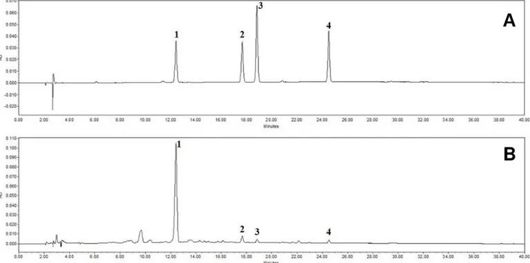
HPLC analysis of FSE reveals that FSE contains the four main chemical
constituents (forsythiaside, phillyrin, pinoresinol, and phylligenin)
The HPLC analysis of FSE revealed four peaks matching those of the commercial standards forsythiaside, phillyrin, pinoresinol, and phylligenin with retention times of approximately 12.5 min, 17.7 min, 18.8 min, and 24.5 min, respectively (Fig 7). The FSE contained
5.996± 0.007 mg/g forsythiaside, 0.422 ± 0.001 mg/g phillyrin, 0.159 ± 0.002 mg/g pinoresinol, and 0.136± 0.000 mg/g phylligenin (Table 2).
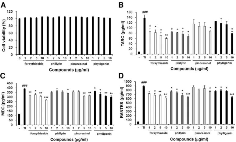
Forsythiaside, phillyrin, pinoresinol, and phylligenin inhibit the production
of chemokines in human keratinocytes
To determine the active components of FSE, we investigated the effect of forsythiaside, phil-lyrin, pinoresinol, and phylligenin on TNF-α/IFN-γ-induced chemokines production Fig 3. FSE reduces infiltration of mast cell in the back skin lesions of NC/Nga mice. (A) The back skin lesions were stained toluidine blue and (B) the number of mast cells in toluidine blue-stained sections were counted (x200). Mast cells were counted at five randomly selected sites of toluidine blue-stained sections. Results are expressed as mean±S.E.M for 7 mice.##p<0.01 and###p<0.001 versus Normal;*p<0.05,**p<0.01, and
***p<0.001 FSE versus Control;§p<0.05,§§p<0.01, and§§§p<0.001 Tacrolimus versus Control. doi:10.1371/journal.pone.0167687.g003
underlying the anti-allergic activity of FSE. In an MTT assay, the treatment of these com-pounds for 24 h had no significant cytotoxic effect on HaCaT cells (Fig 8A). Forsythiaside, phillyrin, pinoresinol, and phylligenin inhibited TNF-α/IFN-γ-induced production of TARC, MDC, and RANTES in a dose-dependent manner (Fig 8B, 8C and 8D). These results indicate that the inhibition effect of FSE in TNF-α/IFN-γ-induced chemokines production is due to the anti-allergic activity of forsythiaside, phillyrin, pinoresinol, and phylligenin.
Discussion
Topical application of FSE in NC/Nga mice with DfE-induced AD ameliorated typical and his-tological changes of AD, such as dermatitis severity score, ear thickness, epidermal hyperplasia, and infiltration of inflammatory cells. Mast cells play a key role in allergic disorders through the release of a wide variety of products, including proteases, lipid mediators, histamine, cyto-kines, chemocyto-kines, and growth factors [26–28]. Histamine, a marker for mast cell degranula-tion, causes vasodiladegranula-tion, increased vascular permeability, and, consequently, recruitment of leukocytes in the acute allergic response of AD [29]. Pro-inflammatory cytokines, such as TNF-α and IL-4, initiate the chronic response of AD via enhancement of T cell activation or B Fig 4. FSE reduces the serum levels of histamine, TNF-α, and total IgE in NC/Nga mice. The concentrations of (A) histamine, (B) TNF-α, and (C) total IgE. Results are expressed as mean±S.E.M for 7 mice.#p<0.05 and##p<0.01 versus Normal;*p<0.05,**p<0.01, and***p<0.001 FSE
versus Control;§p<0.05,§§p<0.01, and§§§p<0.001 Tacrolimus versus Control.
cell survival [30]. After antigen presenting, B cells-produced antigen-specific IgE binds to FcεRI receptors on the mast cell surface, and activated mast cells release allergic mediators to induce allergic inflammation [23]. In AD mice, topical application of FSE reduced the number of mast cells in the skin lesions and the levels of histamine, TNF-α, and IgE in the serum. These results suggest that FSE suppresses allergic inflammation via inhibition of mast cell degranulation, especially the release of histamine. Although elevated serum IgE levels are a typ-ical marker of AD, tacrolimus failed to reduce the serum IgE levels. Similarly, previous studies showed that tacrolimus did not inhibit the elevation of serum IgE levels in mice, although it suppressed the expression of IL-4 mRNA in the skin lesion [19,31]. These results suggest that locally expressed IL-4 does not seem to contribute to the systemic IgE production.
Dysregulation of chemokines, cytokines, and adhesion molecules generally enhances the infiltration of immune cells into the inflammation site in the skin [32]. Th2 chemokines such as TARC and MDC play major roles in the pathogenesis of Th2-dominant skin disorders such as AD [33]. Previous studies have demonstrated that elevated serum levels of TARC and MDC correlate with skin disease severity in AD patients [34], and expression of TARC and MDC is Fig 5. FSE inhibits the expression of chemokines, inflammatory cytokines, and adhesion molecules in the ear tissue of NC/Nga mice. (A) The mRNA expression of TARC, MDC, RANTES, IL-4, TNF-α, ICAM-1, and VCAM-1. (B) Quantitative analysis of PCR bands was performed with the NIH ImageJ program. Results are expressed as mean±S.E.M for 7 mice.#p<0.05,##p<0.01, and###p<0.001 versus Normal;*p<0.05, **p<0.01, and***p<0.001 FSE versus Control;§p<0.05,§§p<0.01, and§§§p<0.001 Tacrolimus versus Control.
increased in the lesional skin of AD patients [35]. RANTES secreted by keratinocytes recruits type 1 T cells or macrophages into skin lesions of AD and is involved in the infiltration and degranulation of eosinophils in the early stage of AD [36]. Adhesion molecules, such as ICAM-1 and VCAM-1, are expressed on keratinocytes, antigen-presenting cells, and endothe-lial cells, and are known to be involved in infiltration and activation of leukocytes into inflam-mation sites [37]. The expression of various adhesion molecules, chemokines, and cytokines on human keratinocytes is induced by pro-inflammatory cytokines such as TNF-α and IFN-γ [38–39]. A previous study showed that VCAM-1 blockade suppressed disease severity and inflammatory cells in an AD model [40]. Accordingly, down-regulation of these chemokines and adhesion molecules could be an effective target for the therapy of AD. Thus, we examined the ability of FSE on the expression of TARC, MDC, RANTES, TNF-α, IL-4, VCAM-1, and ICAM-1 molecules in the lesional skin and found that the expression levels of these proteins suppressed by FSE treatment. In addition, production of TNF-α/IFN-γ-induced TARC, MDC, and RANTES in HaCaT keratinocytes was effectively suppressed in a dose-dependent manner by FSE treatment. These findings suggest that FSE suppresses the expression of chemokines Fig 6. FSE inhibits the production of chemokines in TNF-α-and IFN-γ-treated human keratinocytes. HaCaT cells (3 x 10⁵cells/well) were
seeded into each well of 96-well plates and stimulated with or without FSE (25, 50, 100, 200, and 400μg/mL) for 24 h. (A) Cell viability was measured using the MTT assay. Production levels of TNF-α-and IFN-γ-induced (B) TARC, (C) MDC, and (D) RANTES chemokines. HaCaT cells (3 x 10⁵cells/ well) were seeded into each well of 24-well plates and added with TNF-α(10 ng/mL) and IFN-γ(10 ng/mL) in the presence or absence of FSE (25, 50, 100, 200, and 400μg/mL). Dexamethasone (10−4M) was used as a positive control. The TARC, MDC, and RANTES protein levels were determined
by ELISA after 24 h of cultivation. Results are expressed as mean±S.E.M. of three independent experiments.###p<0.001 versus Normal (untreated
group)*p<0.05,**p<0.01, and***p<0.001 versus Control (TNF-αand IFN-γ-treated group). doi:10.1371/journal.pone.0167687.g006
(TARC, MDC, and RANTES), cytokines (TNF-α and IL-4), and adhesion molecules (VCAM-1 and ICAM-(VCAM-1), thereby inhibiting migration and infiltration of leukocyte to sites of
inflammation.
We found 4 main components in FSE which were forsythiaside, phillyrin, pinoresinol, and phylligenin. This study showed that these constituents decreased TNF-α/IFN-γ-induced pro-duction of TARC, MDC, and RANTES in HaCaT cells. Previous studies reported that these constituents had anti-inflammatory effects [41–44]. These results suggest that forsythiaside, phillyrin, pinoresinol, and phylligenin are the active compounds of FSE. Thus, even though anti-allergic effect of these compounds on AD was not elucidated yet, we assumed that these components might be responsible for the inhibitory effects of FSE on AD-like skin lesions.
Conclusions
Topical application of FSE on lesional skin of DfE-induced AD mice effectively alleviated the development of AD-like lesions by suppressing the expression of chemokines, cytokines, and Fig 7. HPLC analysis of FSE reveals that FSE contains four main chemical constituents. HPLC chromatogram of a standard mixture (A) and
Forsythia suspensa extract (B) at 235 nm. Forsythiaside (1), phillyrin (2), pinoresinol (3), and phylligenin (4) appeared with retention times of
approximately 12.5 min, 17.7 min, 18.8 min, and 24.5 min, respectively. doi:10.1371/journal.pone.0167687.g007
Table 2. Average contents of the reference compounds in Forsythia suspensa extract (n = 3).
Compound Average content (mg/g)
Forsythiaside 5.996±0.007
Phillyrin 0.422±0.001
Pinoresinol 0.159±0.002
Phylligenin 0.136±0.000
adhesion molecules in keratinocytes. Besides, among the components of FSE, forsythiaside, phillyrin, pinoresinol, and phylligenin inhibited the production of TARC, MDC, and RANTES in human keratinocytes. These results suggest thatF. suspensa might be a useful candidate for
treatment of allergic skin inflammatory disorders.
Author Contributions
Conceptualization: YYS TY. Data curation: YYS.
Formal analysis: YYS SJ. Funding acquisition: HKK. Investigation: YYS SJ. Methodology: YYS TY.
Project administration: YYS HKK.
Fig 8. Forsythiaside, phillyrin, pinoresinol, and phylligenin inhibits the production of chemokines in TNF-α-and IFN-γ-treated human keratinocytes. (A) Cell viability measured using the MTT assay. HaCaT cells (3 x 10⁵cells/well) were seeded into each well of 96-well plates and
stimulated with or without compound (1, 5, 10, and 20μg/mL) for 24 h. Production levels of TNF-α-and IFN-γ-induced (B) TARC, (C) MDC, (D) RANTES chemokines. HaCaT cells (3 x 10⁵cells/well) were seeded into each well of 24-well plates and added with TNF-α(10 ng/ml) and IFN-γ(10 ng/
mL) in the presence or absence of compound (1, 5, 10, and 20μg/mL). The TARC, MDC, and RANTES protein levels were determined by ELISA after 24 h of cultivation. Results are expressed as mean±S.E.M. of three independent experiments.###p<0.001 versus Normal (untreated group)
*
p<0.05,**p<0.01, and***p<0.001 versus Control (TNF-αand IFN-γ-treated group). doi:10.1371/journal.pone.0167687.g008
Supervision: YYS. Validation: YYS. Visualization: YYS.
Writing – original draft: YYS SJ.
Writing – review & editing: YYS TY SJ HKK.
References
1. Leung DY, Boguniewicz M, Howell MD, Nomura I, Hamid QA. New insights into atopic dermatitis. J Clin Invest. 2004; 113: 651–657. doi:10.1172/JCI21060PMID:14991059
2. Nedoszytko B, Sokołowska-Wojdyło M, Ruckemann-Dziurdzińska K, Roszkiewicz J, Nowicki RJ. Che-mokines and cytokines network in the pathogenesis of the inflammatory skin diseases: atopic dermati-tis, psoriasis and skin mastocytosis. Postepy Dermatol Alergol. 2014; 31: 84–91. doi:10.5114/pdia. 2014.40920PMID:25097473
3. Vestergaard C, Bang K, Gesser B, Yoneyama H, Matsushima K, Larsen CG. A Th2 chemokine, TARC, produced by keratinocytes may recruit CLA+CCR4+lymphocytes into lesional atopic dermatitis skin. J Invest Dermatol. 2000; 115: 640–646. doi:10.1046/j.1523-1747.2000.00115.xPMID:10998136 4. Sykes L, MacIntyre DA, Yap XJ, Ponnampalam S, Teoh TG, Bennett PR. Changes in the Th1:Th2
cyto-kine bias in pregnancy and the effects of the anti-inflammatory cyclopentenone prostaglandin 15-deoxy-Δ(12,14)-prostaglandin J2. Mediators Inflamm. 2012; 2012:416739. doi:10.1155/2012/416739 PMID:22690041
5. Choi JK, Oh HM, Lee S, Kwon TK, Shin TY, Rho MC, et al. Salvia plebeia suppresses atopic dermatitis-like skin lesions. Am J Chin Med. 2014; 42:967–985. doi:10.1142/S0192415X1450061XPMID: 25004886
6. Pastore S, Mascia F, Giustizieri ML, Giannetti A, Girolomoni G. Pathogenetic mechanisms of atopic dermatitis. Arch Immunol Ther Exp (Warsz). 2000; 48: 497–504. Available:http://www.ncbi.nlm.nih. gov/pubmed/11197604
7. Pivarcsi A, Homey B. Chemokine network in atopic dermatitis: traffic signals of disease. Curr Allergy Asthma Rep. 2005; 5: 284–290. PMID:15967069
8. Čabrijan L, LipozenčićJ. Adhesion molecules in keratinocytes. Clin Dermatol. 2011; 29: 427–431. doi: 10.1016/j.clindermatol.2011.01.012PMID:21679870
9. Qu H, Zhang H, Chai X, Sun W. Isoforsythiaside, an antioxidant and antibacterial phenylethanoid glyco-side isolated from Forsythia suspensa. Bioorg Chem. 2012; 40: 87–91. doi:10.1016/j.bioorg.2011.09. 005PMID:22014602
10. Nishibe S, Okabe K, Tsukamoto H, Sakushima A, Hisada S, Baba H, et al. Studies on the Chinese crude drug “Forsythiae Fructus” VI. The structure and antibacterial activity of suspensaside isolated from Forsythia suspensa. Chem Pharm Bull. 1982; 30: 4548–4553. Available:http://doi.org/10.1248/ cpb.30.4548PMID:7168874
11. Ozaki Y, Rui J, Tang Y, Satake M. Antiinflammatory effect of Forsythia suspensa Vahl and its active fraction. Biol Pharm Bull. 1997; 20: 861–864. Available:http://doi.org/10.1248/bpb.20.861PMID: 9300131
12. Prieto JM, Recio MC, Giner RM, Ma´ñez S, Giner-Larza EM, Rı´os JL. Influence of traditional Chinese anti-inflammatory medicinal plants on leukocyte and platelet functions. J Pharm Pharmacol. 2003; 55: 1275–1282. doi:10.1211/0022357021620PMID:14604471
13. Kim MS, Na HJ, Han SW, Jin JS, Song UY, Lee EJ, et al. Forsythia fructus inhibits the mast-cell-medi-ated allergic inflammatory reactions. Inflammation. 2003; 27: 129–135. PMID:12875366
14. Jeong YI, Hong SH, Cho SH, Lee WJ, Lee SE. Toxoplasma gondii Infection Suppresses House Dust Mite Extract-Induced Atopic Dermatitis in NC/Nga Mice. Allergy Asthma Immunol Res. 2015; 7: 557– 564. doi:10.4168/aair.2015.7.6.557PMID:26333702
15. Carroll CL, Fleischer AB Jr. Tacrolimus: focusing on atopic dermatitis. Drugs Today (Barc). 2006; 42: 431–439.
16. Simpson D, Noble S. Tacrolimus ointment: a review of its use in atopic dermatitis and its clinical poten-tial in other inflammatory skin conditions. Drugs. 2005; 65: 827–858. PMID:15819596
17. Sasayawa T, Hiashi Y, Sakuma S, Hirayama Y, Sasakawa Y, Ohkubo Y, et al. Topical application of FK506 (tacrolimus) ointment inhibits mite antigen-induced dermatitis by local action in NC/Nga mice. Int Arch Allergy Immunol. 2004; 133: 55–63. doi:10.1159/000076128PMID:14726632
18. Hanifin JM, Paller AS, Eichenfield L, Clark RA, Korman N, Weinstein G, et al. Efficacy and safety of tacrolimus ointment treatment for up to 4 years in patients with atopic dermatitis. J Am Acad Dermatol. 2005; 53: S186–S194. doi:10.1016/j.jaad.2005.04.062PMID:16021174
19. Inagaki N, Shiraishi N, Igeta K, Itoh T, Chikumoto T, Nagao M, et al. Inhibition of scratching behavior associated with allergic dermatitis in mice by tacrolimus, but not by dexamethasone. Eur J Pharmacol. 2006; 546:189–196. doi:10.1016/j.ejphar.2006.07.019PMID:16914137
20. Yamamoto M, Haruna T, Yasui K, Takahashi H, Iduhara M, Takaki S, et al. A novel atopic dermatitis model induced by topical application with dermatophagoides farinae extract in NC/Nga mice. Allergol Int. 2007; 56:139–148. doi:10.2332/allergolint.O-06-458PMID:17460441
21. Suto H, Matsuda H, Mitsuishi K, Hira K, Uchida T, Unno T, et al. NC/Nga mice: a mouse model for atopic dermatitis. Int Arch Allergy Immunol. 1999; 120: 70–75. doi:53599PMID:10529609
22. Dilshara MG, Kang CH, Choi YH, Kim GY. Mangiferin inhibits tumor necrosis factor-α-induced matrix metalloproteinase-9 expression and cellular invasion by suppressing nuclear factor-κB activity. BMB Rep. 2015; 48: 559–564. Available:http://dx.doi.org/10.5483/BMBRep.2015.48.10.003PMID: 25739392
23. Metcalfe DD, Baram D, Mekori YA. Mast cells. Physiol Rev. 1997; 77:1033–1079. Available:http:// www.ncbi.nlm.nih.gov/pubmed/9354811PMID:9354811
24. Bardana EJ Jr. Immunoglobulin E- (IgE) and non-IgE-mediated reactions in the pathogenesis of atopic eczema/dermatitis syndrome (AEDS). Allergy. 2004; 59: 25–39. doi:10.1111/j.1398-9995.2004. 00565.xPMID:15245353
25. Albanesi C, Scarponi C, Giustizieri ML, Girolomoni G. Keratinocytes in inflammatory skin diseases. Curr Drug Targets Inflamm Allergy. 2005; 4: 329–334. PMID:16101542
26. Galli SJ, Tsai M, Piliponsky AM. The development of allergic inflammation. Nature, 2008; 454: 445– 454. doi:10.1038/nature07204PMID:18650915
27. Otsuka A, Kabashima K. Mast cells and basophils in cutaneous immune responses. Allergy. 2015; 70: 131–140. doi:10.1111/all.12526PMID:25250718
28. Bulfone-Paus S, Bahri R. Mast Cells as Regulators of T Cell Responses. Front Immunol. 2015; 6:394. doi:10.3389/fimmu.2015.00394PMID:26300882
29. Galli J, Tsai M. IgE and mast cells in allergic disease. Nat Med. 2012; 18: 693–704. doi:10.1038/nm. 2755PMID:22561833
30. Je IG, Choi HG, Kim HH, Lee S, Choi JK, Kim SW, et al. Inhibitory effect of 1,2,4,5-tetramethoxyben-zene on mast cell-mediated allergic inflammation through suppression of IκB kinase complex. Toxicol Appl Pharmacol. 2015; 287: 119–127. doi:10.1016/j.taap.2015.05.006PMID:25981167
31. Lee H, Ha H, Lee JK, Park SJ, Jeong SI, Shin HK. The Leaves of Broussonetia kazinoki Siebold Inhibit Atopic Dermatitis-Like Response on Mite Allergen-Treated NC/Nga Mice. Biomol Ther (Seoul). 2014; 22: 438–444.
32. Seo WY, Youn GS, Choi SY, Park J. Butein, a tetrahydroxychalcone, suppresses pro-inflammatory responses in HaCaT keratinocytes. BMB Rep. 2015; 48: 495–500. doi:10.5483/BMBRep.2015.48.9. 259PMID:25541056
33. Kwon DJ, Bae YS, Ju SM, Goh AR, Youn GS, Choi SY, et al. Casuarinin suppresses TARC/CCL17 and MDC/CCL22 production via blockade of NF-κB and STAT1 activation in HaCaT cells. Biochem Biophys Res Commun. 2012; 417: 1254–1259. doi:10.1016/j.bbrc.2011.12.119PMID:22227193
34. Jahnz-Rozyk K, Targowski T, Paluchowska E, Owczarek W, Kucharczyk A. Serum thymus and activa-tion-regulated chemokine, macrophage-derived chemokine and eotaxin as markers of severity of atopic dermatitis. Allergy. 2005; 60: 685–688. doi:10.1111/j.1398-9995.2005.00774.xPMID:15813816 35. Horikawa T, Nakayama T, Hikita I, Yamada H, Fujisawa R, Bito T, et al. IFN-γ-inducible expression of
thymus and activation-regulated chemokine/CCL17 and macrophage-derived chemokine/CCL22 in epi-dermal keratinocytes and their roles in atopic dermatitis. Int Immunol. 2002; 14: 767–773. PMID: 12096036
36. Kanda N, Watanabe S. Suppressive effects of antimycotics on tumor necrosis factor-alpha-induced CCL27, CCL2, and CCL5 production in human keratinocytes. Biochem Pharmacol. 2006; 72: 463–473. doi:10.1016/j.bcp.2006.05.001PMID:16784723
37. Kwon TR, Mun SK, Oh CT, Hong H, Choi YS, Kim BJ, et al. Therapeutic effects of full spectrum light on the development of atopic dermatitis-like lesions in NC/Nga mice. Photochem Photobiol. 2014; 90: 1160–1169. doi:10.1111/php.12284PMID:24773136
38. Krutmann J, Ko¨ck A, Schauer E, Parlow F, Mo¨ller A, Kapp A, et al. Tumor necrosis factor beta and ultra-violet radiation are potent regulators of human keratinocyte ICAM-1 expression. J Invest Dermatol. 1990; 95:127–131. PMID:1974275
39. Park JH, Kim MS, Jeong GS, Yoon J. Xanthii fructus extract inhibits TNF-α/IFN-γ-induced Th2-chemo-kines production via blockade of NF-κB, STAT1 and p38-MAPK activation in human epidermal keratino-cytes. J Ethnopharmacol. 2015; 171: 85–93. doi:10.1016/j.jep.2015.05.039PMID:26051830 40. Chen L, Lin SX, Amin S, Overbergh L, Maggiolino G, Chan LS. VCAM-1 blockade delays disease
onset, reduces disease severity and inflammatory cells in an atopic dermatitis model. Immunol Cell Biol. 2010; 88: 334–342. doi:10.1038/icb.2009.107PMID:20065994
41. Cheng G, Zhao Y, Li H, Wu Y, Li X, Han Q, Dai C, Li Y. Forsythiaside attenuates lipopolysaccharide-induced inflammatory responses in the bursa of Fabricius of chickens by downregulating the NF-κB sig-naling pathway. Exp Ther Med. 2014; 7: 179–184. doi:10.3892/etm.2013.1378PMID:24348786 42. Zhong WT, Wu YC, Xie XX, Zhou X, Wei MM, Soromou LW, et al. Phillyrin attenuates LPS-induced
pul-monary inflammation via suppression of MAPK and NF-κB activation in acute lung injury mice. Fitotera-pia. 2013; 90:132–139. doi:10.1016/j.fitote.2013.06.003PMID:23751215
43. Jung HW, Mahesh R, Lee JG, Lee SH, Kim YS, Park YK. Pinoresinol from the fruits of Forsythia
kor-eana inhibits inflammatory responses in LPS-activated microglia. Neurosci Lett. 2010; 480: 215–220.
doi:10.1016/j.neulet.2010.06.043PMID:20600612
44. Lim H, Lee JG, Lee SH, Kim YS, Kim HP. Anti-inflammatory activity of phylligenin, a lignan from the fruits of Forsythia koreana, and its cellular mechanism of action. J Ethnopharmacol. 2008; 118: 113– 117. doi:10.1016/j.jep.2008.03.016PMID:18467047