황련해독탕의 피부지방장벽개선을 통한 Th2 분화조절이 아토피피부염 완화에 미치는 효과
손성한1․안상현2․박선영3․김기봉1,4
1부산대학교 한의학전문대학원, 2세명대학교 한의과대학 해부학교실,
3세명대학교 한의과대학 생리학교실, 4부산대학교한방병원 한방소아과
Received: July 25, 2018 ∙ Revised: August 10, 2018 ∙ Accepted: August 11, 2018 Corresponding Author: Kibong Kim
Department of Korean Pediatrics, Pusan National University Korean Medicine Hospital, Geumo-ro 20, Mulgeum-eup, Yangsan-si, Gyeongsangnam-do, 50612, Republic of Korea Tel: +82-55-360-5952 / Fax: +82-55-360-5952
E-mail: [email protected]
ⓒ The Association of Pediatrics of Korean Medicine. All rights reserved. This is an open-access article distributed under the tenus of the Creative Commons Attribution Non-Commercial License (http://creativecommons.org/licenses/by-nc/3.0/), which permits unrestricted non-commercial use, distribution, and reproduction in any medium, provided the original work is properly cited.
Abstract
Hwangnyeonhaedok-tang Extracts Ameliorates Atopic Dermatitis via Epidermal Lipid Barrier Regeneration in NC/Nga Mouse
Son Seong Han1․Ahn Sang Hyun2․Park Sun-Young3․Kim Kibong1,4
1School of Korean Medicine, Pusan National University,
2Department of Anatomy, College of Korean Medicine, Semyung University,
3Department of Physiology, College of Korean Medicine, Semyung University,
4Department of Korean Pediatrics, Pusan National University Korean Medicine Hospital Objectives
Hwangnyeonhaedok-tang is a Korean herbal medical treatment that removes toxic heat, fever and inflammation.
The purpose of this study was to investigate the effect of Hwangnyeonhaedok-tang treatment on the relief of atopic dermatitis (AD) through regeneration of skin lipid barrier.
Methods
Male NC/Nga mice (20 g, 6 week age) were used. Each 10 mice were allocated to the control group (Ctrl), the AD-induced with no treatment group (AE), and the group which induced AD after administering Hwangnyeonhaedok-tang extract (HT). To induce AD-like skin lesions, sodium dodecyl sulfate (SDS) (Sigma-Aldrich, USA) was rubbed on the back of each mouse to remove the lipid lamella of the stratum corneum, and Dermatophagoides (D.) farinae crude extract was applied. HT group was orally administered Hwangnyeonhaedok-tang after induction of AD. IL-4 IL-13, p-IκB, iNOS, Sudan Black B (SB), loricrin, and filaggrin were observed to confirm the effect.
Results
In HT group, AD skin score was decreased by 46%. The cytokine IL-4 and IL-13, which can identify Th2 differentiation, was reduced by 73% and 58% each. Anti-inflammatory effects were observed in p-IκB and iNOS by 69% and 54%, respectively. Finally, SB showed that the regeneration of the lipid layer and the increase of the regeneration power of loricrin and filaggrin were increased by 437% and 464%, respectively.
Conclusions
From the study result, we observed that Hwangnyeonhaedok-tang treatment alleviates AD by decreasing skin score, reducing Th2 differentiation, inducing anti-inflammatory, and increasing skin lipid barrier regeneration. Thus, Hwangnyeonhaedok-tang treatment would be considered as an effective AD relieving treatment.
Key words: Atopic dermatitis, Hwangnyeonhaedok-tang, Lipid barrier, Skin regeneration, Anti-inflammatory
ISSN 1226-8038(Print), 2287-9463(Online), https://doi.org/10.7778/jpkm.2018.32.3.090
Ⅰ. Introduction
아토피피부염 (Atopic Dermatitis, AD)은 특징적인 습진, 피부 건조증, 가려움증을 동반하며 만성적으로 재발하는 피부 염증 질환으로 주로 유아기나 소아기부 터 발병하여 노년기까지 지속된다1). 현대화 및 산업화 가 진행됨에 따라 환경오염의 증가로 AD 환자는 증가 추세이지만2-4) AD에 대한 명확한 발병원인과 기전이 밝혀져 있지 않고 유전적 요인, 생활환경 요인, 면역학 적 요인, 피부장벽 불균형 요인, 심리적 요인 등 다양한 요인의 상호작용에 의해 복합적으로 발병하는 것으로 추정되고 있다5). 특히 유아 및 소아의 아토피는 외부 병원체의 감염으로 인해 Th1과 Th2의 정상적인 비율 을 벗어나 Th2의 수치가 올라가고 IgE 수치의 상승으 로 증상이 더욱 심해지는 것으로 보고되고 있다6,7). 현 재 AD의 치료는 항히스타민제, 국소 스테로이드 외용 제, 경구 스테로이드제를 사용하고 있으며8), 이러한 치 료는 일시적으로 증상을 완화시키기만 할 뿐 근본적인 치료를 할 수 없으므로9,10) 효과적인 치료법이 필요한 실정이다.
한의학에서는 임산부가 임신 중에 알레르기를 유발 할 수 있는 흡입성 항원이나 식이성 항원 물질을 섭취 한 후, 그로 인한 태아의 胎熱을 AD의 원인으로 본다.
따라서 AD를 胎熱과 胎毒의 관점으로 접근을 하고 있 으며, 淸熱, 祛風, 除濕 효능이 있는 약물로 치료하고 있다11). 胎熱이 있는 소아는 內邪, 外邪에 의해 증상이 심해지며 피부증상 외에 喘息, 發作 등 다른 증상으로 도 발전하기도 한다11,12).
황련해독탕 (黃連解毒湯)은 ≪外臺祕要≫에 처음 나오며, ≪東醫寶鑑·雜病編≫에 수록되어 있는 처방 으로, 황련 (黃連, Coptidis Rhizoma), 황금 (黃芩, Scutellariae Radix), 황백 (黃柏, Phellodendri Cortex), 치자 (梔子, Gardeniae Fructus)의 네 가지 약재로 구성되어 있다. 解毒, 淸熱瀉 火, 傷寒大熱을 주로 하며, 發斑, 火毒, 大熱, 疔毒, 實 熱, 膿瘍, 不眠, 煩燥, 咽乾, 口燥 등의 病症에 사용되 어 왔다13).
황련해독탕에 대한 선행 연구로 아토피피부염 개선 과 증상 완화 효과13,14)에 대한 연구결과가 있으나 피부 지질장벽의 재생을 관찰한 연구는 없다.
본 연구는 NC/Nga 생쥐에게 Dermatophagoides (D.) far- inae로 아토피피부염을 유발한 후 황련해독탕을 투여하 였다. External morphology 관찰과 조직화학 및 면역조
직화학적 방법으로 피부 병변의 skin score를 측정하고, Th2 분화와 관련이 있는 cytokine인 IL-4와 IL-13, p-IκB 와 iNOS의 수치와 지방을 염색하는 Sudan Black B (SB), 그리고 loricrin과 filaggrin의 수치로 피부장벽의 재생을 관찰하였다. 이 연구를 통해 황련해독탕이 아 토피유사피부염을 유발한 생쥐에서 피부의 회복과 Th2 분화의 감소, 항염증작용, 그리고 피부장벽의 재 생 효과를 확인하였기에 보고하는 바이다.
Ⅱ. Materials and Methods
1. 실험동물 및 아토피피부염 유발
태령 6주된 NC/Nga 수컷 생쥐 (대한실험동물센터, 한국)를 무균사육장치내에서 2주간 적응시킨 후 체중 20 g된 생쥐를 선별하여 사용하였다. 대조군 (Ctrl), 아 토피피부염 유발군 (AE), 아토피피부염 유발 후 황련해 독탕 투여군 (HT)으로 나누었으며, 각 군에 각 10마리 씩 배정하였다.
AD 유발은 생쥐 등 부위를 면도 후 계면활성제 (surfactant)인 5% sodium dodecyl sulfate (SDS: Sigma, USA) 1 ㎖를 면봉으로 20회 문질러 각질층의 lipid la- mella를 제거한 후 D. farinae crude extract (100 ㎎, Biostir, Japan)을 3주간 주 6회씩 도포하여 유발하였다.
본 연구과정은 부산대학교 IACUC 승인을 받아 시행되 었으며 (IACUC number: PNU-2015-0924), 실험동물의 관리와 사용에 대해서는 NIH 가이드라인에 따라 시행 되었다.
2. 황련해독탕 추출물의 제조, 투여
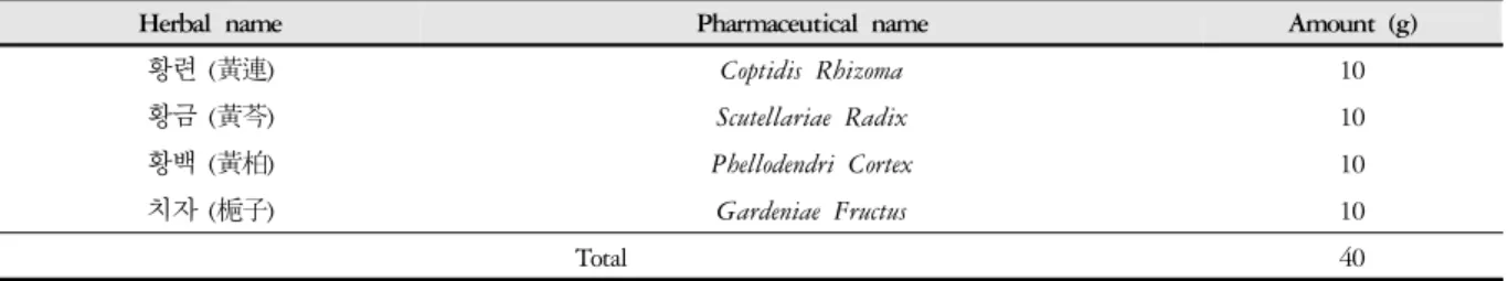
황련해독탕 40 g을 증류수 1000 ㎖에 넣고 3시간 동안 전탕한 후 여과하였다. 그 여액을 rotary evaporator 를 이용하여 50 ㎖로 감압, 농축한 후 동결 건조하여 11 g을 얻었다 (수득률 27.5%). HT군에 AD유발 후 매 일 3주 동안 황련해독탕을 550 ㎎/㎏으로 경구 투여하 였다. 황련해독탕의 구성 및 용량은 표에 별도로 기재 하였다 (Table 1).
3. 피부염의 중증도 평가
등 부분 피부의 형태학적 중증도 (skin score)는 3주 후에 평가되었고 기준선과 비교되었다. 피부 점수 항 목은 (1) 홍반/출혈, (2) 흉터/건조, (3) 부종, (4) 찰상/미 란으로 나누었으며, 항목 당 점수는 0 (없음), 1 (경미), 2 (보통), 3 (심각)으로 나누었다. 각 점수의 합으로 평 가하였다.
4. 피부 조직표본 제작과 조직화학
Sodium pentobarbital 용액으로 마취 후 vascular rinse 와 10% 중성 포르말린 용액 (neutral buffered formalin:
NBF)으로 심장관류고정을 실시하였다. 등쪽 피부를 얻어 10% NBF에 실온에서 24시간 동안 고정한 후 통 상적인 방법으로 paraffin에 포매 후 5 ㎛ 두께로 연속 절편을 만들었다.
조직손상 변화를 관찰하기 위해 Masson trichrome (MT)염색을 실시하였다. 50-60 ℃ Bouin 용액에서 1시 간 동안 매염 처리한 다음 70% 에탄올에서 picric acid 를 제거하였다. Weigert iron hematoxylin으로 10분간 반응시켜 핵을 염색하고, Biebrich scalet-acid fuchsin와 phosphomolybdic-phosphotungstic acid로 각각 15분간, aniline blue에 5분간 처리하여 아교섬유 (청색)를 염색 한 후 관찰하였다.
한편, 각질층 내 지방장벽 변화 관찰을 위해 냉동절 편을 제작하였다. 얻어진 조직은 NBF에 고정한 후 10% formol-calcium에 1주일 동안 oxidation 처리하였 다. 그 후 30% sucrose 용액에 cryoprotection하고 냉동 절편기 (Microm, Germany)로 10 ㎛ 두께의 냉동절편을 제작하였다.
지방조직 분포는 SB 염색법을 통해 조사하였는데, 우선 냉동절편을 absolute propylene glycol로 10분간 탈 수한 후 SB 용액에 10분간 염색하였다. 85% propylene glycol로 3분간 탈색한 후 증류수에 수세하고 nuclear fast red로 1분간 대조 염색하였다. 대조 염색 후 증류수
로 3회 수세한 다음 glycerin jelly로 봉입한 후 광학현미 경으로 관찰하였다.
5. 면역조직화학
Th2 분화조절, 항염증효과 그리고 지방장벽 구조변 화를 조사하기 위해 면역조직화학을 실시하였다. 피부 조직절편을 proteolysis를 위해 proteinase K (20 ㎍/㎖)에 5분간 처리한 후 10% normal rabbit serum에서 2시간 동안 blocking 시켰다. 그리고 1차 항체인 goat anti-IL-4 (1:100, Santa Cruz Biotec, USA), goat anti-IL-13 (1:100, Santa Cruz Biotec), goat anti-p-IκB (1:500, Santa Cruz Biotec), goat anti-iNOS (1:100, Santa Cruz Biotec), goat an- ti-Loricrin (1:100, Santa Cruz Biotec), goat anti-Filaggrin (1:100, Santa Cruz Biotec)에 4 ℃ humidified chamber에 서 72시간 동안 반응시켰다. 그런 다음 2차 항체인 bio- tinylated rabbit anti-goat IgG (1:100, Santa Cruz Biotec) 에 4 ℃에서 48시간 link 시켰고, 그런 다음 avidin bio- tin complex kit (Vector Lab, USA)에 1시간 동안 실온에 서 반응시켰다. 0.05% 3,3'-diaminobenzidine과 0.01%
HCl이 포함된 0.05 M tris-HCl 완충용액 (pH 7.4)에서 발색시킨 다음 hematoxylin으로 대조 염색하였다.
6. 영상분석
조직화학과 면역조직화학 결과는 Image Pro Plus (Media cybernetics, USA)를 이용한 영상분석을 통해 수 치화 (means ± standard error) 했다. 각 군의 표본에서 임의로 선정된 피부점막 10부위를 x200 배율에서 촬영 한 다음 10,000,000 pixels를 영상분석 하였다.
7. 통계처리
통계는 SPSS software (SPSS 23, SPSS Inc., USA)로 이 루어졌으며, one-way ANOVA 시행을 통해 유의성 (P<0.05)을 검증하고 Duncan’s multiple range test로 사 후 검증하였다.
Herbal name Pharmaceutical name Amount (g)
황련 (黃連) Coptidis Rhizoma 10
황금 (黃芩) Scutellariae Radix 10
황백 (黃柏) Phellodendri Cortex 10
치자 (梔子) Gardeniae Fructus 10
Total 40
Table 1. The Amount and Composition of Hwangnyeonhaedok-tang (HHT) Extract
Ⅲ. Results
1. AD 손상 완화
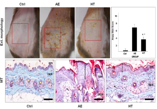
AE군의 대부분의 피부에서 각질층의 탈락과 습진, 홍반, 흉터, 부종, 상피세포 과형성이 관찰되었다. AE 군의 external morphology 관찰에서 skin score로 측정한 결과 Ctrl군과 비교해 1100% 증가한 7.2 ± 0.25로 나타 났다. 이에 반해 HT군은 AE군보다 46% 감소한 3.9 ± 0.28로 관찰되었다 (Fig. 1).
2. Th2 분화 조절
AE군에서 IL-4 수치는 Ctrl군에 비해 633% 증가된 333,405 ± 7,401로 측정되었다. 이에 반해 HT군에서 는 AE군보다 73% 감소한 88,579 ± 4,125로 측정되었 다. IL-13의 수치도 IL-4와 비슷한 양상을 나타내었다.
AE군에서 IL-13 수치는 Ctrl에 비해 1327% 증가한 163,641 ± 3,948로 측정되었다. 이에 반해 HT군에서 는 AE군보다 58% 감소한 68,933 ± 3,277로 측정되었 다 (Fig. 2).
3. 항염증효과
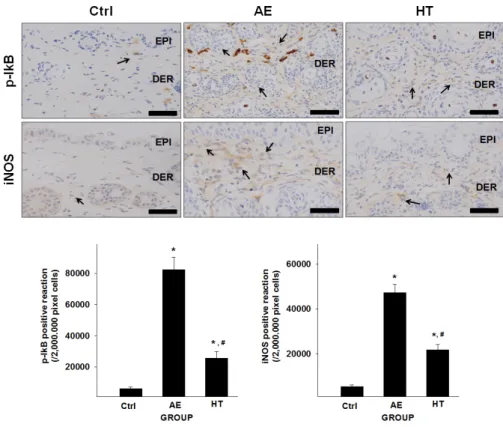
AE군에서 p-IκB 수치는 Ctrl군에 비해 1261% 증가 한 82,338 ± 2,583으로 측정되었다. 이는 NF-κB와 결 합이 끊어져 염증반응이 진행된 것이다. HT군에서는 AE군에 비해 p-IκB가 69% 감소한 21,688 ± 1,374로 측정되었다.
AE군에서 iNOS의 수치는 Ctrl군에 비해 788% 증가 한 47,198 ± 1,168로 측정되었고 HT군에서는 AE군에 비해 54% 감소한 21,688 ± 787로 측정되었다 (Fig. 3).
4. Lipid barrier 재생 효과
SB 염색 결과 AE군은 염증반응으로 인해 지방층 대 부분이 소실된 것을 관찰할 수 있었다. 이에 반해 HT 군에서는 지방층이 회복된 것을 관찰할 수 있었다.
AE군의 각질층 내부에서 관찰된 loricrin은 Ctrl군에 비해 76% 감소된 21,099 ± 933으로 측정되었다. 이에 반해 HT군은 437% 증가한 119,686 ± 2,041로 측정되 었다.
Fig. 1. The mitigative effect of HHT treatment for AD symptoms
The skin features demonstrated visually a marked reduction in HT compared with AE. In Masson trichrome result, hyperplasia and edema decreased in HT compared with AE. (↕: hyperplasia of the epithelium.) Abbreviations; Ctrl: non treatment mice, AE: AD-induced mice, HT: HHT treated mice after AD inducement, DER: dermis, EPI: epidermis, square; AD-induced region, arrow; enlarged capillary, Bar size: 100 μm. *: p<0.05 compared with AD, #: p<0.05 compared with Ctrl.
Fig. 2. Effects of HHT on regeneration of Th2 differentiation (IL-4 and IL-13)
IL-4 positive reaction (↖, arrow indicates dark brown) decreased in HT compared with AD. IL-13-positively reacted cells (↖, arrow indicates dark brown) decreased in HT compared with AE. Abbreviations; Ctrl: non treatment mice, AE: AD-induced mice, HT: HHT treated mice after AD inducement, DER: dermis, EPI: epidermis, Bar size: 100 μm. *: p<0.05 compared with AD, #: p<0.05 compared with Ctrl.
Fig. 3. Effects of HHT on regulation of inflammatory factors (p-IκB and iNOS)
p-IκB positive reaction (↖, arrow indicates dark brown) decreased in HT compared with AE. iNOS-positively reacted cells (↖, arrow indicates dark brown) decreased in HT compared with AE. Abbreviations; Ctrl: non treatment mice, AE: AD-induced mice, HT: HHT treated mice after AD inducement, DER: dermis, EPI: epidermis, Bar size: 100 μm. *: p<0.05 compared with AD, #: p<0.05 compared with Ctrl.
각질세포사이 keratin을 응집하는 matrix protein인 filaggrin은 AE군에서 75% 감소된 20,984 ± 939로 측 정되었다. 이에 반해 HT군은 464% 증가한 118,313 ± 3,152로 측정되었다 (Fig. 4).
Ⅳ. Discussion
AD는 영아기부터 노년기까지 만성적인 재발과 증 상 악화로 인해 삶의 질의 저하를 가져오는 것으로 알 려져 있다15). 다양한 원인으로 아토피피부염이 유발된 다고 보고되고 있지만 현재까지는 증상 완화나 증상이 악화되는 것을 방지할 뿐 근본적인 치료법이 개발되지 않아 치료제의 연구 및 개발이 필요한 실정이다5). 현재 AD의 치료법은 항히스타민제, 국소 스테로이드 외용 제, 경구 스테로이드제를 사용하고 있지만 이는 근본 적인 치료보다는 일시적인 증상 완화를 통해 AD 환자 의 불편함을 감소시키는 정도이다16). 항히스타민제는
AD의 주요 증상인 소양감을 억제하여 불편함을 감소 시켜 환자에게 만족감을 줄 수 있으나, 필요시에 반복 적으로 투여해야 하며, 졸음이 오거나 자통 (刺通)을 유발하거나 소화불량 등의 부작용이 나타나고 있다
17,18)
. 스테로이드제는 세포 염증반응을 억제하여 효과 적인 AD 완화제로 국소, 외용, 경구 등 다양한 방법으 로 오래전부터 사용되고 있다8,19). 하지만 스테로이드 제는 즉각적인 완화 효과는 좋으나 사용을 중단하면 다시 재발하게 되며, 장기적으로 사용 시 피부가 약화 되고 위축되어 AD 증상이 더욱 심하게 나타나게 된다.
또한 투여기간이 길수록 동량의 약물에 대한 효과가 점차 줄어들어 더욱 높은 농도 혹은 강력한 스테로이 드제를 투여해야 하는 부작용이 나타난다9,10). 이번 연 구 또한 현재 사용되고 있는 치료제를 대체하고 효과 적인 근본치료를 위해 시행되었다.
본 연구는 D. farinae로 아토피유사피부염을 유발한 NC/Nga 생쥐에서 황련해독탕으로 external morphology 의 완화, 항염증작용 그리고 피부 지질장벽 재생 효과를 확인하고자 시행하였다. 실험동물로는 6주령 NC/Nga Fig. 4. Effects of HHT on regeneration of lipid barrier (sudan black B, loricrin and filaggrin)
Loricrin positive reaction (↖, arrow indicates dark brown) increased in HT compared with AE. Filaggrin-positively reacted cells (↖, arrow indicates dark brown) increased in HT compared with AE. Abbreviations; Ctrl: non treatment mice, AE: AD-induced mice, HT: HHT treated mice after AD inducement, SC: stratum corneum, Bar size: 100 μm. *: p<0.05 compared with AD, #: p<0.05 compared with Ctrl.
수컷 생쥐를 사용하였고 아토피유사피부염을 유발시 키기 위해 SDS로 등 부위의 lipid lamella를 제거하여 피부장벽의 기능을 손상시킨 후 D. farinae를 3주간 도 포하였다. 이는 이전의 연구와 마찬가지로 SDS를 통해 AD가 유발되기 쉬운 상태로 만든 후, 항원의 도포로 Th1과 Th2의 균형을 깨어 Th2가 우세한 상황으로 만 든 것이다20).
피부는 인체 외부를 감싸는 큰 기관이며, 중요한 기 능은 외부 환경으로부터 1차적인 물리적 장벽으로 몸 을 보호하는 역할을 하는 것이다20). 피부는 크게 표피 층과 진피층으로 나누어지고 표피층은 주로 각질형성 세포로 구성되어 있으며 분화도에 따라 기저층, 유극 층, 과립층, 각질층의 네 가지 층으로 나뉜다21). 1차적 인 물리적 장벽 역할을 하는 피부장벽은 표피층의 각 질세포의 분화과정으로 형성되며 수분 항상성과 물리 적 장벽으로 외부 병원체에 대한 방어를 한다22). 각질 세포의 분화는 기저층에서 분열된 각질세포가 상층으 로 이동함에 따라 다양한 단백질의 영향을 받아 진행 된다. 대표적인 단백질로는 loricrin, filaggrin, involucrin, small proline-rich protein (SPRR) 등이 있다22). Loricrin 은 loricrin끼리 혹은 SPRRs와 결합하여 dimer 형태로 각질세포막을 강화하는 역할을 하는데 AD 환자의 경 우에는 과발현된 IL-4와 IL-13이 STAT-6 경로를 활성 화하여 loricrin의 발현을 감소시켜 각질세포막이 약화 되는 것이 확인되었다23,24). Filaggrin은 AD 환자의 대 표적인 유전인자로 각질세포 외막과 keratin을 서로 연 결해주어 견고한 층을 형성해주는 역할을 하는데 변이 된 filaggrin은 AD 환자에게 피부장벽 손상을 유발하며 결과적으로 피부 염증반응을 일으킨다25,26).
이 연구에서는 피부 중증도 완화부터 Th2 분화조절, 항염증작용, 피부 지질장벽 재생을 관찰했다. External morphology 관찰은 피부 중증도 평가로 각질층의 탈락, 습진, 홍반, 흉터, 부종, 상피세포 과형성을 skin score로 객관화하여 평가하였는데 AE군에 비해 HT군에서 유 의미한 감소를 확인하였다 (Fig. 1). 이 결과는 이번 연 구에서 면역조직화학적으로 관찰한 항목들 (IL-4, IL-13, p-IκB, iNOS, filaggrin, loricrin)의 수치변화로 AD가 완 화되는 것을 육안적으로 확인하는 방법이었다. IL-4, IL-13, p-IκB, iNOS와 같은 염증을 촉진시키는 매개물 질은 혈관투과성 증가, 홍반, 부종 등의 반응을 촉진하
는데27-29), 황련해독탕이 복합적으로 염증반응을 완화
시키고 지질장벽을 재생하여 skin score를 낮춘 결과라 생각된다.
Th2 cytokine은 직접적으로 피부장벽 손상 후 분화 된 각질세포의 주요 단백질인 filaggrin, loricrin의 발현 을 억제하여 피부의 회복률을 저하시킨다30,31). Th cell 을 Th2 cell로 분화시키는 데 관련 있는 IL-4와 이와 밀 접한 연관이 있는 IL-13의 수치 증가는 AD에서 과도한 염증반응을 유발한다28,29). 본 연구에서는 HT군에서 IL-4와 IL-13의 수치가 유의미하게 감소한 것을 확인하 였다 (Fig. 2). Th2 cytokine은 알러지 반응을 유발할 뿐 아니라 선천면역을 저하시킨다. 그리고 최근 연구에서 Th2 면역반응으로부터 생성된 cytokine이 직접적으로 피부장벽 기능을 저하시킨다고 보고되었다32). 따라서 IL-4, IL-13의 감소는 AD의 Th1/Th2 비율을 정상화 시키며 피부장벽 회복률을 높일 수 있을 것이라 생각 한다.
NF-κB는 세포염증반응에서 핵심적인 자리에 있는 regulator로 IκB와 결합을 이룬 상태로 세포질 내에 비 활성 상태로 존재한다33). 세포가 손상이나 스트레스를 받으면 IκB kinase인 IKK (IKK α)에 의해 NF-κB가 IκB 로 분리되고 핵 안으로 들어가 염증반응을 일으킨다34). 본 연구에서는 HT군에서 p-IκB 수치가 감소되었는데 (Fig. 3), 이는 Free p-IκB가 줄어든 것으로 AE군에 비해 IκB가 NF-κB에 결합되어 있는 수가 많다는 것을 의미 한다. 따라서 황련해독탕의 투여가 NF-κB와 IκB 결합 을 끊어지지 않게 하여 염증반응을 억제하는 것으로 생각된다.
그리고 NF-κB에 의해 생성된 전염증성 효소에 의해 전사되는 iNOS는 nitric oxide (NO)를 과도하게 생성하 여 산화적 스트레스를 일으켜 조직손상과 염증반응을
유발한다35,36). iNOS 또한 HT군에서 수치가 감소했는
데 (Fig. 3), 이는 상위 조절자인 NF-κB에 영향을 받는 하위 유전자로 IKK에 의해 인산화된 p-IκB의 수치가 줄어들어 NF-κB의 발현이 줄어든 결과이다33). 따라서 황련해독탕이 세포염증반응에 전반적으로 작용하여 세포염증반응을 줄인다고 생각된다.
마지막으로 SB 염색을 통해 지방층의 회복을 확인 했으며, 과립층에서 각질세포막을 구성하는 주요 단백 질인 loricrin의 수치 증가를 확인할 수 있었다. 또한 keratin을 응집시켜 각질층을 강화시키고 효소에 의해 free amino acid로 분해되어 물과 결합하여 각질층의 항 상성을 유지시키는 filaggrin의 수치 증가를 관찰하였 다 (Fig. 4). 앞서 Th2 cytokine의 과다발현은 직접적으 로 피부장벽을 손상시킨 후 filaggrin, loricrin의 발현을 억제한다고 하였는데, HT군의 filaggrin과 loricrin의 수
치 증가는 황련해독탕의 Th2 분화 억제와 항염증작용 에 의한 결과라 생각된다.
결과적으로 황련해독탕 투여를 통해 아토피유사피 부염 병변을 완화시키고, IL-4, IL-13이 감소되어 Th2 분화를 억제하였다. p-IκB와 iNOS 수치 감소로 항염증 작용이 있는 것을 확인하였으며, 각질층의 지질 증가 와 loricrin, filaggrin 증가로 피부장벽이 재생되는 것을 확인하였다.
이번 연구는 황련해독탕의 아토피피부염 치료 가능 성에 대한 근거로, 동물실험을 통한 황련해독탕의 항 염증작용과 지질장벽 재생 효과에 대한 유의미한 결과 를 얻었으나, 황련해독탕을 실제 아토피피부염 치료에 적용하기 위해서는 임상적인 안정성과 유효성 검증이 필요할 것으로 생각된다.
V. Conclusion
황련해독탕의 피부장벽 재생을 통한 아토피피부염 개선 효과를 확인하기 위해 본 연구는 태령 6주의 수컷 NC/Nga 생쥐에 약물과 D.farinae를 처리하여 아토피유 사피부염을 유발 후 황련해독탕을 투여하여 생쥐의 피 부표면 완화, Th2 분화 조절, 항염증작용, 지질장벽 재 생 정도를 skin score, 면역조직화학적 관찰을 통해 다 음과 같은 결과를 얻었다.
1. 각질층의 skin score 측정 결과 황련해독탕 투여군 에서 유의하게 완화되었다.
2. 피부조직절편의 IL-4, IL-13 양성반응은 황련해독 탕 투여군에서 유의하게 감소되었다.
3. 피부조직절편의 p-IκB, iNOS 양성반응은 황련해 독탕 투여군에서 유의하게 감소되었다.
4. 피부조직절편의 지방층, loricrin, filaggrin 양성반 응은 황련해독탕 투여군에서 유의하게 증가하였다.
Acknowledgement
본 연구는 2018년도 정부 (교육부)의 재원으로 한국 연구재단의 지원을 받아 수행된 기초연구사업임 (No.
NRF-2016R1D1A1B03930474).
References
1. Engel-Yeger B, Mimouni D, Rozenman D, Shani-Adir A. Sensory processing patterns of adults with atopic dermatitis. J Eur Acad Dermatol Venereol. 2011;25(2):
152-6.
2. Oh JW, Pyun BY, Choung JT, Ahn KM, Kim CH, Song SW, Son JA, Lee SY, Lee SI. Epidemiological change of atopic dermatitis and food allergy in school-aged children in Korea between 1995 and 2000.
J Korean Med Sci. 2004;19(5):716-23.
3. Jee HM, Kim KW, Kim CS, Sohn MH, Shin DC, Kim KE. Prevalence of asthma, rhinitis and eczema in Korean children using the International Study of Asthma and Allergies in Childhood (ISAAC) questionnaires. Pediatr Allergy Respir Dis. 2009;19(2):
165-72.
4. Ahn KM, Kim JH, Kwon HJ, Chae YM, Hahm MI, Lee KJ, Park YM, Lee SY, Han MY, Kim WK. The prevalence of symptoms of asthma, allergic rhino- conjunctivitis, and eczema in Korean children: nation- wide cross-sectional survey using complex sampling design. J Korean Med Assoc. 2011;54(7):769-78.
5. Kabashima K. New concept of the pathogenesis of atopic dermatitis: interplay among the barrier, allergy, and pruritus as a trinity. J Dermatol Sci. 2013;70(1):3-11.
6. Rousset F, Robert J, Andary M, Bonnin JP, Souillet G, Chrétien I, Brière F, Pène J, de Vries JE. Shifts in interleukin-4 and interferon-gamma production by T cells of patients with elevated serum IgE levels and the modulatory effects of these lymphokines on sponta- neous IgE synthesis. J Allergy Clin Immunol. 1991;87(1 Pt1):58-69.
7. van der Kleij HP, Kraneveld AD, van Houwelingen AH, Kool M, Weitenberg AC, Redegeld FA, Nijkamp FP. Murine model for non-IgE-mediated asthma.
Inflammation. 2004;28(3):115-25.
8. Park Y. Status of clinical practice on diagnosis and management of atopic dermatitis in Korea: a ques- tionnaire survey of physicians. Allergy Asthma Respir Dis. 2013;1(3):257-65.
9. Callen J, Chamlin S, Eichenfield LF, Ellis C, Girardi M, Goldfarb M, Hanifin J, Lee P, Margolis D, Paller AS, Piacquadio D, Peterson W, Kaulback K, Fennerty M, Wintroub BU. A systematic review of the safety of topical therapies for atopic dermatitis. Br J Dermatol.
2007;156(2):203-21.
10. Pariser D. Topical corticosteroids and topical calcineurin inhibitors in the treatment of atopic dermatitis: focus on percutaneous absorption. Am J Ther. 2009;16(3):264-73.
11. Im GM, Jeong HW, Kim HS, Jeong WY. Oriental medical approach on the allergic disease. J Physiol Pathol Korean Med. 2002;16(5):831-9.
12. Schneider L, Tilles S, Lio P, Boguniewicz M, Beck L, LeBovidge J, Novak N, Bernstein D, Blessing-Moore J, Khan D, Lang D, Nicklas R, Oppenheimer J, Portnoy J, Randolph C, Schuller D, Spector S, Tilles S, Wallace D. Atopic dermatitis: a practice parameter update 2012.
J Allergy Clin Immunol. 2013;131(2):295-9.
13. Ki HP, Jang SI, Yun YG. Ameliorative effects of Hwangnyeonhaedok-tang on atopic dermatitis. Kor J Herbology. 2013;21(1):80-90.
14. Kim BA, Kim MS, Kang BM, Byeon SH, Park IH, Park JH, Jung JW, Ahn EM, Jung HA, Jang JH, Bae W, Lee HY, Choi PN, Park CI. Inhibitory studies of Hwangryunhaedok-tang on development of atopic der- matitis in Nc/Nga mice. Kor J Herbology. 2008;23(2):
59-65.
15. Wahlgren CF. Itch and atopic dermatitis: an overview.
J Dermatol. 1999;26(11):770-9.
16. Baron ED, Barzilai D, Johnston G, Kawashima M, Takigawa M, Nakagawa H, Graham-Browh R, Stevens SR. Atopic dermatitis management: comparing the treat- ment patterns of dermatologists in Japan, U.S.A. and U.K. Br J Dermatol. 2002;147(4):710-5.
17. Drake LA, Fallon JD, Sober A. Relief of pruritus in patients with atopic dermatitis after treatment with topical doxepin cream. The Doxepin Study Group. J Am Acad Dermatol. 1994;31(4):613-6.
18. Bonnel RA, La Grenade L, Karwoski CB, Beitz JG.
Allergic contact dermatitis from topical doxepin: Food and Drug Administration's postmarketing surveillance experience. J Am Acad Dermatol. 2003;48(2):294-6.
19. Lassus A. Clinical comparison of alclometasone dipropio-
nate cream 0.05% with hydrocortisone butyrate cream 0.1% in the treatment of atopic dermatitis in children.
J Int Med Res. 1983;11(5):315-9.
20. Steinert PM, Cantieri JS, Teller DC, Lonsdale-Eccles JD, Dale BA. Characterization of a class of cationic proteins that specifically interact with intermediate filaments. Proc Natl Acad Sci USA. 1981;78(7):4097-101.
21. Kim HJ, Shin JU, Lee KH. Atopic dermatitis and skin barrier dysfunction. Allergy Asthma Respir Dis. 2013;
1(1):20-8.
22. Steinert PM, Marekov LN. The proteins elafin, filaggrin, keratin intermediate filaments, loricrin, and small pro- line-rich proteins 1 and 2 are isodipeptide cross-linked components of the human epidermal cornified cell envelope. J Biol Chem. 1995;270(30):17702-11.
23. Steinett PM, Marekov LN. Direct evidence that in- volucrin is a major early isopeptide cross-linked compo- nent of the keratinocyte cornified cell envelope. J Biol Chem. 1997;272(3):2021-30.
24. Kim BE, Leung DY, Boguniewicz M, Howell MD.
Loricrin and involucrin expression is down-regulated by Th2 cytokines through STAT-6. Clin Immunol.
2008;126(3):332-7.
25. Smith FJ, Irvine AD, Terron-Kwiatkowski A, Sandilands A, Campbell LE, Zhao Y, Liao H, Evans AT, Goudie DR, Lewis-Jones S, Arseculeratne G, Munro CS, Sergeant A, O'Regan G, Bale SJ, Compton JG, DiGiovanna JJ, Presland RB, Fleckman P, McLean WH. Loss-of-function mutations in the gene encoding filaggrin cause ichthyosis vulgaris. Nat Genet. 2006;38(3):337-42.
26. O'Regan GM, Irvine AD. The role of filaggrin in the atopic diathesis. Clin Exp Allergy. 2010;40(7):965-72.
27. Cha HY, Ahn SH, Cheon JH, Park IS, Kim JT, Kim K. Hataedock treatment has preventive therapeutic ef- fects in atopic dermatitis-induced NC/Nga mice under high-fat diet conditions. Evid Based Complement Alternat Med. 2016;2016:1739760.
28. Bruch-Gerharz D, Fehsel K, Suschek C, Michel G, Ruzicka T, Kolb-Bachofen V. A proinflammatory activity of interleukin 8 in human skin: expression of the inducible nitric oxide synthase in psoriatic lesions and cultured keratinocytes. J Exp Med. 1996;184(5):2007-12.
29. Bruch-Gerharz D, Ruzicka T, Kolb-Bachofen V. Nitric
oxide and its implications in skin homeostasis and disease - a review. Arch Dermatol Res. 1998;290(12):643-51.
30. Hatano Y, Terashi H, Arakawa S, Katagiri K.
Interleukin-4 suppresses the enhancement of ceramide synthesis and cutaneous permeability barrier functions induced by tumor necrosis factor-α and interferon-γ in human epidermis. J Invest Dermatol. 2005;124(4):
786-92.
31. Albanesi C, Fairchild HR, Madonna S, Scarponi C, De Pità O, Leung DY, Howell MD. IL-4 and IL-13 negatively regulate TNF-α-and IFN-γ-induced β-defensin ex- pression through STAT-6, suppressor of cytokine signal- ing (SOCS)-1, and SOCS-3. J Immunol. 2007;179(2):
984-92.
32. Kubo A, Nagao K, Amagai M. Epidermal barrier dysfunc- tion and cutaneous sensitization in atopic diseases. J
Clin Invest. 2012;122(2):440-7.
33. Chung HY, Cesari M, Anton S, Marzetti E, Giovannini S, Seo AY, Carter C, Yu BP, Leeuwenburgh C. Molecular inflammation: underpinnings of aging and age-related diseases. Ageing Res Rev. 2009;8(1):18-30.
34. Baeuerle PA, Baltimore D. NF-kappa B: ten years after.
Cell. 1996;87(1):13-20.
35. Aktan F. iNOS-mediated nitric oxide production and its regulation. Life Sci. 2004;75(6):639-53.
36. Aktan F, Henness S, Roufogalis BD, Ammit AJ.
Gypenosides derived from Gynostemma pentaphyllum suppress NO synthesis in murine macrophages by inhibit- ing iNOS enzymatic activity and attenuating NF-κ B-mediated iNOS protein expression. Nitric Oxide.
2003;8(4):235-42.